



RLX-33

Based on 1 Customer Validation
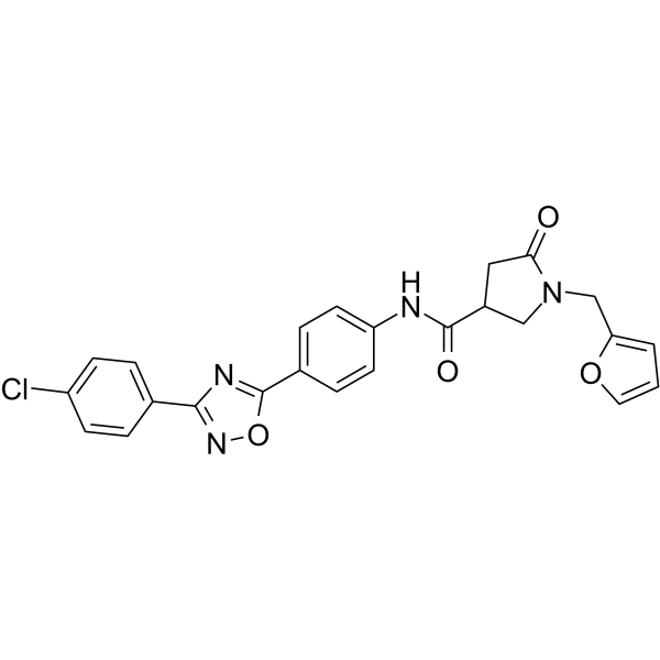
RLX-33 is a potent, selective and blood-brain barrier (BBB) penetrant relaxin family peptide 3 (RXFP3) antagonist, also blocks relaxin-3-induced ERK1/2 phosphorylation, with IC50 values of 2.36 μM for RXFP3, 7.82 and 13.86 μM for ERK1 and ERK2 phosphorylation, respectively. RLX-33 can block the stimulation of food intake induced by the RXFP3-selective agonist R3/I5 in rats. RLX-33 can be used for the research of metabolic syndrome.
For research use only. We do not sell to patients.
- Purity : 99.79%
- CAS No.: 2784577-71-3
- Formula: C24H19ClN4O4
- Molecular Weight:462.89
-
Storage:Powder -20°C, 3 years , 4°C, 2 years ; In solvent -80°C, 6 months , -20°C, 1 month

To place orders, for customer services and technical support, please contact: MedChemExpress USA
Tel: 609-228-6898 E-mail: [email protected] [email protected]


-
 Biological Activity
-
Chemical Information
-
Solvent & Solubility
- Protocols
- Purity & Documentation
- References
-
 Help & FAQs


-
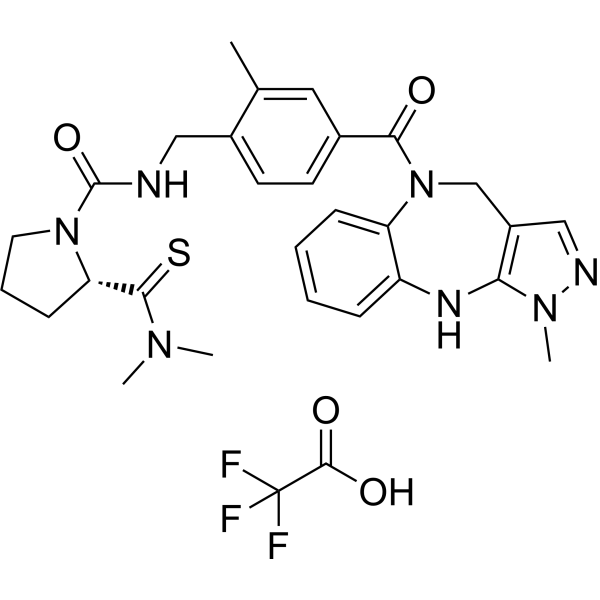
HY-124733A
LIT-001Oxytocin Receptor Agonist -
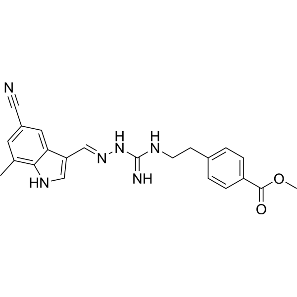
HY-145278
RXFP3/4 agonist 2RXFP3/4 Agonist -
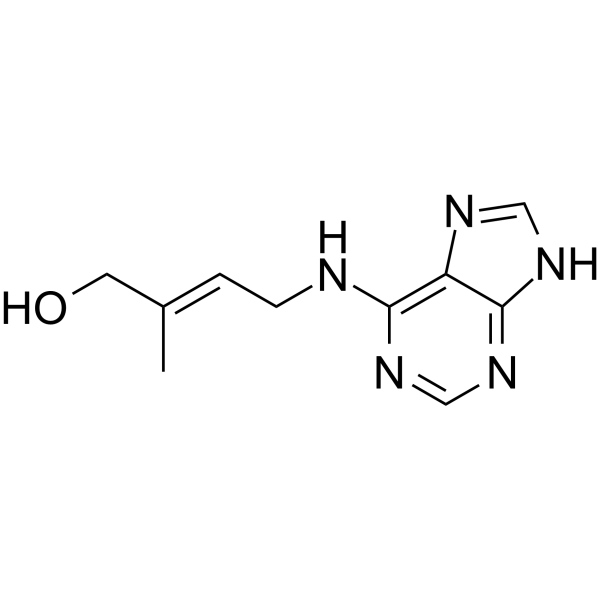
HY-19700
trans-ZeatinCytokinin -
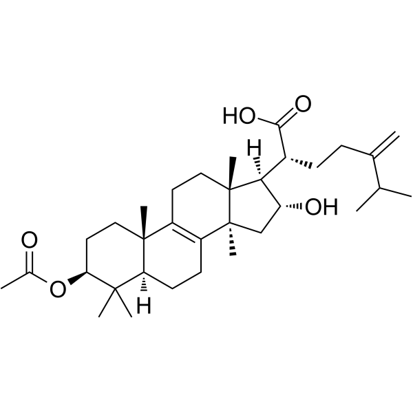
HY-N0371
Pachymic acidTriterpenoid Compound -
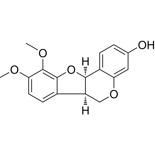
HY-N2484
MethylnissolinOsteoclast Inhibitor -
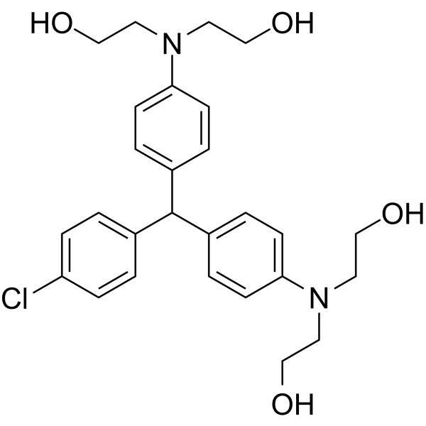
HY-104047
LM22B-10TrkB/TrkC Activator -
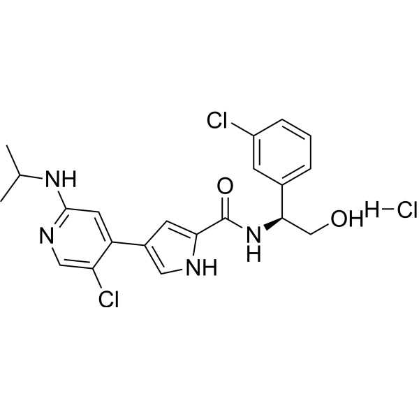
HY-15816A
Ulixertinib hydrochlorideERK1/2 Inhibitor -
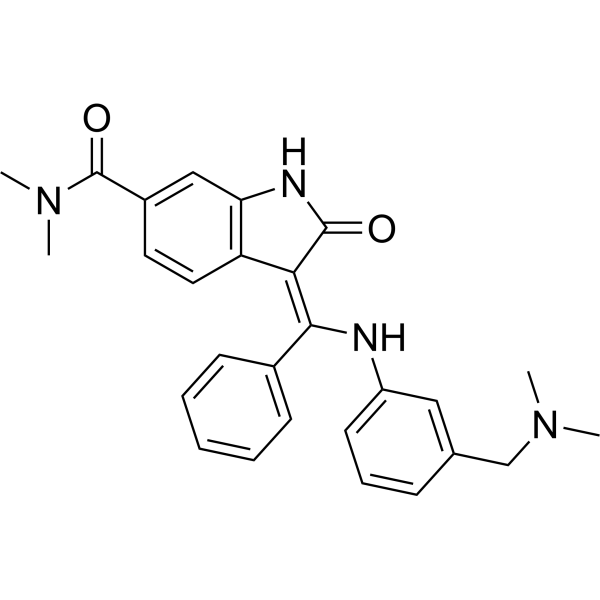
HY-12056
BIX02189MEK5 Inhibitor -
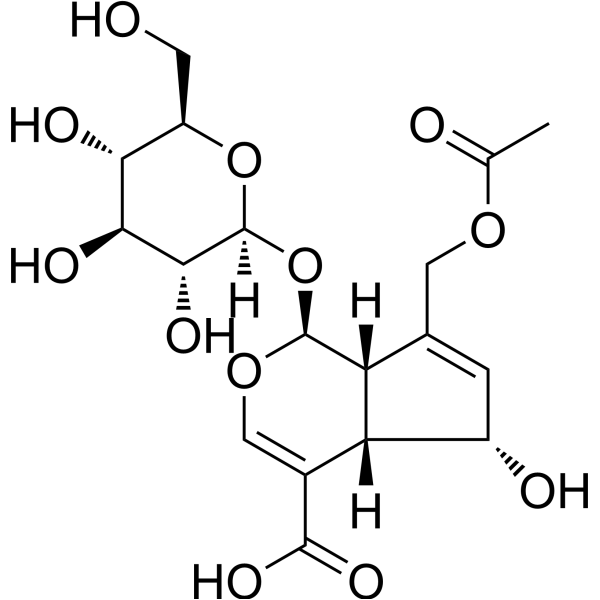
HY-N6246
Asperulosidic AcidNF-κB Inhibitor -
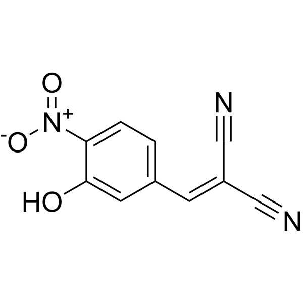
HY-108330
AG126Tyrosine Kinase Inhibitor -
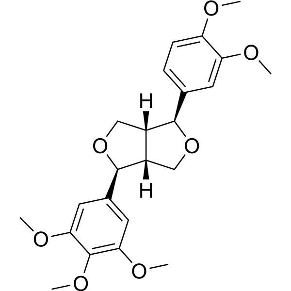
HY-N1374
MagnolinERK Inhibitor -
HY-N0590
CorynoxeineERK Inhibitor


Biological Activity
Description
IC50 & Target
IC50: 2.36 μM (RXFP3), 7.82 μM (ERK1 phosphorylation), 13.86 μM (ERK2 phosphorylation)[1]
In Vivo
RLX-33 (10 mg/kg; IP, single dosage) exhibits a good brain penetration and highly protein-bound in rats plasma[1].
Pharmacokinetic Parameters of RLX-33 in male Wistar rats (IP, 10 mg/kg)[1].
| plasma | brain | |
| Cmax (ng/mL) | 1401 | 1552 |
| tmax (h) | 0.5 | 2.0 |
| t1/2 (h) | 1.9 | 4.9 |
| AUCinf (ng/mL·h) | 5352 | 12519 |
| CL_F (mL/min/kg) | 43.8 |
MedChemExpress (MCE) has not independently confirmed the accuracy of these methods. They are for reference only.
-
Animal Model:Male Wistar rats (intracerebroventricular administration of R3/I5 stimulated food intake[1]
-
Dosage:10 mg/kg
-
Administration:IP, single dosage
-
Result:Attenuated the R3/I5-induced increase in food intake.
-
Animal Model:Male Wistar rats[1]
-
Dosage:10 mg/kg
-
Administration:IP, single dosage (Pharmacokinetic Analysis)
-
Result:Exhibited a good brain penetration and highly protein-bound, with protein binding of 99.8% in rat plasma.


Chemical Information
-
CAS No. 2784577-71-3
-
Appearance Solid
-
Molecular Weight 462.89
-
Formula C24H19ClN4O4
-
Color White to light yellow
-
SMILES
O=C(C(C1)CN(CC2=CC=CO2)C1=O)NC3=CC=C(C4=NC(C5=CC=C(Cl)C=C5)=NO4)C=C3
-
Shipping
Room temperature in continental US; may vary elsewhere.
-
Storage
Powder -20°C 3 years 4°C 2 years In solvent -80°C 6 months -20°C 1 month


Solvent & Solubility
In Vitro:
DMSO : 125 mg/mL (270.04 mM; Need ultrasonic; Hygroscopic DMSO has a significant impact on the solubility of product, please use newly opened DMSO)
Please refer to the solubility information to select the appropriate solvent. Once prepared, please aliquot and store the solution to prevent product inactivation from repeated freeze-thaw cycles.
Storage method and period of stock solution: -80°C, 6 months; -20°C, 1 month. When stored at -80°C, please use it within 6 months. When stored at -20°C, please use it within 1 month.
Please refer to the solubility information to select the appropriate solvent. Once prepared, please aliquot and store the solution to prevent product inactivation from repeated freeze-thaw cycles.
Storage method and period of stock solution: -80°C, 6 months; -20°C, 1 month. When stored at -80°C, please use it within 6 months. When stored at -20°C, please use it within 1 month.
Concentration (start) × Volume (start) = Concentration (final) × Volume (final)
In Vivo:
Select the appropriate dissolution method based on your experimental animal and administration route.
- For the following dissolution methods, please ensure to first prepare a clear stock solution using an In Vitro approach and then sequentially add co-solvents:
- To ensure reliable experimental results, the clarified stock solution can be appropriately stored based on storage conditions. As for the working solution for In Vivo experiments, it is recommended to prepare freshly and use it on the same day.
- The percentages shown for the solvents indicate their volumetric ratio in the final prepared solution. If precipitation or phase separation occurs during preparation, heat and/or sonication can be used to aid dissolution.
In Vivo Dissolution Calculator
Please enter the basic information of animal experiments:
-
-
-
-
Recommended: Prepare an additional quantity of animals to account for potential losses during experiments.
Please enter your animal formula composition:
-
%DMSO +
Recommended: Keep the proportion of DMSO in working solution below 2% if your animal is weak.
-
%+
-
+%Tween-80 + +
-
%Saline +
The co-solvents required include: DMSO, . All of co-solvents are available by MedChemExpress (MCE). , Tween 80. All of co-solvents are available by MedChemExpress (MCE).
Working solution concentration: 0.22 mg/mL
Method for preparing stock solution: mg drug dissolved in μL DMSO. Stock solution concentration: mg/mL.
1. Take μL DMSO stock solution;
2. Add μL .
μL , mix evenly;
3. Then add μL Tween 80, mix evenly;
4. Then add μL
Please ensure that the stock solution in the first step is dissolved to a clear state, and add co-solvents in sequence. You can use ultrasonic heating (ultrasonic cleaner, recommended frequency 20-40 kHz), vortexing, etc. to assist dissolution.


Purity & Documentation
-
Data Sheet (271 KB)
-
SDS (393 KB)
- English - EN (393 KB)
- Français - FR (393 KB)
- Deutsch - DE (393 KB)
- Norwegian - NO (393 KB)
- Español - ES (393 KB)
- Swedish - SV (393 KB)
- Italian - IT (393 KB)
- Korean - KR (393 KB)
- Portuguese - PT (393 KB)
-
Handling Instructions (2659 KB)
References


Complete Stock Solution Preparation Table
Please refer to the solubility information to select the appropriate solvent. Once prepared, please aliquot and store the solution to prevent product inactivation from repeated freeze-thaw cycles.
Storage method and period of stock solution: -80°C, 6 months; -20°C, 1 month. When stored at -80°C, please use it within 6 months. When stored at -20°C, please use it within 1 month.
| Optional Solvent | Concentration Solvent Mass | 1 mg | 5 mg | 10 mg | 25 mg |
|---|---|---|---|---|---|
| DMSO | 1 mM | 2.1603 mL | 10.8017 mL | 21.6034 mL | 54.0085 mL |
| 5 mM | 0.4321 mL | 2.1603 mL | 4.3207 mL | 10.8017 mL | |
| 10 mM | 0.2160 mL | 1.0802 mL | 2.1603 mL | 5.4009 mL | |
| 15 mM | 0.1440 mL | 0.7201 mL | 1.4402 mL | 3.6006 mL | |
| 20 mM | 0.1080 mL | 0.5401 mL | 1.0802 mL | 2.7004 mL | |
| 25 mM | 0.0864 mL | 0.4321 mL | 0.8641 mL | 2.1603 mL | |
| 30 mM | 0.0720 mL | 0.3601 mL | 0.7201 mL | 1.8003 mL | |
| 40 mM | 0.0540 mL | 0.2700 mL | 0.5401 mL | 1.3502 mL | |
| 50 mM | 0.0432 mL | 0.2160 mL | 0.4321 mL | 1.0802 mL | |
| 60 mM | 0.0360 mL | 0.1800 mL | 0.3601 mL | 0.9001 mL | |
| 80 mM | 0.0270 mL | 0.1350 mL | 0.2700 mL | 0.6751 mL | |
| 100 mM | 0.0216 mL | 0.1080 mL | 0.2160 mL | 0.5401 mL |


RLX-33 Related Classifications
HY-150700 Related Classifications
 Powered by Bioz