



ZL-1211
ZL-1211 is an anti-CLDN18.2 monoclonal antibody. ZL-1211 exhibits enhanced binding affinity to CD16A. ZL-1211 induces ADCC-mediated cell lysis and triggers the production of IFNγ, TNFα and IL6. ZL-1211 promotes antibody-dependent cellular cytotoxicity. ZL-1211 exerts anti-tumor activity in a mouse xenograft model of gastric cancer. ZL-1211 can be used for the research of gastric cancer.
For research use only. We do not sell to patients.
-
Storage:
Please store the product under the recommended conditions in the Certificate of Analysis.


To place orders, for customer services and technical support, please contact: MedChemExpress USA
Tel: 609-228-6898 E-mail: [email protected] [email protected]


-
 Biological Activity
-
Chemical Information
- Protocols
- Purity & Documentation
- References
-
 Help & FAQs




Biological Activity
Description
Isotype
Human IgG1 kappa
Recommend Isotype Controls
Species Reactivity
Human
IC50 & Target
[1]|
IL-6 |
In Vitro
ZL-1211 (1 h) specifically binds to CLDN18.2 on the surface of CHO cells and NUGC4 cells with overexpressed CLDN18.2[1].
ZL-1211 (serial dilution concentrations; 4-5 h) potently induces ADCC against CLDN18.2-overexpressing NUGC4-hCLDN18.2 cells via PBMC effector cells[1].
ZL-1211 (at serially diluted concentrations) potently induces ADCC effects against MIA-PaCa2-hCLDN18.2 cell clones with high, medium, and low CLDN18.2 expression via purified NK cells[1].
ZL-1211 (0.1 μg/mL; 20 min) potently induces complement-dependent cytotoxicity (CDC) against MIA-PaCa2-hCLDN18.2 cell clones with high and moderate CLDN18.2 expression via serum complement[1].
ZL-1211 (at serially diluted concentrations) potently induces antibody-dependent cell-mediated cytotoxicity (ADCC) against gastric cancer cell lines with high (SNU601), moderate (SNU620), and low (KATOIII) endogenous CLDN18.2 expression via purified NK cells at E:T ratios of 1:1 and 3:1. Its efficacy is significantly superior to that of clinical benchmark analogs, and it shows no activity against CLDN18.2-negative SNU5 cells[1].
ZL-1211 (0.01 μg/mL; 3 days) activates NK cells co-cultured with SNU601 gastric cancer cells, as evidenced by a significant increase in intracellular perforin levels compared with the control hIgG1[1].
MedChemExpress (MCE) has not independently confirmed the accuracy of these methods. They are for reference only.
-
Cell Line:CLDN18.2-overexpressing NUGC4-hCLDN18.2, peripheral blood mononuclear cell (PBMC) effector cells
-
Concentration:serially diluted concentrations (cytotoxicity assessment)
-
Incubation Time:4-5 hours (incubation at 37°C)
-
Result:Induced robust ADCC-mediated tumor cell death, with greater potency than ZL-1211 wild-type Fc and the benchmark analog.
-
Cell Line:CLDN18.2-high, -medium, -low MIA-PaCa2-hCLDN18.2
-
Concentration:0.1 μg/mL (CDC induction in high/medium clones)
-
Incubation Time:20 minutes (pre-incubation at room temperature); 30 minutes (incubation at 37°C with complement)
-
Result:Induced CDC-mediated tumor cell death in CLDN18.2-high and -medium clones at 0.1 μg/mL, while the benchmark analog required a much higher concentration to induce CDC.
In Vivo
ZL-1211 (10 mg/kg; i.p.; once weekly) exhibits significant antitumor efficacy against CLDN18.2-low-expressing KATOIII gastric cancer tumors in Balb/c nude mice[1].
ZL-1211 (5 mg/kg; i.p.; once weekly) exerts NK cell-dependent antitumor efficacy against CLDN18.2-highly expressing SNU601 gastric cancer in Balb/c nude mice[1].
ZL-1211 (50 mg/kg; i.p.; three times per week) exerts dose-dependent anti-tumor efficacy against CLDN18.2-expressing gastric cancer PDX models in Balb/c nude mice, while enhancing NK cell infiltration into responsive tumors[1].
MedChemExpress (MCE) has not independently confirmed the accuracy of these methods. They are for reference only.
-
Animal Model:Balb/c nude mice (7-9 weeks old, female)[1]
-
Dosage:0.1 mg/kg; 1 mg/kg; 10 mg/kg
-
Administration:i.p.; once weekly
-
Result:Suppressed SNU620 tumor growth at all tested doses.
Achieved significant tumor inhibition at 0.1 mg/kg, 1 mg/kg, and 10 mg/kg doses at day 42, with similar inhibition to benchmark antibody at 10 mg/kg despite having ~twice lower serum exposure than the benchmark.
-
Animal Model:Balb/c nude mice (7-9 weeks old, female)[1]
-
Dosage:10 mg/kg
-
Administration:i.p.; once weekly
-
Result:Significantly suppressed KATOIII tumor growth, with smaller mean tumor volumes compared to the benchmark antibody at day 40.
Did not inhibit growth of CLDN18.2-negative SNU5 tumors.
-
Animal Model:Balb/c nude mice; NOD.SCID mice; NCG mice[1]
-
Dosage:5 mg/kg
-
Administration:i.p.; once weekly
-
Result:Significantly suppressed SNU601 tumor growth in Balb/c nude mice, with mean tumor volumes ~50% lower than control at day 60.
Showed minimal to no tumor growth inhibition in NOD.SCID mice and NCG mice, correlating with tumor NK cell levels.
-
Animal Model:Balb/c nude mice[1]
-
Dosage:50 mg/kg
-
Administration:i.p.; three times weekly
-
Result:Induced tumor growth inhibition (TGI) in 3 of 7 PDX models: GA0006 (TGI = 56.6%), GA6831 (TGI = 56.0%), and GA2419 (TGI = 40.3%).
No significant TGI was observed in CLDN18.2-negative PDX models.
Increased NK cell density significantly in treated tumors compared to control in the responder PDX group.
- Cell Cytotoxicity Assay
- Mammalian live/dead viability and cytotoxicity staining
- Patient-Derived Xenograft (PDX)
- Protein Extraction
- Splenic/Portal-Vein Liver Metastasis Xenograft
- Subcutaneous Cell-Line-Derived Xenograft
- Orthotopic Cell-Line Xenograft
- Cell Viability Determination by MTT Colorimetric Assay
- Intraperitoneal/Peritoneal Dissemination Xenograft
Gene ID
Accession
Target
CLDN18.2
Conjugated
Unconjugated
Reconsititution
The product can be reconstituted/diluted with sterile PBS or saline.
Format
-
Product Image
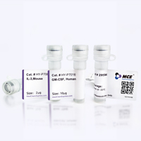
Application
ELISA, FACS, Functional assay


Chemical Information
-
Formulation
Please refer to the lot-specific COA for specific buffer information.
-
Storage
Please store the product under the recommended conditions in the Certificate of Analysis.


Purity & Documentation
References

Calculators
Concentration (start) × Volume (start) = Concentration (final) × Volume (final)


ZL-1211 Related Classifications
HY-P992487 Related Classifications
 Powered by Bioz