



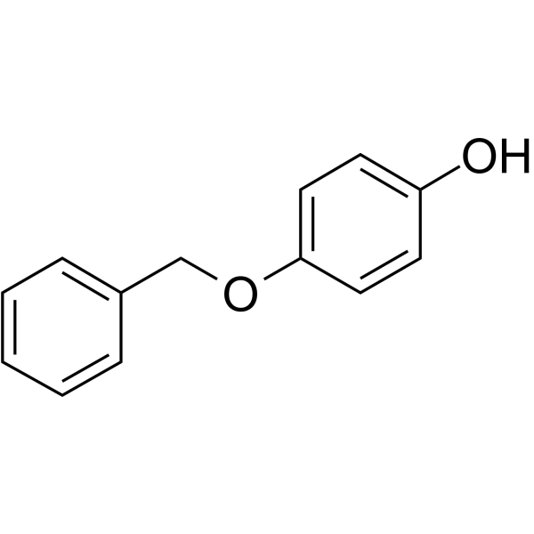
Monobenzone
Based on 2 publication(s) in Google Scholar
Monobenzone is a potent skin depigmenting agent. Monobenzone induces depigmentation and exhibits good potential for vitiligo research. Monobenzone is a potent inhibitor of RNR (Ribonucleotide reductase) enzyme activity by targeting RRM2 (a regulatory small subunit M2 of RNR) protein, and thus has significant anti-leukemia efficacy in vitro and in vivo. Monobenzone inhibits acute myeloid leukemia (AML) cells proliferation and DNA synthesis, induces cell cycle arrest, and Apoptosis .
Nur für Forschungszwecke. Wir verkaufen nicht an Patienten.
- Reinheit : 99.98%
- CAS. Nr.: 103-16-2
- Formel: C13H12O2
- Molecular Weight:200.24
-
Speicherung:Powder -20°C, 3 years , 4°C, 2 years ; In solvent -80°C, 2 years , -20°C, 1 year

To place orders, for customer services and technical support, please contact: MedChemExpress USA
Tel: 609-228-6898 E-mail: [email protected] [email protected]


-
 Biologische Aktivität
-
Chemical Information
-
Lösungsmittel & Löslichkeit
- Protokoll
- Reinheit & Dokumentation
- Verweise
-
 Help & FAQs


-
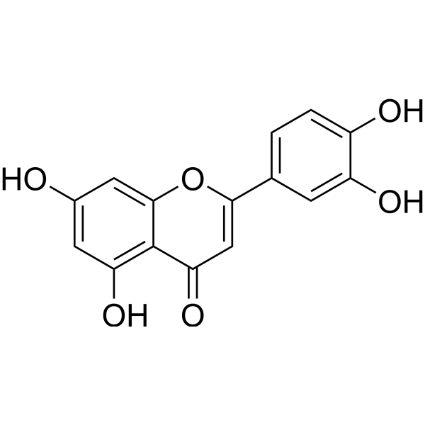
HY-N0162
LuteolinNrf2 Inhibitor -

HY-30272R
Monobenzone (Standard)Skin Depigmenting Agent -
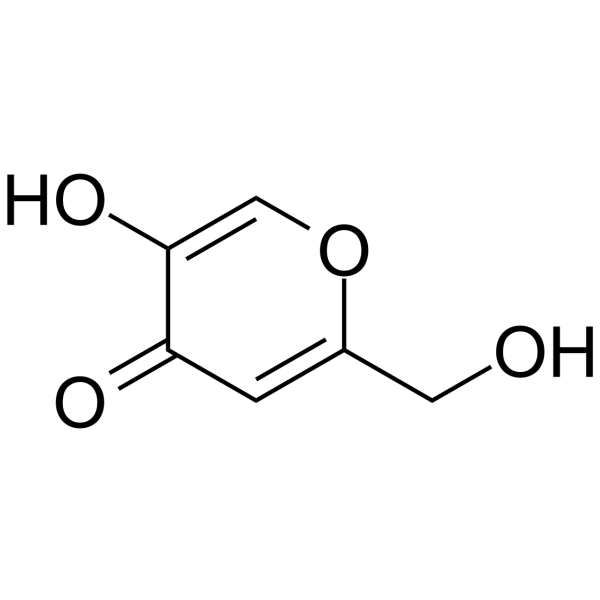
HY-W050154
Kojic acidTyrosinase Inhibitor -
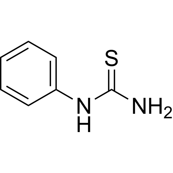
HY-Y0351
PhenylthioureaPhenoloxidase Inhibitor -
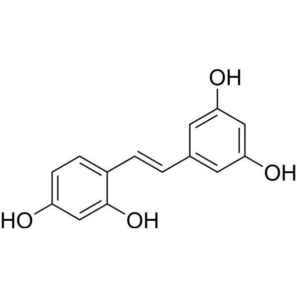
HY-N1430
OxyresveratrolAutophagy Inducer -
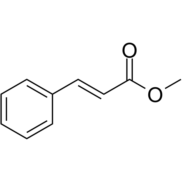
HY-W017212
Methyl cinnamateTyrosinase Inhibitor -
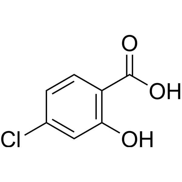
HY-W016867
4-Chlorosalicylic acid -
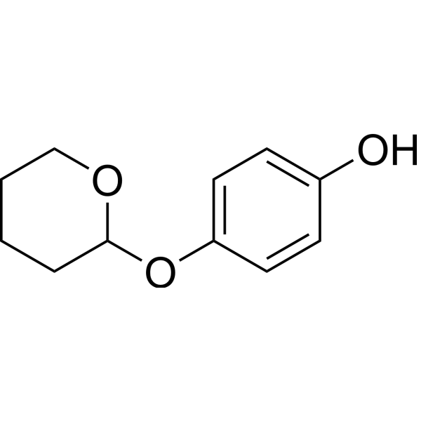
HY-B1461
DeoxyarbutinTyrosinase Inhibitor -

HY-W050154R
Kojic acid (Standard)Tyrosinase Inhibitor -

HY-W016867R
4-Chlorosalicylic acid (Standard) -

HY-W017212R
Methyl cinnamate (Standard)Tyrosinase Inhibitor -

HY-N1430R
Oxyresveratrol (Standard)Autophagy Inducer
Publications Citing Use of MedChemExpress (MCE) Monobenzone
More

Biologische Aktivität
Beschreibung
In Vitro
Monobenzone (2.5- 40 μM, 24-48 h) inhibits AML cells proliferation with IC50s of 6-18 μM and causes dose dependent DNA synthesis inhibition, induces cell cycle arrest and apoptosis in KG-1A, MOLM13, NB4 cells[2].
Monobenzone (0-10 μM, 24-48 h) blocks DNA damage repair in KG-1A, MOLM13, NB4 cells[2].
Monobenzone (0-100 μM, 72 h) overcomes the drug resistance to Ara-C (Cytarabine (HY-13605)), DOX (Doxorubicin(HY-15142)) and HU (Hydroxyurea(HY-B0313)) in KG-1A-ARAC, KG-1A-DOX, and KG-1A-HU cells[2].
Monobenzone (0-100 μM, 72 h) with the Bcl-2 inhibitor ABT-737 (HY-50907) synergistically inhibits cell the growth with an IC50 of 2.52 μM in MOLM13 cells and an IC50 of 9.69 μM in KG-1A cells compared with monobenzone alone[2].
MedChemExpress (MCE) has not independently confirmed the accuracy of these methods. They are for reference only.
-
Cell Line:Ara-C-, DOX-, and HU-resistant KG-1A cell lines (KG-1A-ARAC, KG-1A-DOX, and KG-1A-HU)
-
Concentration:3, 10, 30, 100 μM
-
Incubation Time:72 h
-
Result:Inhibited the growth of KG-1A, KG-1A-ARAC, KG-1A-DOX, and KG-1A-HU cells in a dose-dependent manner with IC50 values of 13, 16, 24, 37 μM.
-
Cell Line:KG-1A, MOLM13, NB4 cells (AML cells)
-
Concentration:0.25, 1, 4, 16, 64, 256, 1024 μM
-
Incubation Time:72 h
-
Result:Exerted a strong, dose-dependent inhibitory effect on the proliferation of treated AML cells, with IC50 values of approximately 6-18 μM.
-
Cell Line:KG-1A, MOLM13, NB4 cells
-
Concentration:2.5, 5, 10 μM
-
Incubation Time:24 h
-
Result:Induced cell cycle arrest at the S phase.
dNTP precursors significantly reversed the cell cycle arrest in S phase, suggesting that RNR (Ribonucleotide reductase) is the active target in the treated cells, resulting in the inhibition of DNA synthesis and cell proliferation.
-
Cell Line:KG-1A, MOLM13, NB4 cells
-
Concentration:0, 1.25, 2.5, 5, 10μM
-
Incubation Time:24, 48 h
-
Result:Increased r-H2Ax dose-dependently, blocking DNA damage repair in the treated cells
In Vivo
Monobenzone (100 - 200 mg/kg, i.p., daily, 2 weeks) effectively inhibits AML cell (MOLM13 cells) xenograft growth in nude mice [2].
MedChemExpress (MCE) has not independently confirmed the accuracy of these methods. They are for reference only.
-
Animal Model:Rag1 knockout and healthy C57BL/6 mice [1].
-
Dosage:40%, 60% Monobenzone cream
-
Administration:daily, 30-50 days
-
Result:Developed depigmentation on exposed site and non-exposed site.
Developed depigmentation restricted only in the drug exposure area in Rag1 KO mice.
Induced depigmentation in the mice also indicated loss of melanocytes in the lesional skin and CD8+ T infiltration in the perilesional skin.
-
Animal Model:Two hundred microlitres of PBS containing 2 *105 MOLM13 cells were injected subcutaneously into 4-week-old male nude mice[2].
-
Dosage:100, 200 mg/kg
-
Administration:i.p., daily, 2 weeks
-
Result:Significantly reduced AML cell growth in nude mice compared with the solvent control.
Inhibited AML cell growth in vivo with relatively low toxicity.
- EdU Incorporation Assay (Click Chemistry-Based DNA Synthesis Measurement)
- Detection of Apoptosis
- TUNEL staining for apoptotic DNA fragmentation
- Flow cytometric DNA-content cell-cycle staining
- Annexin V plus membrane-impermeant dye apoptosis staining
- BrdU Incorporation Assay
- Apoptosis Solutions
- Protocol for Cell Cycle
- Cell Viability Determination by MTT Colorimetric Assay


Chemical Information
-
CAS. Nr. 103-16-2
-
Appearance Solid
-
Molecular Weight 200.24
-
Formel C13H12O2
-
Color Off-white to light yellow
-
SMILES
OC1=CC=C(C=C1)OCC2=CC=CC=C2
-
Versand
Room temperature in continental US; may vary elsewhere.
-
Speicherung
Powder -20°C 3 years 4°C 2 years In solvent -80°C 2 years -20°C 1 year


Publications (2)
-
Journal Impact Factor
-
Most Recent
-
ACS Nano
Single-Molecule-Based, Label-Free Monitoring of Molecular Glue Efficacies for Promoting Protein-Protein Interactions Using YaxAB Nanopores. [Abstract]2024 Nov 12;18(45):31451-31465. PMID: 39482865 -
J Nat Med
Sappanone A exerted promising therapeutic effects in vitiligo through Wnt5a-mediated noncanonical Wnt signaling. [Abstract]2026 Jul;80(4):1152-1167. PMID: 42230481


Lösungsmittel & Löslichkeit
In Vitro:
Methanol : 400 mg/mL (1997.60 mM; Need ultrasonic)
DMSO : ≥ 100 mg/mL (499.40 mM; Hygroscopic DMSO has a significant impact on the solubility of product, please use newly opened DMSO)
* "≥" means soluble, but saturation unknown.
Please refer to the solubility information to select the appropriate solvent. Once prepared, please aliquot and store the solution to prevent product inactivation from repeated freeze-thaw cycles.
Storage method and period of stock solution: -80°C, 2 years; -20°C, 1 year. When stored at -80°C, please use it within 2 years. When stored at -20°C, please use it within 1 year.
Please refer to the solubility information to select the appropriate solvent. Once prepared, please aliquot and store the solution to prevent product inactivation from repeated freeze-thaw cycles.
Storage method and period of stock solution: -80°C, 2 years; -20°C, 1 year. When stored at -80°C, please use it within 2 years. When stored at -20°C, please use it within 1 year.
Konzentration (Stammlösung) × Volumen (Stammlösung) = Konzentration (Ziellösung) × Volumen (Ziellösung)
In Vivo:
Select the appropriate dissolution method based on your experimental animal and administration route.
- For the following dissolution methods, please ensure to first prepare a clear stock solution using an In Vitro approach and then sequentially add co-solvents:
- To ensure reliable experimental results, the clarified stock solution can be appropriately stored based on storage conditions. As for the working solution for In Vivo experiments, it is recommended to prepare freshly and use it on the same day.
- The percentages shown for the solvents indicate their volumetric ratio in the final prepared solution. If precipitation or phase separation occurs during preparation, heat and/or sonication can be used to aid dissolution.
Add each solvent one by one: 10% DMSO 40% PEG300 5% Tween-80 45% Saline
Solubility: ≥ 2.08 mg/mL (10.39 mM); Clear solution
This protocol yields a clear solution of ≥ 2.08 mg/mL (saturation unknown).
Taking 1 mL working solution as an example, add 100 μL DMSO stock solution (20.8 mg/mL) to 400 μL PEG300, and mix evenly; then add 50 μL Tween-80 and mix evenly; then add 450 μL Saline to adjust the volume to 1 mL.
Preparation of Saline: Dissolve 0.9 g sodium chloride in ddH₂O and dilute to 100 mL to obtain a clear Saline solution.
Add each solvent one by one: 10% DMSO 90% (20% SBE-β-CD in Saline)
Solubility: ≥ 2.08 mg/mL (10.39 mM); Clear solution
This protocol yields a clear solution of ≥ 2.08 mg/mL (saturation unknown).
Taking 1 mL working solution as an example, add 100 μL DMSO stock solution (20.8 mg/mL) to 900 μL 20% SBE-β-CD in Saline, and mix evenly.
Preparation of 20% SBE-β-CD in Saline (4°C, storage for one week): 2 g SBE-β-CD powder is dissolved in 10 mL Saline, completely dissolve until clear.
In Vivo Dissolution Calculator
Please enter the basic information of animal experiments:
-
-
-
-
Recommended: Prepare an additional quantity of animals to account for potential losses during experiments.
Please enter your animal formula composition:
-
%DMSO +
Recommended: Keep the proportion of DMSO in working solution below 2% if your animal is weak.
-
%+
-
+%Tween-80 + +
-
%Saline +
The co-solvents required include: DMSO, . All of co-solvents are available by MedChemExpress (MCE). , Tween 80. All of co-solvents are available by MedChemExpress (MCE).
Working solution concentration: 0.22 mg/mL
Method for preparing stock solution: mg drug dissolved in μL DMSO. Stock solution concentration: mg/mL.
1. Take μL DMSO stock solution;
2. Add μL .
μL , mix evenly;
3. Then add μL Tween 80, mix evenly;
4. Then add μL
Please ensure that the stock solution in the first step is dissolved to a clear state, and add co-solvents in sequence. You can use ultrasonic heating (ultrasonic cleaner, recommended frequency 20-40 kHz), vortexing, etc. to assist dissolution.


Reinheit & Dokumentation
-
Data Sheet (279 KB)
-
SDS (393 KB)
- English - EN (393 KB)
- Français - FR (393 KB)
- Deutsch - DE (393 KB)
- Norwegian - NO (393 KB)
- Español - ES (393 KB)
- Swedish - SV (393 KB)
- Italian - IT (393 KB)
- Korean - KR (393 KB)
- Portuguese - PT (393 KB)
-
Handling Instructions (2659 KB)
Verweise
[1]. Zhu Y, et al. A mouse model of vitiligo induced by monobenzone. Exp Dermatol. 2013 Jul;22(7):499-501. doi: 10.1111/exd.12184. [Content Brief]
[2]. Dong J, et al. Identification of Monobenzone as a Novel Potential Anti-Acute Myeloid Leukemia Agent That Inhibits RNR and Suppresses Tumour Growth in Mouse Xenograft Model. Cancers (Basel). 2022 Sep 27;14(19):4710. [Content Brief]


Complete Stock Solution Preparation Table
Please refer to the solubility information to select the appropriate solvent. Once prepared, please aliquot and store the solution to prevent product inactivation from repeated freeze-thaw cycles.
Storage method and period of stock solution: -80°C, 2 years; -20°C, 1 year. When stored at -80°C, please use it within 2 years. When stored at -20°C, please use it within 1 year.
| Optional Solvent | Concentration Solvent Mass | 1 mg | 5 mg | 10 mg | 25 mg |
|---|---|---|---|---|---|
| DMSO / Methanol | 1 mM | 4.9940 mL | 24.9700 mL | 49.9401 mL | 124.8502 mL |
| 5 mM | 0.9988 mL | 4.9940 mL | 9.9880 mL | 24.9700 mL | |
| 10 mM | 0.4994 mL | 2.4970 mL | 4.9940 mL | 12.4850 mL | |
| 15 mM | 0.3329 mL | 1.6647 mL | 3.3293 mL | 8.3233 mL | |
| 20 mM | 0.2497 mL | 1.2485 mL | 2.4970 mL | 6.2425 mL | |
| 25 mM | 0.1998 mL | 0.9988 mL | 1.9976 mL | 4.9940 mL | |
| 30 mM | 0.1665 mL | 0.8323 mL | 1.6647 mL | 4.1617 mL | |
| 40 mM | 0.1249 mL | 0.6243 mL | 1.2485 mL | 3.1213 mL | |
| 50 mM | 0.0999 mL | 0.4994 mL | 0.9988 mL | 2.4970 mL | |
| 60 mM | 0.0832 mL | 0.4162 mL | 0.8323 mL | 2.0808 mL | |
| 80 mM | 0.0624 mL | 0.3121 mL | 0.6243 mL | 1.5606 mL | |
| 100 mM | 0.0499 mL | 0.2497 mL | 0.4994 mL | 1.2485 mL |


Monobenzone Related Classifications
HY-30272 Related Classifications
 Powered by Bioz