



Elrelesertib
Based on 13 publication(s) in Google Scholar
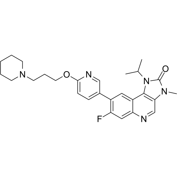
Elrelesertib (AZD1390) is an orally active, brain-penetrant ATM kinase inhibitor with IC50 values of 0.78 nM. Elrelesertib blocks ATM-dependent DNA damage response, inhibits ATM autophosphorylation and downstream Chk2, Rad50 phosphorylation, and accumulates at DNA breaks. Elrelesertib acts as a radiosensitizer, induces apoptosis, genomic instability, G2-M cell cycle arrest, ROS elevation, and mitochondrial membrane potential alteration. Elrelesertib has low efflux liability against P-gp and BCRP. Elrelesertib can be used for the research of central nervous system malignancies, glioblastoma multiforme, glioma, lung cancer brain metastases, and breast cancer.
Nur für Forschungszwecke. Wir verkaufen nicht an Patienten.
- Reinheit : 99.22%
- CAS. Nr.: 2089288-03-7
- Formel: C27H32FN5O2
- Molecular Weight:477.57
-
Speicherung:Powder -20°C, 3 years , 4°C, 2 years ; In solvent -80°C, 1 year , -20°C, 6 months

To place orders, for customer services and technical support, please contact: MedChemExpress USA
Tel: 609-228-6898 E-mail: [email protected] [email protected]


-
 Biologische Aktivität
-
Chemical Information
-
Lösungsmittel & Löslichkeit
- Protokoll
- Reinheit & Dokumentation
- Verweise
-
 Help & FAQs


-
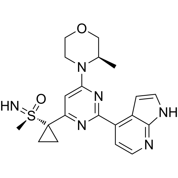
HY-19323
CeralasertibATR Inhibitor -
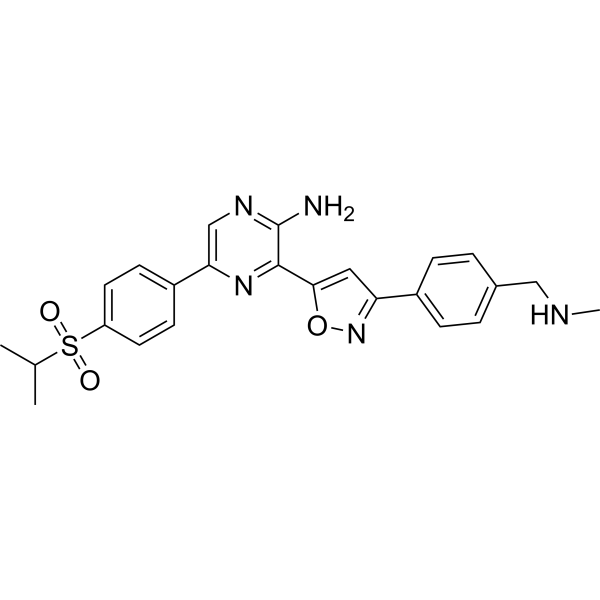
HY-13902
BerzosertibATR Inhibitor -
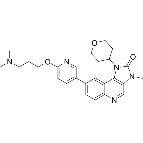
HY-100016
AZD0156ATM Inhibitor -
HY-12016
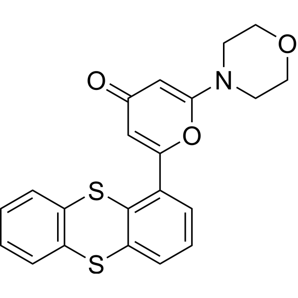
KU-55933ATM Inhibitor -
HY-10162
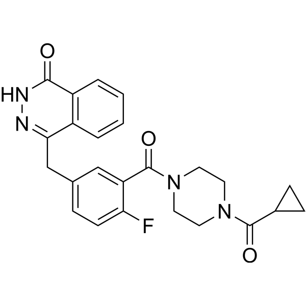
OlaparibPARP Inhibitor -
HY-111783
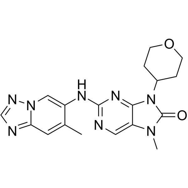
AZD-7648DNA-PK Inhibitor -
HY-101566
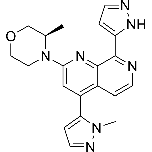
ElimusertibATR Inhibitor -
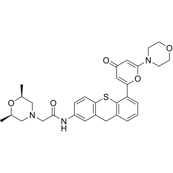
HY-12061
KU-60019ATM/ATR Inhibitor -
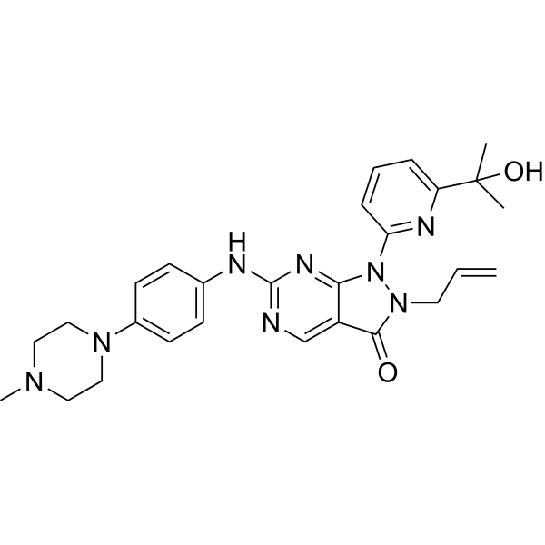
HY-10993
AdavosertibWee1 Inhibitor -
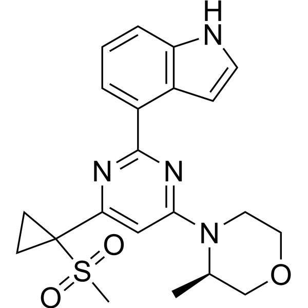
HY-15557
AZ20ATR Inhibitor -
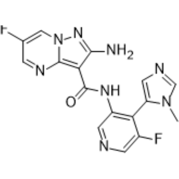
HY-111451
TuvusertibATR Inhibitor -
HY-101570
NedisertibDNA-PK Inhibitor
Publications Citing Use of MedChemExpress (MCE) Elrelesertib
More- Signal Transduct Target Ther. 2025 Jun 13;10(1):185. [Abstract]
- Nat Nanotechnol. 2021 Jul;16(7):830-839. [Abstract]
- Sci Adv. 2026 May 22;12(21):eaec2231. [Abstract]
- EMBO J. 2023 Mar 15;42(6):e112094. [Abstract]
- Int J Radiat Oncol Biol Phys. 2023 Mar 15;115(4):957-971. [Abstract]
- J Med Chem. 2024 May 9;67(9):7620-7634. [Abstract]
- Mol Cancer Ther. 2026 Jul 15:10.1158/1535-7163.MCT-26-0182.
- CNS Neurosci Ther. 2024 Apr;30(4):e14696. [Abstract]
- Cancers (Basel). 2026 Jul 23;18(15):2381. [Abstract]
- Shock. 2023 Jul 1;60(1):100-109. [Abstract]
- EANM Innovation. 2026 May 19;3:100274.
- University of Califomia San Francisco. 2026.
- bioRxiv. 2025 Jul 31.
 Customer Validation & Images

-
WB
-
Cell Imaging/Staining
-
In Vivo Efficacy Study
-
IP
-
Others


Biologische Aktivität
Beschreibung
IC50 & Target
[1]|
ATM 0.78 nM (IC50) |
Chk2 |
Cellular Effect
|
Cell Line

|
Type | Value | Description | References |
|---|---|---|---|---|
| NCI-H2228 | IC50 |
0.78 nM
|
Inhibition of cellular ATM activity in human NCI-H2228 p53 mutant lung cancer cells, measured via high-content immunofluorescence imaging assay of DDR biomarkers with 1-hour pre-incubation and analysis up to 48 hours post-irradiation.
Inhibition of cellular ATM activity in human NCI-H2228 p53 mutant lung cancer cells, measured via high-content immunofluorescence imaging assay of DDR biomarkers with 1-hour pre-incubation and analysis up to 48 hours post-irradiation.
|
29938225 |
| MCF7 | IC50 |
16.143 μM
|
Reduction in cell viability against human breast cancer MCF-7 cells incubated for 12 hrs by WST-1 assay.
Reduction in cell viability against human breast cancer MCF-7 cells incubated for 12 hrs by WST-1 assay.
|
39681282 |
| MDA-MB-231 | IC50 |
5.524 μM
|
Reduction in cell viability against human breast cancer MDA-MB-231 cells incubated for 12 hrs by WST-1 assay.
Reduction in cell viability against human breast cancer MDA-MB-231 cells incubated for 12 hrs by WST-1 assay.
|
39681282 |
| MCF7 | IC50 |
10.685 μM
|
Reduction in cell viability against human breast cancer MCF-7 cells incubated for 24 hrs by WST-1 assay.
Reduction in cell viability against human breast cancer MCF-7 cells incubated for 24 hrs by WST-1 assay.
|
39681282 |
| MDA-MB-231 | IC50 |
2.563 μM
|
Reduction in cell viability against human breast cancer MDA-MB-231 cells incubated for 24 hrs by WST-1 assay.
Reduction in cell viability against human breast cancer MDA-MB-231 cells incubated for 24 hrs by WST-1 assay.
|
39681282 |
In Vitro
Elrelesertib (10-100 nM; 1 h pre-incubation, 1 h/6 h post-irradiation, 6 h post-washout) dose-dependently modulates ATM pathway activity in NCI-H2228 p53 mutant lung cancer cells, with pathway reactivation observed 6 hours after washout[1].
Elrelesertib (0.125-1250 nM; 1 h pre-incubation, 1/6/24/48 h post-irradiation) inhibits DDR biomarkers in NCI-H2228 p53G12C mutant lung cancer cells, with an XC50 of 2.7 nM for discrete phospho-ATM foci, and dose-dependently increases micronuclei formation when combined with radiation[1].
Elrelesertib (10 nM; 1 h pre-incubation, 12-14 days post-irradiation) potently radiosensitizes p53 mutant GBM and lung cancer cell lines more effectively than p53 wild-type cell lines in clonogenic survival assays[1].
Elrelesertib (10 nM; 1 h pre-incubation, 5 days post-irradiation) potently radiosensitizes p53 mutant GBM cell lines more effectively than p53 wild-type GBM cell lines in a high-throughput Live/Dead antiproliferation assay[1].
MedChemExpress (MCE) has not independently confirmed the accuracy of these methods. They are for reference only.
-
Cell Line:LN18 glioblastoma (GBM) cells (p53 mutant)
-
Concentration:3, 10, 300 nM
-
Incubation Time:1 h (pre-incubation); 4 h (post-irradiation)
-
Result:Dose-dependently inhibited ATM autophosphorylation (phospho-ATM Ser1981) at 4 hours post-treatment, with strong inhibition observed at 3 nM.
Did not affect phosphorylation of non-ATM pathway markers including KAP1, CDK1, or p53.
-
Cell Line:NCI-H2228 lung cancer cells (p53 mutant)
-
Concentration:10, 100 nM
-
Incubation Time:1 h (pre-incubation); 1, 6 h (post-irradiation); 6 h (post-washout)
-
Result:Dose-dependently inhibited phospho-KAP1 Ser824 and phospho-Chk2 Thr68 at 1 and 6 hours post-treatment.
Began to rise phospho-Chk2 levels at 6 hours after washout.
-
Cell Line:NCI-H2228 lung cancer cells (p53 mutant)
-
Concentration:0.125, 0.3125, 1, 3, 12.5, 31.25, 100, 300, 1250 nM
-
Incubation Time:1 h (pre-incubation); 1, 6, 24, 48 h (post-irradiation)
-
Result:Achieved XC50 values of 0.78 nM for inhibition of total nuclear phospho-ATM and γH2AX staining, and 2.7 nM for inhibition of discrete nuclear phospho-ATM foci.
Dose-dependently increased micronuclei formation when combined with radiation.
-
Cell Line:NCI-H2228 lung cancer cells (p53 mutant)
-
Concentration:0.125, 0.3125, 1, 3, 12.5, 31.25,100 nM
-
Incubation Time:1 h (pre-incubation); 24, 48 h (post-irradiation)
-
Result:Combined with 2 Gy IR, caused a dose-dependent increase in G2 phase accumulation at 24 hours.
Combined with 2 Gy IR, caused a dose-dependent increase in sub-G1 population at 48 hours, indicating induction of apoptosis.
-
Cell Line:GBM and lung cancer cell lines (LN18, T98G, NCI-H2228, A172, DDBRTG-05MG, U87MG)
-
Concentration:10 nM
-
Incubation Time:1 h (pre-incubation); 12-14 days (post-irradiation)
-
Result:Radiosensitized p53 mutant cell lines more potently than p53 wild-type lines.
-
Cell Line:GBM cell lines (LN18, T98G, HS683T, SW1088, SW1783, DDBRTG-05MG, CCF-STTG1, A172, U87-MG)
-
Concentration:10 nM
-
Incubation Time:1 h (pre-incubation); 5 days (post-irradiation)
-
Result:Radiosensitized p53 mutant cell lines more potently than p53 wild-type lines.
-
Cell Line:human breast cancer cell lines MCF-7, MDA-MB-231, human non-transformed epithelial cell line RPE-1
-
Concentration:0.2, 0.4, 0.6, 0.8, 1 μM (MDA-MB-231, RPE-1); 1, 2, 3, 4, 5 μM (MCF-7)
-
Incubation Time:24 h
-
Result:Caused a dose-dependent reduction in cell proliferation in all three cell lines over 24 h.
Induced significant effects at the highest three concentrations for each cell line compared to controls.
-
Cell Line:human breast cancer cell lines MCF-7, MDA-MB-231, human non-transformed epithelial cell line RPE-1
-
Concentration:0.2, 0.6, 1 μM (MDA-MB-231, RPE-1); 1, 3, 5 μM (MCF-7)
-
Incubation Time:24 h
-
Result:Caused a dose-dependent increase in the proportion of cells in the G2-M phase and a corresponding decrease in the S phase in all three cell lines compared to untreated controls.
Indicated cell cycle arrest at the G2-M checkpoint.
-
Cell Line:human breast cancer cell lines MCF-7, MDA-MB-231, human non-transformed epithelial cell line RPE-1
-
Concentration:0.2, 0.6, 1 μM (RPE-1); 0.2, 0.65, 1 μM (MDA-MB-231); 1, 3, 5 μM (MCF-7)
-
Incubation Time:24 h
-
Result:Caused a dose-dependent increase in early and late apoptotic cells, as well as necrotic cells, in all three cell lines compared to DMSO-treated controls.
Induced statistically significant increases in apoptotic cell percentage in breast cancer cell lines.
Parmacokinetics
| Species | Dose | Route | CL | Vss | T1/2 | Bioavailability |
|---|---|---|---|---|---|---|
| Rat[1] | 10 mg/kg | p.o. | 16.3 mL/min/kg | 3.0 L/kg | 2.4 h | 74 % |
In Vivo
Elrelesertib (2-20 mg/kg; p.o.; single dose) inhibits IR-induced ATM pathway activation in a dose- and time-dependent manner and induces apoptosis in a mouse model of intracranial lung cancer brain metastasis, with free brain exposure correlating with pharmacodynamic activity[1].
Elrelesertib (2-20 mg/kg; p.o.; q.d; 4 days) combined with daily 2.5 Gy IR provides dose-dependent tumor growth inhibition and survival benefit in a mouse model of lung cancer brain metastasis established via intracarotid artery injection[1].
Elrelesertib (5-20 mg/kg; p.o.; q.d) combined with stereotactic IR significantly improves survival in an immunocompetent mouse model of intracranial glioblastoma, with a dose-dependent efficacy response observed[1].
Elrelesertib (20 mg/kg; p.o.; q.d; 5 days) combined with daily 2 Gy IR produces robust tumor growth inhibition and survival benefit in a panel of subcutaneous glioblastoma PDX models, with p53 mutant models showing particularly strong responses[1].
MedChemExpress (MCE) has not independently confirmed the accuracy of these methods. They are for reference only.
-
Animal Model:Immunodeficient nude mice (6-8 weeks old)[1]
-
Dosage:5, 20 mg/kg
-
Administration:p.o.; q.d for 4 days; b.i.d for 4 days
-
Result:Showed marginal tumor growth inhibition (TGI) at 5 mg/kg q.d plus IR.
Produced superior TGI and significantly improved survival compared to IR alone at 20 mg/kg q.d or b.i.d plus IR.
Induced a statistically significant increase in the apoptotic biomarker cleaved caspase-3 (CC3) compared to IR alone.
-
Animal Model:Immunodeficient nude mice (6-8 weeks old)[1]
-
Dosage:5, 15 mg/kg
-
Administration:p.o.; q.d for 5 days
-
Result:The triplet combination of 15 mg/kg q.d, 2 Gy ×5 IR, and 25 mg/kg TMZ q.d was well-tolerated, provided additive TGI and survival benefit over the 15 mg/kg plus IR doublet arm.
-
Animal Model:Immunodeficient nude mice (6-8 weeks old)[1]
-
Dosage:2, 5, 20 mg/kg
-
Administration:p.o.; q.d for 4 days
-
Result:Produced dose-dependent TGI and survival benefit compared to IR alone.
Showed the most pronounced efficacy in reducing tumor growth and extending survival at 20 mg/kg dose.
-
Animal Model:C57 Black 6 mice[1]
-
Dosage:5, 20 mg/kg
-
Administration:p.o.; q.d for the duration of IR dosing
-
Result:Significantly improved survival compared to IR or Elrelesertib alone at 20 mg/kg q.d combined with two 5-Gy IR fractions.
Significantly improved survival compared to IR alone at 5 mg/kg q.d combined with ten 2-Gy IR fractions, though efficacy was reduced compared to the 20 mg/kg dose.
-
Animal Model:NMRI nude mice (female, 7-11 weeks old)[1]
-
Dosage:20 mg/kg
-
Administration:p.o.; q.d for 5 days
-
Result:Produced robust tumor growth inhibition and survival benefit across the PDX panel.
Showed particularly strong responses in p53 mutant models (ST108, ST112), while the least responsive model was p53 wild-type (ST2473).
Several p53 wild-type models also showed meaningful responses to the combination.
-
Animal Model:Immunodeficient nude mice[1]
-
Dosage:2, 5, 20 mg/kg
-
Administration:p.o.; single dose
-
Result:Inhibited IR-induced phospho-ATM (Ser1981) in a dose- and time-dependent manner, with 20 mg/kg producing the highest and most sustained inhibition (peaking at approximately 95% inhibition at 3 hours post-IR).
Reduced IR-induced phospho-Rad50 (Ser635) by up to 86% and induced statistically significant increases in apoptotic biomarker CC3 compared to IR alone.
Free brain concentrations peaked within 1 hour of dosing and correlated with ATM inhibition activity.
- Western Blot
- Mitochondrial membrane-potential fluorescent assay
- Annexin V plus membrane-impermeant dye apoptosis staining
- BrdU Incorporation Assay
- ROS/oxidative-stress fluorescent staining
- Protocol for Cell Cycle
- Breast Cancer Modeling
- Kinase activity and phosphorylation assays
- Detection of Apoptosis
- TUNEL staining for apoptotic DNA fragmentation
- Flow cytometric DNA-content cell-cycle staining
- Fluorescent plasma-membrane potential dye assay
- Mitochondrial membrane-potential and mitochondrial mass staining
- Apoptosis Solutions
- Protocol for Kinase activity and phosphorylation assays
- Genotoxicity/Mutagenicity Study
Clinical Trial
| NCT Number | Sponsor | Condition | Start Date |
Phase
|
|---|---|---|---|---|
| NCT01329991 | 2011-05 | PHASE1 |


Chemical Information
-
CAS. Nr. 2089288-03-7
-
Appearance Solid
-
Molecular Weight 477.57
-
Formel C27H32FN5O2
-
Color White to off-white
-
SMILES
O=C(N1C(C)C)N(C)C2=C1C3=CC(C4=CC=C(OCCCN5CCCCC5)N=C4)=C(F)C=C3N=C2
-
Synonyms
AZD1390
-
Versand
Room temperature in continental US; may vary elsewhere.
-
Speicherung
Powder -20°C 3 years 4°C 2 years In solvent -80°C 1 year -20°C 6 months


Publications (13)
-
Journal Impact Factor
-
Most Recent
-
Signal Transduct Target Ther
Dual ENPP1/ATM depletion blunts DNA damage repair boosting radioimmune efficacy to abrogate triple-negative breast cancer. [Abstract]2025 Jun 13;10(1):185. PMID: 40506449
Elrelesertib purchased from MedChemExpress. Usage Cited in: Signal Transduct Target Ther. 2025 Jun 13;10(1):185. [Abstract]
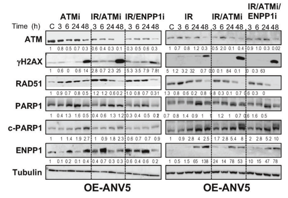
Immunoblot analysis was performed on cell lysates extracted from ANV5-OE cells at different time points after treatment with IR (2 Gy), IR/ENPP1i (5 µM), IR/ATMi (AZD1390: 5 µM), or triple therapy to detect the protein expression levels of γHA2X, PARP1, cleaved PARP1 (c-PARP), ENPP1, RAD51, GAPDH, and Tubulin.
Elrelesertib purchased from MedChemExpress. Usage Cited in: Signal Transduct Target Ther. 2025 Jun 13;10(1):185. [Abstract]
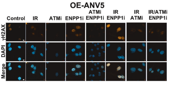
The number of positive cells labeled with anti-γH2AX antibody was quantitatively assessed. Cells were incubated with ENPP1i (5 µM) and ATMi (AZD1390: 5 µM) for 24 h.
Elrelesertib purchased from MedChemExpress. Usage Cited in: Signal Transduct Target Ther. 2025 Jun 13;10(1):185. [Abstract]
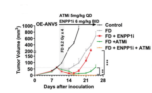
After orthotopic transplantation of OE-ANV5 cells, mice were treated with FD (6.2 Gy × 4) alone, or in combination with ENPP1i (6 mg/kg, twice daily), ATMi (AZD1390: 5 mg/kg, once daily), or triple therapy (n = 8 mice per group), and changes in tumor volume were observed.
-
Nat Nanotechnol
Therapeutically reprogrammed nutrient signalling enhances nanoparticulate albumin bound drug uptake and efficacy in KRAS-mutant cancer. [Abstract]2021 Jul;16(7):830-839. PMID: 33958764 -
Sci Adv
53BP1 orchestrates sequence feature of RAG targets to balance DNA repair outcomes during V(D)J recombination. [Abstract]2026 May 22;12(21):eaec2231. PMID: 42172328 -
EMBO J
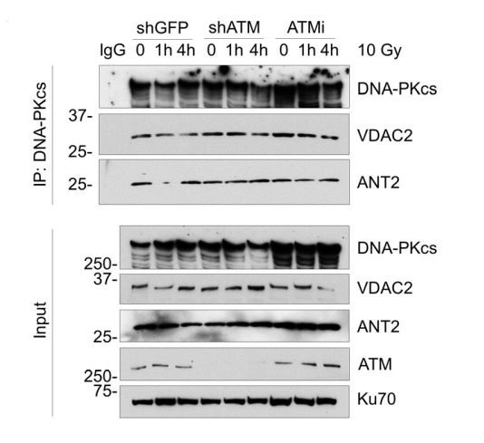
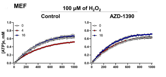
DNA-PKcs and ATM modulate mitochondrial ADP-ATP exchange as an oxidative stress checkpoint mechanism. [Abstract]2023 Mar 15;42(6):e112094. PMID: 36727301
Elrelesertib purchased from MedChemExpress. Usage Cited in: EMBO J. 2023 Mar 15;42(6):e112094. [Abstract]
HeLa cells expressing Flag‐tagged VDAC2 and V5‐tagged ANT2 were subjected to Co‐IP using anti‐Flag antibody and analyzed by western blot as indicated. ANT2, but not VDAC2, temporarily dissociated from DNA‐PKcs after treatment of 10 Gy IR. ATM knockdown and inhibition (AZD1390, 10 nM) disrupted the dissociation.
Elrelesertib purchased from MedChemExpress. Usage Cited in: EMBO J. 2023 Mar 15;42(6):e112094. [Abstract]
Primary mouse embryonic fibroblasts (MEF) RPE-1 were analyzed for ADP-ATP exchange activity at different time points (up to 16 hours) in response to 100 μM H₂O₂. The ATM inhibitor (AZD1390, 10 nM) was also included in the parallel analysis.
-
Int J Radiat Oncol Biol Phys
Radiation-Induced Remodeling of the Tumor Microenvironment Through Tumor Cell-Intrinsic Expression of cGAS-STING in Esophageal Squamous Cell Carcinoma. [Abstract]2023 Mar 15;115(4):957-971. PMID: 36368436 -
J Med Chem
Discovery of a Meisoindigo-Derived PROTAC as the ATM Degrader: Revolutionizing Colorectal Cancer Therapy via Synthetic Lethality with ATR Inhibitors. [Abstract]2024 May 9;67(9):7620-7634. PMID: 38634707 -
-
CNS Neurosci Ther
AZD1390, an ataxia telangiectasia mutated inhibitor, attenuates microglia-mediated neuroinflammation and ischemic brain injury. [Abstract]2024 Apr;30(4):e14696. PMID: 38668740 -
Cancers (Basel)
Molecular Characterization of PARP Inhibitor Response Reveals Co-Targeting Strategies in Advanced Prostate Cancer. [Abstract]2026 Jul 23;18(15):2381. PMID: 42588601 -
Shock
ATAXIA TELANGIECTASIA MUTATED PROTECTS AGAINST LIPOPOLYSACCARIDE-INDUCED BLOOD-BRAIN BARRIER DISRUPTION BY REGULATING ATK/DRP1-MEDIATED MITOCHONDRIAL HOMEOSTASIS. [Abstract]2023 Jul 1;60(1):100-109. PMID: 37141173 -
-
-


Lösungsmittel & Löslichkeit
In Vitro:
DMSO : 5 mg/mL (10.47 mM; ultrasonic and warming and heat to 60°C; Hygroscopic DMSO has a significant impact on the solubility of product, please use newly opened DMSO)
Please refer to the solubility information to select the appropriate solvent. Once prepared, please aliquot and store the solution to prevent product inactivation from repeated freeze-thaw cycles.
Storage method and period of stock solution: -80°C, 1 year; -20°C, 6 months. When stored at -80°C, please use it within 1 year. When stored at -20°C, please use it within 6 months.
Please refer to the solubility information to select the appropriate solvent. Once prepared, please aliquot and store the solution to prevent product inactivation from repeated freeze-thaw cycles.
Storage method and period of stock solution: -80°C, 1 year; -20°C, 6 months. When stored at -80°C, please use it within 1 year. When stored at -20°C, please use it within 6 months.
Konzentration (Stammlösung) × Volumen (Stammlösung) = Konzentration (Ziellösung) × Volumen (Ziellösung)
In Vivo:
For the following dissolution methods, please prepare the working solution directly:
It is recommended to prepare fresh solutions and use them promptly within a short period of time.
The percentages shown for the solvents indicate their volumetric ratio in the final prepared solution. If precipitation or phase separation occurs during preparation, heat and/or sonication can be used to aid dissolution.


Reinheit & Dokumentation
-
Data Sheet (300 KB)
-
SDS (393 KB)
- English - EN (393 KB)
- Français - FR (393 KB)
- Deutsch - DE (393 KB)
- Norwegian - NO (393 KB)
- Español - ES (393 KB)
- Swedish - SV (393 KB)
- Italian - IT (393 KB)
- Korean - KR (393 KB)
- Portuguese - PT (393 KB)
-
Handling Instructions (2659 KB)
Verweise


Complete Stock Solution Preparation Table
Please refer to the solubility information to select the appropriate solvent. Once prepared, please aliquot and store the solution to prevent product inactivation from repeated freeze-thaw cycles.
Storage method and period of stock solution: -80°C, 1 year; -20°C, 6 months. When stored at -80°C, please use it within 1 year. When stored at -20°C, please use it within 6 months.
| Optional Solvent | Concentration Solvent Mass | 1 mg | 5 mg | 10 mg | 25 mg |
|---|---|---|---|---|---|
| DMSO | 1 mM | 2.0939 mL | 10.4697 mL | 20.9393 mL | 52.3483 mL |
| 5 mM | 0.4188 mL | 2.0939 mL | 4.1879 mL | 10.4697 mL | |
| 10 mM | 0.2094 mL | 1.0470 mL | 2.0939 mL | 5.2348 mL |


Elrelesertib Related Classifications
HY-109566 Related Classifications
 Powered by Bioz