



Ampiroxicam
Based on 2 publication(s) in Google Scholar
Ampiroxicam is an orally active prodrug of non-steroidal anti-inflammatory drug (NSAID). Ampiroxicam inhibits paw swelling in adjuvant-induced arthritis and acute inflammation models, and suppresses phenylbenzoquinone-induced stretching responses in mice. Ampiroxicam is rapidly and completely converted to Piroxicam (HY-B0253) via non-specific esterases and first-pass metabolism. Ampiroxicam induces contact hypersensitivity and photosensitivity reactions through photoproducts generated by UVA. Ampiroxicam can be used in research related to photosensitivity, adjuvant-induced arthritis, rheumatism, osteoarthritis and other inflammatory diseases.
For research use only. We do not sell to patients.
- Purity : 98.73%
- CAS No.: 99464-64-9
- Formula: C20H21N3O7S
- Molecular Weight:447.46
-
Storage:Powder -20°C, 3 years , 4°C, 2 years ; In solvent -80°C, 2 years , -20°C, 1 year

To place orders, for customer services and technical support, please contact: MedChemExpress USA
Tel: 609-228-6898 E-mail: [email protected] [email protected]


-
 Biological Activity
-
Chemical Information
-
Solvent & Solubility
- Protocols
- Purity & Documentation
- References
-
 Help & FAQs


-
HY-B0261
MeloxicamCOX Inhibitor -
HY-B0253
PiroxicamCOX Inhibitor -
HY-B0367
LornoxicamCOX Inhibitor -
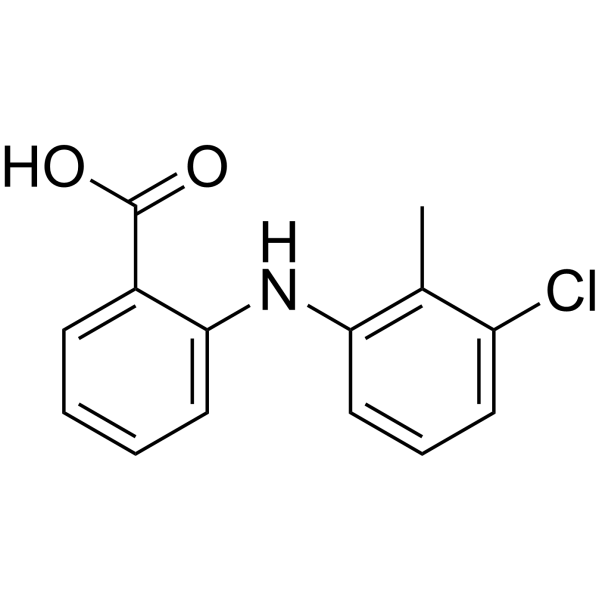
HY-B0335
Tolfenamic AcidCOX-2 Inhibitor -
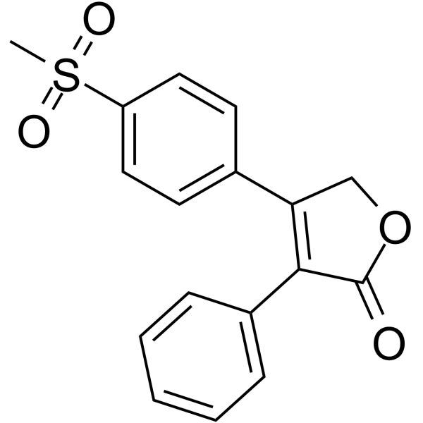
HY-17372
Rofecoxib -
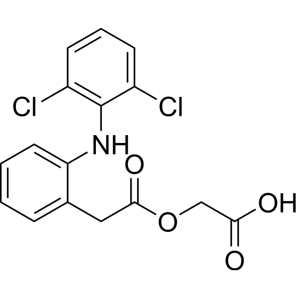
HY-B0634
AceclofenacNSAID -

HY-17484R
Ampiroxicam (Standard)COX Inhibitor -
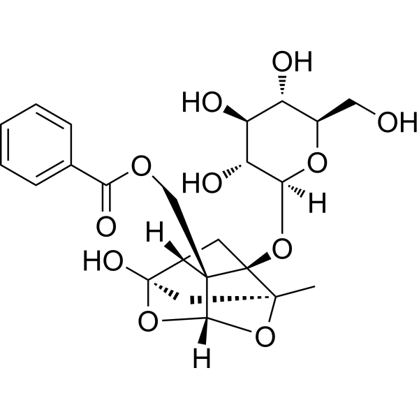
HY-N0293
PaeoniflorinHSP Activator -
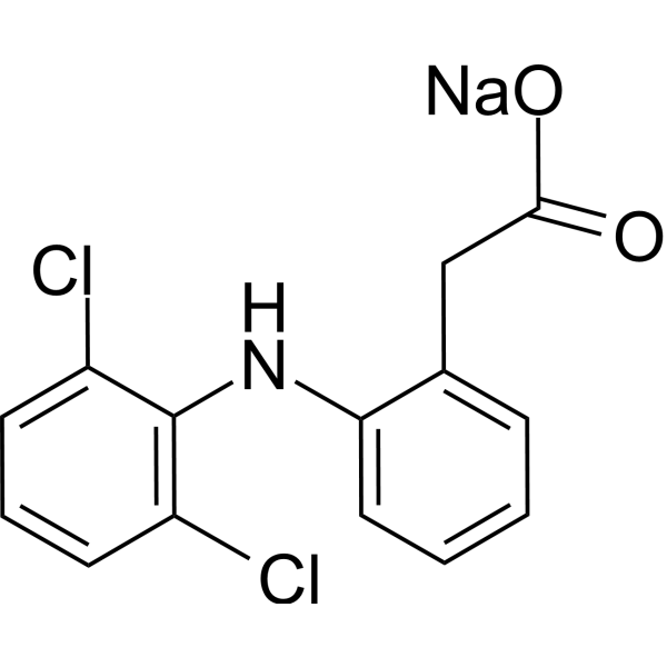
HY-15037
Diclofenac SodiumCOX Inhibitor -
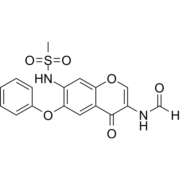
HY-17009
IguratimodAnti-rheumatic Agent -

HY-B0261R
Meloxicam (Standard)COX Inhibitor -

HY-N0293R
Paeoniflorin (Standard)HSP Activator
Publications Citing Use of MedChemExpress (MCE) Ampiroxicam
More

Biological Activity
Description
IC50 & Target
|
COX |
Parmacokinetics
| Species | Dose | Route | AUC | Bioavailability | Cmax | Tmax |
|---|---|---|---|---|---|---|
| Rat[2] | 10 mg/kg | p.o. | / | / | 26 μg/mL | 4 h |
| Rat[2] | 3.2 mg/kg | p.o. | 126 μg·h/mL | 91 % | 12 μg/mL | 2 h |
| Rat[2] | 3.2 mg/kg | i.v. | 125 μg·h/mL | 91 % | / | / |
| Dog[2] | 1.0 mg/kg | p.o. | 99 μg·h/mL | 72 % | 2.5 μg/mL | 1.5 h |
| Dog[2] | 1.0 mg/kg | i.v. | 108 μg·h/mL | 78 % | / | / |
| Monkey[2] | 5.0 mg/kg | p.o. | 207 μg·h/mL | 52 % | 16 μg/mL | 2 h |
| Monkey[2] | 5.0 mg/kg | i.v. | 280 μg·h/mL | 54 % | / | / |
In Vivo
Ampiroxicam (1-4.1 mg/kg; p.o.; single dose) inhibits the stretching response in fasted CF-1 mice induced by benzoquinone, with an MPE50 of 2.0 mg/kg[2].
Ampiroxicam (4.3-41 mg/kg; p.o.; single daily dose for 5 days) inhibits carrageenan-induced hind paw edema in fasted Sprague-Dawley rats[2].
MedChemExpress (MCE) has not independently confirmed the accuracy of these methods. They are for reference only.
-
Animal Model:Hartley (female, 380-450 g, contact hypersensitivity model via adjuvant patch test modified maximization method)[1]
-
Dosage:1% (UVA-irradiated, sensitization); 1% (UVA-irradiated, challenge); 1% (thiosalicylate challenge)
-
Administration:topical occlusive patch; daily for 3 consecutive days (primary sensitization); topical occlusive patch; single dose on day 9 (repeat sensitization); topical open application; single dose on day 21 (challenge)
-
Result:Induced a mean total skin reaction score of 3.3 (5/5 animals positive) when challenged with UVA-irradiated 1% ampiroxicam.
Induced a mean total skin reaction score of 4.7 (5/5 animals positive) when challenged with 1% thiosalicylate.
Induced a mean total skin reaction score of 4.0 (4/5 animals positive) with UVA (25.5 J/cm2)-irradiated 1% ampiroxicam challenge.
Showed no positive reactions to non-irradiated 1% ampiroxicam, UVA-irradiated 1% piroxicam, or non-irradiated 1% piroxicam.
-
Animal Model:CF-1 (fasted overnight, N ≥5/group)[2]
-
Dosage:1.0-4.1 mg/kg
-
Administration:oral gavage; single dose
-
Result:Determined an MPE50 of 2.0 mg/kg (95% confidence interval 1.0-4.1 mg/kg) for suppression of phenylbenzoquinone-induced stretching.
-
Animal Model:Sprague-Dawley (fasted, N ≥6/group)[2]
-
Dosage:4.3-41 mg/kg
-
Administration:oral; single dose; oral; daily; 5 days
-
Result:Determined an ED50 of 28 mg/kg (95% confidence interval 21-41 mg/kg) for single oral dose administration.
Determined an ED50 of 7.8 mg/kg (95% confidence interval 4.3-28 mg/kg) for 5 consecutive daily oral doses.


Chemical Information
-
CAS No. 99464-64-9
-
Appearance Solid
-
Molecular Weight 447.46
-
Formula C20H21N3O7S
-
Color White to off-white
-
SMILES
O=C(OC(OC(C1=CC=CC=C12)=C(C(NC3=NC=CC=C3)=O)N(C)S2(=O)=O)C)OCC
-
Synonyms
CP 65703
-
Shipping
Room temperature in continental US; may vary elsewhere.
-
Storage
Powder -20°C 3 years 4°C 2 years In solvent -80°C 2 years -20°C 1 year


Publications (2)
-
Journal Impact Factor
-
Most Recent
-
ACS Environ Au
Machine Learning-Assisted Recognition of Environmental Sulfur-Containing Chemicals in Nontargeted Mass Spectrometry Analysis of Inadequate Mass Resolution. [Abstract]2025 Aug 5;5(6):573-582. PMID: 41277996 -
Anal Chem
Exposome-Scale Investigation of Cl-/Br-Containing Chemicals Using High-Resolution Mass Spectrometry, Multistage Machine Learning, and Cloud Computing. [Abstract]2025 Jun 3;97(21):11099-11109. PMID: 40401576


Solvent & Solubility
In Vitro:
DMSO : 50 mg/mL (111.74 mM; Need ultrasonic; Hygroscopic DMSO has a significant impact on the solubility of product, please use newly opened DMSO)
Please refer to the solubility information to select the appropriate solvent. Once prepared, please aliquot and store the solution to prevent product inactivation from repeated freeze-thaw cycles.
Storage method and period of stock solution: -80°C, 2 years; -20°C, 1 year. When stored at -80°C, please use it within 2 years. When stored at -20°C, please use it within 1 year.
Please refer to the solubility information to select the appropriate solvent. Once prepared, please aliquot and store the solution to prevent product inactivation from repeated freeze-thaw cycles.
Storage method and period of stock solution: -80°C, 2 years; -20°C, 1 year. When stored at -80°C, please use it within 2 years. When stored at -20°C, please use it within 1 year.
Concentration (start) × Volume (start) = Concentration (final) × Volume (final)
In Vivo:
Select the appropriate dissolution method based on your experimental animal and administration route.
- For the following dissolution methods, please ensure to first prepare a clear stock solution using an In Vitro approach and then sequentially add co-solvents:
- To ensure reliable experimental results, the clarified stock solution can be appropriately stored based on storage conditions. As for the working solution for In Vivo experiments, it is recommended to prepare freshly and use it on the same day.
- The percentages shown for the solvents indicate their volumetric ratio in the final prepared solution. If precipitation or phase separation occurs during preparation, heat and/or sonication can be used to aid dissolution.
Add each solvent one by one: 10% DMSO 40% PEG300 5% Tween-80 45% Saline
Solubility: ≥ 2.5 mg/mL (5.59 mM); Clear solution
This protocol yields a clear solution of ≥ 2.5 mg/mL (saturation unknown).
Taking 1 mL working solution as an example, add 100 μL DMSO stock solution (25.0 mg/mL) to 400 μL PEG300, and mix evenly; then add 50 μL Tween-80 and mix evenly; then add 450 μL Saline to adjust the volume to 1 mL.
Preparation of Saline: Dissolve 0.9 g sodium chloride in ddH₂O and dilute to 100 mL to obtain a clear Saline solution.
In Vivo Dissolution Calculator
Please enter the basic information of animal experiments:
-
-
-
-
Recommended: Prepare an additional quantity of animals to account for potential losses during experiments.
Please enter your animal formula composition:
-
%DMSO +
Recommended: Keep the proportion of DMSO in working solution below 2% if your animal is weak.
-
%+
-
+%Tween-80 + +
-
%Saline +
The co-solvents required include: DMSO, . All of co-solvents are available by MedChemExpress (MCE). , Tween 80. All of co-solvents are available by MedChemExpress (MCE).
Working solution concentration: 0.22 mg/mL
Method for preparing stock solution: mg drug dissolved in μL DMSO. Stock solution concentration: mg/mL.
1. Take μL DMSO stock solution;
2. Add μL .
μL , mix evenly;
3. Then add μL Tween 80, mix evenly;
4. Then add μL
Please ensure that the stock solution in the first step is dissolved to a clear state, and add co-solvents in sequence. You can use ultrasonic heating (ultrasonic cleaner, recommended frequency 20-40 kHz), vortexing, etc. to assist dissolution.


Purity & Documentation
-
Data Sheet (286 KB)
-
SDS (393 KB)
- English - EN (393 KB)
- Français - FR (393 KB)
- Deutsch - DE (393 KB)
- Norwegian - NO (393 KB)
- Español - ES (393 KB)
- Swedish - SV (393 KB)
- Italian - IT (393 KB)
- Korean - KR (393 KB)
- Portuguese - PT (393 KB)
-
Handling Instructions (2659 KB)
References
[1]. Sasaki T, et al. Antigenic characterization in ampiroxicam-induced photosensitivity using an in vivo model of contact hypersensitivity. J Dermatol Sci. 1999;21(3):170-175. [Content Brief]
[2]. Carty TJ, et al. Ampiroxicam, an anti-inflammatory agent which is a prodrug of piroxicam. Agents Actions. 1993;39(3-4):157-165. [Content Brief]
[3]. Ogiso T, et al. Pharmacokinetic drug interactions between ampiroxicam and sulfaphenazole in rats. Biol Pharm Bull. 1999;22(2):191-196. [Content Brief]
[4]. Kurumaji Y. Ampiroxicam-induced photosensitivity. Contact Dermatitis. 1996 Apr;34(4):298-9. [Content Brief]


Complete Stock Solution Preparation Table
Please refer to the solubility information to select the appropriate solvent. Once prepared, please aliquot and store the solution to prevent product inactivation from repeated freeze-thaw cycles.
Storage method and period of stock solution: -80°C, 2 years; -20°C, 1 year. When stored at -80°C, please use it within 2 years. When stored at -20°C, please use it within 1 year.
| Optional Solvent | Concentration Solvent Mass | 1 mg | 5 mg | 10 mg | 25 mg |
|---|---|---|---|---|---|
| DMSO | 1 mM | 2.2348 mL | 11.1742 mL | 22.3484 mL | 55.8709 mL |
| 5 mM | 0.4470 mL | 2.2348 mL | 4.4697 mL | 11.1742 mL | |
| 10 mM | 0.2235 mL | 1.1174 mL | 2.2348 mL | 5.5871 mL | |
| 15 mM | 0.1490 mL | 0.7449 mL | 1.4899 mL | 3.7247 mL | |
| 20 mM | 0.1117 mL | 0.5587 mL | 1.1174 mL | 2.7935 mL | |
| 25 mM | 0.0894 mL | 0.4470 mL | 0.8939 mL | 2.2348 mL | |
| 30 mM | 0.0745 mL | 0.3725 mL | 0.7449 mL | 1.8624 mL | |
| 40 mM | 0.0559 mL | 0.2794 mL | 0.5587 mL | 1.3968 mL | |
| 50 mM | 0.0447 mL | 0.2235 mL | 0.4470 mL | 1.1174 mL | |
| 60 mM | 0.0372 mL | 0.1862 mL | 0.3725 mL | 0.9312 mL | |
| 80 mM | 0.0279 mL | 0.1397 mL | 0.2794 mL | 0.6984 mL | |
| 100 mM | 0.0223 mL | 0.1117 mL | 0.2235 mL | 0.5587 mL |


Ampiroxicam Related Classifications
HY-17484 Related Classifications
 Powered by Bioz