



TH588 hydrochloride
Based on 2 publication(s) in Google Scholar
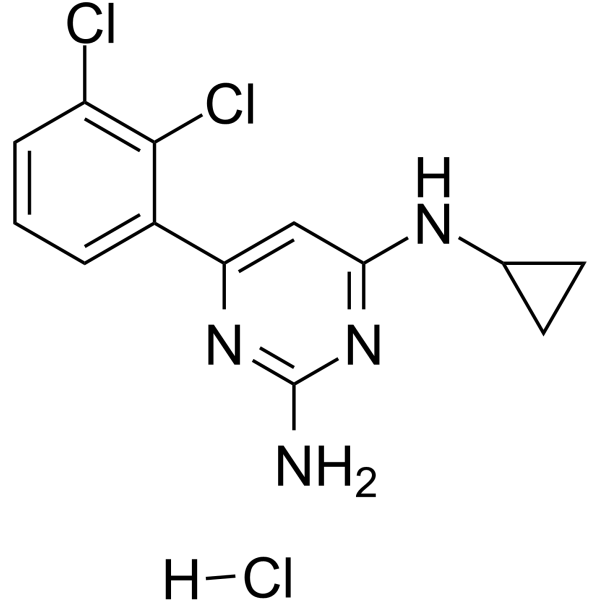
TH588 hydrochloride is a highly selective human MTH1 inhibitor (IC50=5 nM) with additional microtubule-targeting properties. TH588 hydrochloride interferes with mitotic progression, induces genomic 8-oxodG formation, DNA damage and cell cycle arrest, and exhibits significant cytotoxicity. Combined with phenethyl isothiocyanate, TH588 hydrochloride enhances ROS-mediated effects and effectively inhibits the growth of visceral metastases of malignant melanoma in mice. TH588 hydrochloride is widely applicable to research related to cancers, pancreatic cancer, leukemia, lymphoma, malignant melanoma and lung cancer.
For research use only. We do not sell to patients.
- CAS No.: 1640282-30-9
- Formula: C13H13Cl3N4
- Molecular Weight:331.63
-
Storage:
Please store the product under the recommended conditions in the Certificate of Analysis.

To place orders, for customer services and technical support, please contact: MedChemExpress USA
Tel: 609-228-6898 E-mail: [email protected] [email protected]


-
 Biological Activity
-
Chemical Information
- Protocols
- Purity & Documentation
- References
-
 Help & FAQs


-
HY-13323
CX-5461RNA Polymerase I Inhibitor -
HY-12484
BMH-21DNA Intercalator/Pol I Elongation Inhibitor -
HY-19620
BranaplamSMN2 Splicing Modulator -
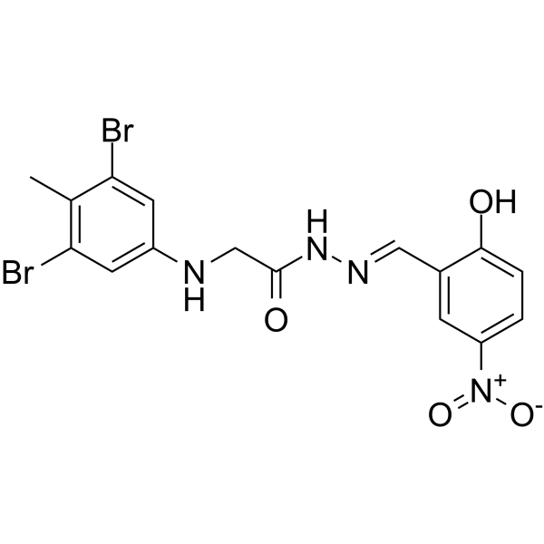
HY-15586
L67DNA Ligase Inhibitor -
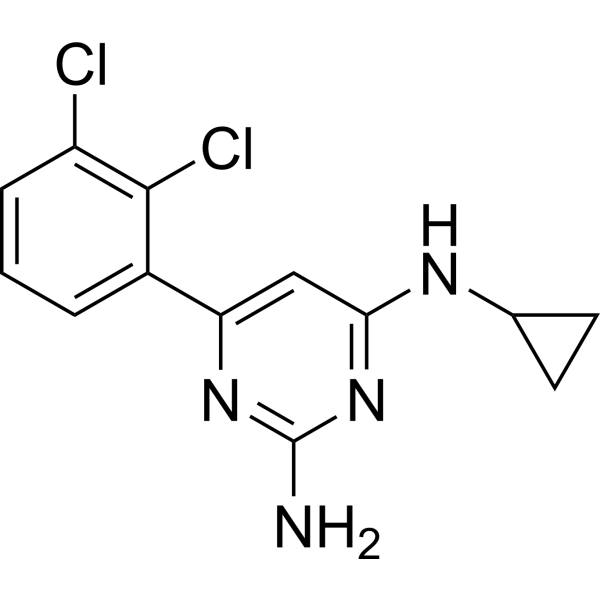
HY-12814
TH588MTH1 (NUDT1) Inhibitor -
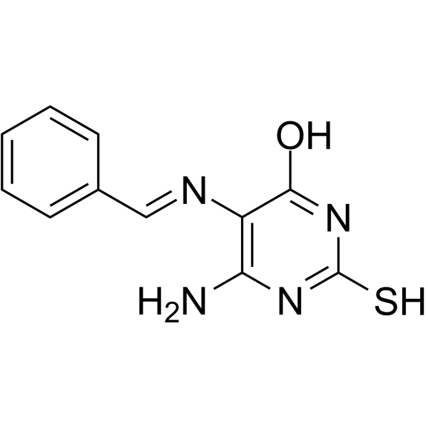
HY-15588
L189DNA Ligase Inhibitor -
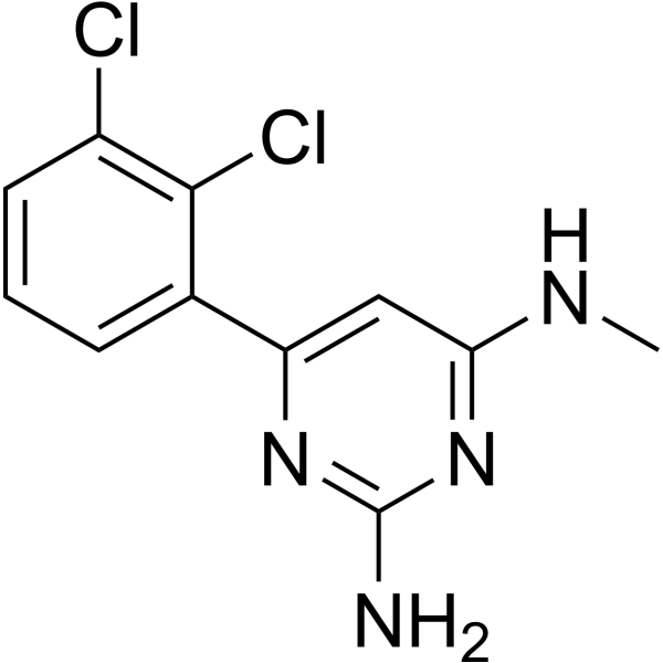
HY-16965
TH287MTH1 Inhibitor -
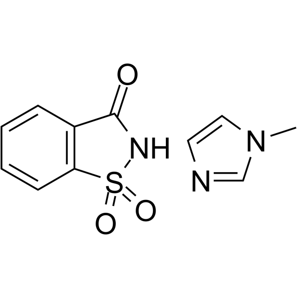
HY-112060
Saccharin 1-methylimidazoleDNA/RNA Synthesis Activator -
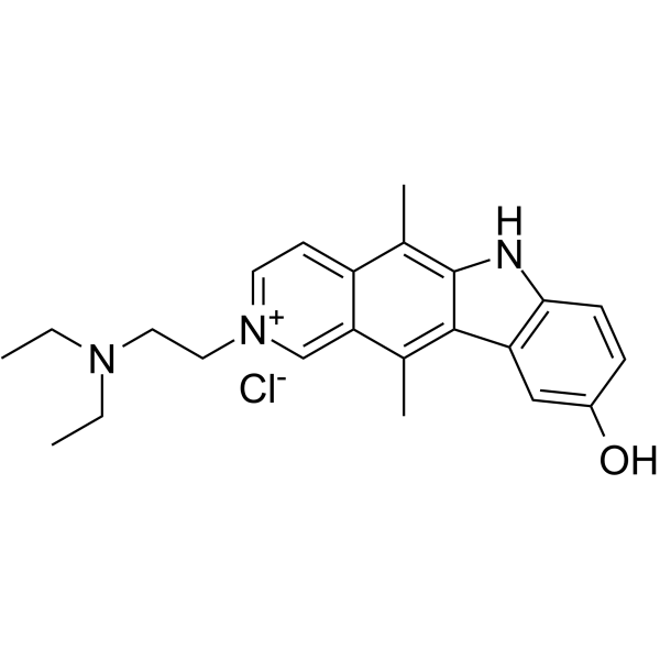
HY-U00337
Datelliptium chloride -

HY-13323R
CX-5461 (Standard)Reference Standard -

HY-12814R
TH588 (Standard)MTH1 (NUDT1) Inhibitor -
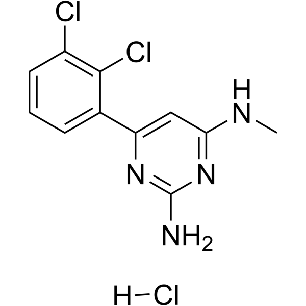
HY-16965A
TH287 hydrochlorideMTH1 Inhibitor
Publications Citing Use of MedChemExpress (MCE) TH588 hydrochloride
MoreAll DNA/RNA Synthesis Isoforms
More

Biological Activity
Description
In Vitro
TH588 hydrochloride (1-2 µM; 72 h) reduces viability of doxycycline-induced A2780 Pol δ replacement cells, with significantly greater cytotoxicity in cells expressing the error-prone or double mutant Pol δ variants[1].
TH588 hydrochloride (1 µM; 24 h) induces cleaved PARP signalling in doxycycline-induced A2780 Pol δ replacement cells, with significantly higher levels in cells expressing the error-prone Pol δ variant[1].
TH588 hydrochloride (2 µM; 24 h) induces caspase-mediated apoptosis in doxycycline-induced A2780 Pol δ replacement cells, with significantly higher apoptosis in cells expressing the error-prone Pol δ variant[1].
TH588 hydrochloride (5 µM; 16 h) triggers mitotic DNA replication in U2OS cells when pre-incubated with RO3306, resulting in a significant increase in mitotic cells with EdU foci[1].
TH588 hydrochloride (0.1-10 µM; 72 h) reduces viability of U2OS cells in a dose-dependent manner, and this cytotoxicity is completely prevented by co-treatment with reversine[1].
TH588 hydrochloride (2-6 μM; 5 days) concentration-dependently inhibits the growth of human pancreatic cancer Panc-1 cells over 5 days, and this effect is independent of oxidative stress[2].
TH588 hydrochloride (1-3 μM; 7 days) significantly enhances H2O2-induced growth inhibition of human pancreatic cancer Panc-1 cells over 7 days, despite having no effect on cell growth when used alone[2].
TH588 hydrochloride (1-3 μM; 5 days) significantly enhances doxorubicin-induced growth inhibition of human pancreatic cancer MIAPaCa-2 cells over 5 days[2].
TH588 hydrochloride (1-3 μM; 5 days) significantly enhances PEITC-induced growth inhibition of human pancreatic cancer MIAPaCa-2 cells over 5 days, with the combination showing greater efficacy than other ROS inducer-TH588 pairings[2].
TH588 hydrochloride (2 μM; 2 days), combined with 4 μM PEITC, induces caspase-dependent cell death in human pancreatic cancer MIAPaCa-2 cells over 2 days[2].
TH588 hydrochloride (0.2-0.6 μM; 5 days) significantly enhances PEITC-induced growth inhibition of human leukemia NB4 cells over 5 days, regardless of relatively low MTH1 mRNA expression[2].
TH588 hydrochloride (0.2-0.6 μM; 5 days) significantly enhances PEITC-induced growth inhibition of human lymphoma BALM3 cells over 5 days[2].
TH588 hydrochloride (1-2 μM; 56 hours) induces nuclear formation of 8-oxo-dG and pH2AX (markers of oxidative DNA damage) in human pancreatic cancer MIAPaCa-2 cells after 56 hours of incubation[2].
TH588 hydrochloride (0.4 μM; 56 hours), combined with 2 μM PEITC, cooperatively induces nuclear formation of 8-oxo-dG and γH2AX foci in human pancreatic cancer MIAPaCa-2 cells after 56 hours of incubation[2].
TH588 hydrochloride (1-8 µM; 2 hours) concentration-dependently disrupts centrosome separation, spindle bipolarity, and chromosome congression in unsynchronized mitotic H460 human lung cancer cells, with 4 µM inducing monopolar spindles in over 50% of mitotic cells[3].
TH588 hydrochloride (4-6 µM; 24 hours) induces G2/M phase cell cycle arrest in H460 human lung cancer cells, with 4 µM increasing G2/M phase cells to ~42% and 6 µM increasing G2/M phase cells to ~35%[3].
TH588 hydrochloride (4-7 µM; 48 total hours) concentration-dependently induces G1 arrest and prevents cell cycle re-entry in H460 human lung cancer cells, with 7 µM causing G1 arrest in ~70% of cells[3].
TH588 hydrochloride (6 µM; 48 total hours) shows that TP53 and USP28 are required for TH588-induced G1 arrest in H460 human lung cancer cells, with TP53 knockout reducing 6 µM TH588-induced G1 arrest from ~40% to ~10%[3].
TH588 hydrochloride (2-8 µM; 24 hours) concentration-dependently increases p53 levels in H460 human lung cancer cells, and this activation is dependent on USP28, as USP28-knockout clones show no p53 induction[3].
MedChemExpress (MCE) has not independently confirmed the accuracy of these methods. They are for reference only.
-
Cell Line:U2OS cells
-
Concentration:5 µM
-
Incubation Time:16 h
-
Result:Induced EdU foci in mitotic cells, indicating mitotic DNA replication.
Showed significantly higher percentage of mitotic cells with EdU foci in TH588-treated cells compared to control cells, though lower than the positive control aphidicolin-treated cells.
-
Cell Line:U2OS cells
-
Concentration:0.1-10 µM
-
Incubation Time:72 h
-
Result:Reduced cell viability in a dose-dependent manner, with viability dropping to ~15% at 10 µM TH588.
Showed co-treatment with reversine completely abrogated TH588-induced cytotoxicity, maintaining viability at ~100% across all TH588 concentrations tested.
-
Cell Line:human pancreatic cancer MIAPaCa-2 cells
-
Concentration:1-3 μM
-
Incubation Time:5 days
-
Result:Significantly enhanced doxorubicin-induced growth inhibition of MIAPaCa-2 cells.\nHardly affected MIAPaCa-2 cell growth when used alone at 1 μM.
Significantly enhanced PEITC-induced growth inhibition.
Formed the most effective growth-inhibiting combination with PEITC among tested ROS inducer pairings.
-
Cell Line:unsynchronized H460 human lung cancer cells
-
Concentration:1-8 µM
-
Incubation Time:2 hours
-
Result:Caused concentration-dependent decreases in centrosome separation, with reduced separation at all tested concentrations.
Induced monopolar spindles and uncongressed chromosomes in over 50% of mitotic cells at 4 µM; higher concentrations increased the proportion of these abnormalities.
Did not affect centrosome duplication or the localization of aurora kinase A, polo-like kinase 1, or kinesin family member 23.
-
Cell Line:USP28-knockout, TP53-knockout, and control H460 human lung cancer cells
-
Concentration:4-7 µM
-
Incubation Time:3 days followed by 12 days drug-free
-
Result:USP28-knockout clones showed significantly increased colony formation at all tested concentrations compared to controls, with 7 µM treatment yielding ~40% of DMSO control colonies (compared to ~5% for controls).
TP53-knockout clones also showed increased colony formation at all concentrations, with 7 µM treatment yielding ~5% of DMSO control colonies (compared to ~5% for controls, with three TP53 knockout clones showing stronger rescue than controls).
-
Cell Line:H460 human lung cancer cells
-
Concentration:4-7 µM
-
Incubation Time:48 total hours (final 24 hours including EdU)
-
Result:Increased the proportion of G1-arrested, EdU-negative cells in a concentration-dependent manner: 4 µM induced ~20% G1 arrest, 5 µM ~40%, 6 µM ~60%, and 7 µM ~70%.
Saw over 98% of DMSO-treated cells were EdU-positive and re-entered the cell cycle, while TH588 treatment also induced a small sub-G1 apoptotic population at higher concentrations.
-
Cell Line:USP28-knockout, TP53-knockout, and control H460 human lung cancer cells
-
Concentration:6 µM
-
Incubation Time:48 total hours (final 24 hours including EdU)
-
Result:TP53-knockout clones showed a marked reduction in G1-arrested, EdU-negative cells (from ~40% in controls to ~10%) and a corresponding increase in EdU-positive cells (from ~55% in controls to ~85%).
USP28-knockout clones showed a weaker but significant reduction in G1-arrested cells (from ~40% in controls to ~30%) and increase in EdU-positive cells (from ~55% in controls to ~65%).
In Vivo
MedChemExpress (MCE) has not independently confirmed the accuracy of these methods. They are for reference only.
-
Animal Model:5-6 weeks female SCID mice (SW480 xenograft cancer model)[1]
-
Dosage:30 mg/kg
-
Administration:Subcutaneous injection (s.c.); once daily for 35 days
-
Result:Reduced tumour growth in SW480 xenograft cancer model.
- Flow cytometric DNA-content cell-cycle staining
- Cell Cytotoxicity Assay
- Mammalian live/dead viability and cytotoxicity staining
- BrdU Incorporation Assay
- ROS/oxidative-stress fluorescent staining
- Protocol for Cell Cycle
- Genotoxicity/Mutagenicity Study
- Cell Viability Determination by MTT Colorimetric Assay


Chemical Information
-
CAS No. 1640282-30-9
-
Molecular Weight 331.63
-
Formula C13H13Cl3N4
-
SMILES
NC1=NC(C2=CC=CC(Cl)=C2Cl)=CC(NC3CC3)=N1.[H]Cl
-
Shipping
Room temperature in continental US; may vary elsewhere.
-
Storage
Please store the product under the recommended conditions in the Certificate of Analysis.


Publications (2)
-
Journal Impact Factor
-
Most Recent
-
Biochim Biophys Acta Mol Basis Dis
MTH1 inhibition synergizes with ROS-inducing agents to trigger cervical cancer cells undergoing parthanatos. [Abstract]2024 Apr 22;1870(5):167190. PMID: 38657912 -
J Mol Med (Berl)
2019 Aug;97(8):1183-1193. PMID: 31201471


Purity & Documentation
References
[1]. Rudd SG, et al. MTH1 Inhibitor TH588 Disturbs Mitotic Progression and Induces Mitosis-Dependent Accumulation of Genomic 8-oxodG. Cancer Res. 2020;80(17):3530-3541. [Content Brief]
[2]. Ikejiri F, et al. TH588, an MTH1 inhibitor, enhances phenethyl isothiocyanate-induced growth inhibition in pancreatic cancer cells. Oncol Lett. 2018;15(3):3240-3244. [Content Brief]
[3]. Gul N, et al. The MTH1 inhibitor TH588 is a microtubule-modulating agent that eliminates cancer cells by activating the mitotic surveillance pathway. Sci Rep. 2019;9(1):14667. Published 2019 Oct 11. [Content Brief]

Calculators
Concentration (start) × Volume (start) = Concentration (final) × Volume (final)


TH588 hydrochloride Related Classifications
HY-12814A Related Classifications
 Powered by Bioz