



Platycodin D2
Based on 3 publication(s) in Google Scholar
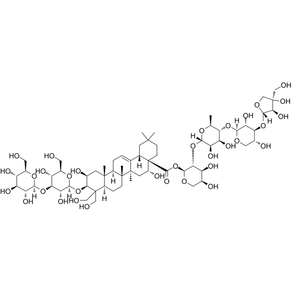
Platycodin D2 is an orally active triterpenoid saponin found in Platycodon grandiflorum. Platycodin D2 induces mitophagy in cancer cells through NIX, thereby activating the P21/CyclinA2 pathway and promoting cell senescence. Platycodin D2 induces mitochondrial dysfunction, enhances autophagy, inhibits hepatocellular carcinoma cell proliferation, and exhibits anti-tumor activity against multiple cancer cell types. Platycodin D2 promotes mRNA expression of T-bet, GATA-3, Th1 cytokines IL-2 and IFN-γ, and Th2 cytokines IL-4 and IL-10, enhances splenocyte proliferation, and acts as a vaccine adjuvant with low rabbit red blood cell hemolytic activity. Platycodin D2 induces mitochondrial ROS production, incomplete autophagy, and ferroptosis to inhibit breast cancer cell proliferation. Platycodin D2 can be used for the research of cancer, inflammation and immunology.
Nur für Forschungszwecke. Wir verkaufen nicht an Patienten.
- Reinheit : 99.84%
- CAS. Nr.: 66663-90-9
- Formel: C63H102O33
- Molecular Weight:1387.46
-
Speicherung:Powder -20°C, 3 years , 4°C, 2 years ; In solvent -80°C, 6 months , -20°C, 1 month

To place orders, for customer services and technical support, please contact: MedChemExpress USA
Tel: 609-228-6898 E-mail: [email protected] [email protected]


-
 Biologische Aktivität
-
Chemical Information
-
Lösungsmittel & Löslichkeit
- Protokoll
- Reinheit & Dokumentation
- Verweise
-
 Help & FAQs


Publications Citing Use of MedChemExpress (MCE) Platycodin D2
More

Biologische Aktivität
Beschreibung
IC50 & Target
[2]|
IL-2 |
IL-4 |
IL-10 |
Cellular Effect
|
Cell Line

|
Type | Value | Description | References |
|---|---|---|---|---|
| HCT-15 | IC50 |
5.7 μM
Compound: 8, deapio-platycodin D
|
Antiproliferative activity against human HCT15/CL02 cells after 48 hrs by sulforhodamine B assay
Antiproliferative activity against human HCT15/CL02 cells after 48 hrs by sulforhodamine B assay
|
[PMID: 20939516] |
| HCT-15 | IC50 |
9.6 μM
Compound: 8, deapio-platycodin D
|
Antiproliferative activity against human HCT15 cells after 48 hrs by sulforhodamine B assay
Antiproliferative activity against human HCT15 cells after 48 hrs by sulforhodamine B assay
|
[PMID: 20939516] |
| MES-SA | IC50 |
3.5 μM
Compound: 8, deapio-platycodin D
|
Antiproliferative activity against human MESSA cells after 48 hrs by sulforhodamine B assay
Antiproliferative activity against human MESSA cells after 48 hrs by sulforhodamine B assay
|
[PMID: 20939516] |
| MES-SA/Dx5 | IC50 |
5.2 μM
Compound: 8, deapio-platycodin D
|
Antiproliferative activity against human MESSA/DX5 cells after 48 hrs by sulforhodamine B assay
Antiproliferative activity against human MESSA/DX5 cells after 48 hrs by sulforhodamine B assay
|
[PMID: 20939516] |
In Vitro
Platycodin D2 (1-100 μM; 48 h) specifically inhibits proliferation of Huh-7, MHCC97H, HCCLM3, HepG-2, SK-Hep1, and Huh-6 HCC cells with an IC50 of 10.2-12.7 μM, while having no significant cytotoxic effect on THLE-2 and L02 normal liver cells (IC50 >200 μM)[1].
Platycodin D2 (10 μM; 48 h) does not significantly induce apoptosis in Huh-7 or HCCLM3 HCC cells[1].
Platycodin D2 (10 μM; 24-48 h) induces robust autophagy in Huh-7 and HCCLM3 HCC cells, as evidenced by increased LC3 puncta formation, elevated autophagic flux, autophagolysosome formation, and altered expression of autophagy-related proteins, without affecting apoptosis-related proteins[1].
Platycodin D2 (5, 10 μM; 48 h) induces mitochondrial dysfunction in Huh-7 and HCCLM3 HCC cells, evidenced by increased ROS production, reduced mitochondrial membrane potential, and selective mitophagy via LC3 co-localization with damaged mitochondria[1].
Platycodin D2 (10 μM; 48 h) induces mitophagy in HCCLM3 HCC cells via NIX, as silencing NIX abrogates PD2's effects on autophagy, cell viability, mitochondrial function, and downstream P21/CyclinA2 expression[1].
Platycodin D2 (10 μM; 48 h) induces G2/M phase arrest and senescence in Huh-7 and HCCLM3 HCC cells, evidenced by altered cell cycle distribution, upregulated SASP gene expression, shifted expression of senescence-related proteins, increased β-galactosidase activity, reduced Lamin B1 levels, and nuclear γ-H2A.X aggregation[1].
Platycodin D2 (3.906-125 μg/mL; 30 min) exhibits haemolytic activity against 0.5% rabbit red blood cell suspensions with an HD50 of 18.57 μg/mL[2].
Platycodin D2 (0.0016-1.0 μg/mL; 16 h) significantly enhances mRNA expression of Th1 (IL-2, IFN-γ, T-bet) and Th2 (IL-4, IL-10, GATA-3) cytokines and transcription factors in Con A-stimulated naive ICR mouse splenocytes[2].
Platycodin D2 (5-50 μM; 48 h) potently inhibits the proliferation of MCF7, SKBR3, and Hs578T breast cancer cells with IC50 values of 14.62 μM, 29.60 μM, and 5.24 μM, respectively, after 48 h of incubation[3].
Platycodin D2 (5-50 μM; 48 h) blocks autophagy flux in MCF7, SKBR3, and Hs578T breast cancer cells by increasing LC3II/I and p62 expression while decreasing Syntaxin 17, SNAP29, VAMP8, and Lamp2b expression after 48 h of incubation[3].
Platycodin D2 (5-50 μM; 48 h) induces ferroptosis in MCF7, SKBR3, and Hs578T breast cancer cells by decreasing SLC7A11, GSH and GPX4 expression after 48 h of incubation[3].
Platycodin D2 (5-50 μM; 48 h) increases intracellular ferrous ion levels in MCF7, SKBR3, and Hs578T breast cancer cells after 48 h of incubation, a hallmark of ferroptosis[3].
Platycodin D2 (5-50 μM; 48 h) upregulates mitochondrial outer membrane protein TOM20 expression in MCF7, SKBR3, and Hs578T breast cancer cells after 48 h of incubation, indicating mitochondrial damage[3].
Platycodin D2 (5-50 μM; 48 h) increases intracellular MDA levels in MCF7, SKBR3, and Hs578T breast cancer cells after 48 h of incubation, indicating enhanced lipid peroxidation associated with ferroptosis[3].
Platycodin D2 (5-50 μM; 48 h) decreases intracellular SOD levels in MCF7, SKBR3, and Hs578T breast cancer cells after 48 h of incubation, reducing antioxidant capacity and promoting ferroptosis[3].
Platycodin D2 (5-50 μM; 48 h) increases mitochondrial ROS production and reduces mitochondrial membrane potential in MCF7, SKBR3, and Hs578T breast cancer cells after 48 h of incubation, contributing to mitochondrial damage[3].
MedChemExpress (MCE) has not independently confirmed the accuracy of these methods. They are for reference only.
-
Cell Line:Huh-7, MHCC97H, HCCLM3, HepG-2, SK-Hep1, Huh-6 hepatocellular carcinoma (HCC) cells, THLE-2, L02 normal liver cells
-
Concentration:1, 5, 10, 20, 50, 100 μM
-
Incubation Time:48 h
-
Result:Significantly reduced viability of all tested HCC cell lines, with IC50 values of 10.2 μM (Huh-7), 11.5 μM (MHCC97H), 10.4 μM (HCCLM3), 11.2 μM (HepG-2), 12.7 μM (SK-Hep1), and 11.8 μM (Huh-6).
Had no obvious inhibitory effect on normal THLE-2 and L02 liver cells, with IC50 values >200 μM for both.
-
Cell Line:Huh-7, HCCLM3 HCC cells
-
Concentration:10 μM
-
Incubation Time:24 h; 48 h
-
Result:Significantly increased the number of EGFP-LC3 puncta per cell (20 puncta for HCCLM3, 60 puncta for Huh-7, compared to near-zero in controls).
Showed significantly more red fluorescence than green fluorescence via mRFP-GFP-LC3 staining, indicating increased autophagic flux.
Revealed abundant autophagolysosomes in PD2-treated cells via transmission electron microscopy.
Increased LC3-II and Beclin 1 expression, and decreased P62 expression via Western blotting, while apoptosis-related proteins (Cleaved-caspase3, PARP, Cleaved-PARP) showed no significant changes.
-
Cell Line:Huh-7, HCCLM3 HCC cells
-
Concentration:10 μM
-
Incubation Time:48 h
-
Result:Induced G2/M phase arrest in HCC cells, with G2/M phase percentage increasing.
Showed significant upregulation of SASP genes (IL-6, IL-8, MMP3, TGF-β, IGFBP3, CXCL-1) in both cell lines via Real Time qPCR.
Revealed upregulated P21 and γ-H2A.X, and downregulated CDK1, CyclinA2, E2F, and p-RB via Western blotting.
Showed a significant increase in β-galactosidase positive cells, reduced Lamin B1 fluorescence and nuclear aggregation of γ-H2A.X in PD2-treated cells via β-galactosidase staining and immunofluorescence.
-
Cell Line:MCF7, SKBR3, Hs578T
-
Concentration:5, 10, 20 μM (MCF7, Hs578T); 25, 50 μM (SKBR3)
-
Incubation Time:48 h
-
Result:Upregulated the expression of LC3II/I and p62 proteins in all three breast cancer cell lines.
Downregulated the expression of Syntaxin 17, SNAP29, VAMP8, and Lamp2b proteins in all three breast cancer cell lines.\nDownregulated the expression of ferroptosis-related proteins SLC7A11 and GPX4 in all three breast cancer cell lines.\nSignificantly upregulated the expression of TOM20 in all three breast cancer cell lines.
In Vivo
Platycodin D2 (25-100 μg; s.c.; on Day 1 and Day 15) elicits balanced Th1 and Th2 immune responses in OVA (HY-W250978)-immunized ICR mice, significantly enhancing splenocyte proliferation, OVA-specific antibody titers at tested doses[2].
Platycodin D2 (2.5-5 mg/kg; i.g.; once every 2 days; 5 total doses) significantly inhibits breast cancer tumor growth in nude mice, with the 5 mg/kg dose reducing tumor volume by 80.5%, and mediates this effect via autophagy flux blockage and ferroptosis[3].
MedChemExpress (MCE) has not independently confirmed the accuracy of these methods. They are for reference only.
-
Animal Model:BALB/c nude mice (female, 5-6 weeks old, injected with 5×10⁶ HCCLM3 cells in the right limb)[1]
-
Dosage:5 mg/kg; 10 mg/kg
-
Administration:intratumoral injection; once every 3 days; 4 doses
-
Result:Significantly inhibited tumor growth compared to the control group.
Reduced mean tumor volume to ~100 mm3 by week 4 at 5 mg/kg dose.
Reduced mean tumor volume to near 0 mm3 by week 4 at 10 mg/kg dose.
Maintained higher body weights than the control and 5-FU groups throughout the study.
Dose-dependently increased mean density of LC3-II, NIX, and P21 in tumor tissues.
Dose-dependently decreased mean density of Ki67 and Cyclin A2 in tumor tissues.
-
Animal Model:OVA-immunized ICR mice (female, 6 weeks old, 18-22 g)[2]
-
Dosage:25 μg; 50 μg; 75 μg; 100 μg
-
Administration:s.c.; on Day 1 and Day 15
-
Result:Significantly increased Con A- and LPS-stimulated splenocyte proliferation at 25 μg and 50 μg compared to the OVA control group (P<0.01 or P<0.001).
Significantly enhanced OVA-induced splenocyte proliferation at all four tested doses compared to the OVA control group (P<0.05 or P<0.01).
Significantly enhanced serum OVA-specific IgG, IgG1, IgG2a, and IgG2b antibody titers at all four tested doses compared to the OVA control group (P<0.01 or P<0.001).
Resulted in significantly higher IgG and IgG1 titers at 75 μg and 100 μg than the Alum-treated group (P<0.05).
-
Animal Model:BALB/c nude mice (female, 4-5 weeks old, subcutaneous MCF7 cell implantation tumor-bearing model)[3]
-
Dosage:2.5 mg/kg; 5 mg/kg
-
Administration:p.o.; once every 2 days; 5 total doses
-
Result:Reduced tumor volume by 80.5% at 5 mg/kg compared to control.
Reduced tumor volume by 65.8% at 2.5 mg/kg compared to control.
Did not affect mouse body weight.
Downregulated ferroptosis-related proteins SLC7A11 and GPX4 in tumor tissues.
Upregulated autophagy-related proteins LC3II and p62 in tumor tissues.
Increased ROS levels in tumor tissues.
Increased lipid peroxidation in tumor tissues.
- RNA extraction experimental
- Somatic Cell Culture
- Liver Cancer Modeling
- Breast Cancer Modeling
- Senescence-associated β-galactosidase staining
- Mitochondrial membrane-potential and mitochondrial mass staining
- Autophagy
- Lysosome and acidic-vesicle live-cell staining
- Ferroptosis Solutions
- Mitophagy Solutions
- MTT Cell Proliferation Assay
- Macroautophagy Solutions
- RT-PCR
- Naïve CD4+ T-cell subset differentiation/polarization
- BrdU Incorporation Assay
- ROS/oxidative-stress fluorescent staining
- CFSE Dye Dilution Proliferation Assay
- Dye-dilution cell tracking and proliferation staining
- CCK-8/WST-8 Cell Proliferation Assay
- Protocol for Cell Cycle
- Cell Counting-Based Growth Curve Assay
- Research Protocol for Inflammation-related Diseases
- Colony Formation (Clonogenic) Assay
- Real Time qPCR (Q-PCR)
- Cell Viability Determination by MTT Colorimetric Assay
- EdU Incorporation Assay (Click Chemistry-Based DNA Synthesis Measurement)
- Ki-67 Immunostaining Proliferation Assay
- PCNA Immunodetection Proliferation Assay
- Protocol for Cell Counting and Cell Density Analysis


Chemical Information
-
CAS. Nr. 66663-90-9
-
Appearance Solid
-
Molecular Weight 1387.46
-
Formel C63H102O33
-
Color Off-white to light yellow
-
SMILES
O[C@H]([C@@H]([C@@H](O[C@@]1([H])[C@@H]([C@H]([C@H](O)CO1)O[C@@]2([H])[C@@H]([C@](CO)(O)CO2)O)O)[C@H](C)O3)O)[C@]3([H])O[C@H]([C@H]([C@@H](O)CO4)O)[C@@H]4OC([C@]56[C@](CC(C)(C)CC6)([H])C7=CC[C@@]([C@@]8([C@@](C(CO)([C@@H](O[C@@]9([H])[C@@H]([C@H]([C@H](O)[C@@H](CO)O9)O[C@]%10([H])O[C@@H]([C@@H](O)[C@H](O)[C@H]%10O)CO)O)[C@@H](O)C8)CO)([H])CC%11)C)([H])[C@]%11(C)[C@]7(C)C[C@H]5O)=O
-
Structure Classification
-
Versand
Room temperature in continental US; may vary elsewhere.
-
Speicherung
Powder -20°C 3 years 4°C 2 years In solvent -80°C 6 months -20°C 1 month


Publications (3)
-
Journal Impact Factor
-
Most Recent
-
Phytother Res
Platycodin D2 Mediates Incomplete Autophagy and Ferroptosis in Breast Cancer Cells by Regulating Mitochondrial ROS. [Abstract]2025 Feb;39(2):581-592. PMID: 39581858 -
Cancer Cell Int
Platycodin D2 enhances P21/CyclinA2-mediated senescence of HCC cells by regulating NIX-induced mitophagy. [Abstract]2024 Feb 19;24(1):79. PMID: 38374035 -
Molecules
Characterization of Saponins from Various Parts of Platycodon grandiflorum Using UPLC-QToF/MS. [Abstract]2021 Dec 24;27(1):107. PMID: 35011337


Lösungsmittel & Löslichkeit
In Vitro:
DMSO : 100 mg/mL (72.07 mM; Need ultrasonic; Hygroscopic DMSO has a significant impact on the solubility of product, please use newly opened DMSO)
Please refer to the solubility information to select the appropriate solvent. Once prepared, please aliquot and store the solution to prevent product inactivation from repeated freeze-thaw cycles.
Storage method and period of stock solution: -80°C, 6 months; -20°C, 1 month. When stored at -80°C, please use it within 6 months. When stored at -20°C, please use it within 1 month.
Please refer to the solubility information to select the appropriate solvent. Once prepared, please aliquot and store the solution to prevent product inactivation from repeated freeze-thaw cycles.
Storage method and period of stock solution: -80°C, 6 months; -20°C, 1 month. When stored at -80°C, please use it within 6 months. When stored at -20°C, please use it within 1 month.
Konzentration (Stammlösung) × Volumen (Stammlösung) = Konzentration (Ziellösung) × Volumen (Ziellösung)
In Vivo:
Select the appropriate dissolution method based on your experimental animal and administration route.
- For the following dissolution methods, please ensure to first prepare a clear stock solution using an In Vitro approach and then sequentially add co-solvents:
- To ensure reliable experimental results, the clarified stock solution can be appropriately stored based on storage conditions. As for the working solution for In Vivo experiments, it is recommended to prepare freshly and use it on the same day.
- The percentages shown for the solvents indicate their volumetric ratio in the final prepared solution. If precipitation or phase separation occurs during preparation, heat and/or sonication can be used to aid dissolution.
Add each solvent one by one: 10% DMSO 40% PEG300 5% Tween-80 45% Saline
Solubility: ≥ 2.5 mg/mL (1.80 mM); Clear solution
This protocol yields a clear solution of ≥ 2.5 mg/mL (saturation unknown).
Taking 1 mL working solution as an example, add 100 μL DMSO stock solution (25.0 mg/mL) to 400 μL PEG300, and mix evenly; then add 50 μL Tween-80 and mix evenly; then add 450 μL Saline to adjust the volume to 1 mL.
Preparation of Saline: Dissolve 0.9 g sodium chloride in ddH₂O and dilute to 100 mL to obtain a clear Saline solution.
Add each solvent one by one: 10% DMSO 90% (20% SBE-β-CD in Saline)
Solubility: ≥ 2.5 mg/mL (1.80 mM); Clear solution
This protocol yields a clear solution of ≥ 2.5 mg/mL (saturation unknown).
Taking 1 mL working solution as an example, add 100 μL DMSO stock solution (25.0 mg/mL) to 900 μL 20% SBE-β-CD in Saline, and mix evenly.
Preparation of 20% SBE-β-CD in Saline (4°C, storage for one week): 2 g SBE-β-CD powder is dissolved in 10 mL Saline, completely dissolve until clear.
In Vivo Dissolution Calculator
Please enter the basic information of animal experiments:
-
-
-
-
Recommended: Prepare an additional quantity of animals to account for potential losses during experiments.
Please enter your animal formula composition:
-
%DMSO +
Recommended: Keep the proportion of DMSO in working solution below 2% if your animal is weak.
-
%+
-
+%Tween-80 + +
-
%Saline +
The co-solvents required include: DMSO, . All of co-solvents are available by MedChemExpress (MCE). , Tween 80. All of co-solvents are available by MedChemExpress (MCE).
Working solution concentration: 0.22 mg/mL
Method for preparing stock solution: mg drug dissolved in μL DMSO. Stock solution concentration: mg/mL.
1. Take μL DMSO stock solution;
2. Add μL .
μL , mix evenly;
3. Then add μL Tween 80, mix evenly;
4. Then add μL
Please ensure that the stock solution in the first step is dissolved to a clear state, and add co-solvents in sequence. You can use ultrasonic heating (ultrasonic cleaner, recommended frequency 20-40 kHz), vortexing, etc. to assist dissolution.


Reinheit & Dokumentation
-
Data Sheet (292 KB)
-
SDS (251 KB)
- English - EN (251 KB)
- Français - FR (251 KB)
- Deutsch - DE (251 KB)
- Norwegian - NO (251 KB)
- Español - ES (251 KB)
- Swedish - SV (251 KB)
- Italian - IT (251 KB)
- Korean - KR (251 KB)
- Portuguese - PT (251 KB)
-
Handling Instructions (2659 KB)
Verweise
[1]. Sun L, et al. Platycodin D2 enhances P21/CyclinA2-mediated senescence of HCC cells by regulating NIX-induced mitophagy. Cancer Cell Int. 2024;24(1):79. Published 2024 Feb 19. [Content Brief]
[2]. Xie Y, et al. Platycodin D2 is a potential less hemolytic saponin adjuvant eliciting Th1 and Th2 immune responses. Int Immunopharmacol. 2008;8(8):1143-1150. [Content Brief]
[3]. Li Y, et al. Platycodin D2 Mediates Incomplete Autophagy and Ferroptosis in Breast Cancer Cells by Regulating Mitochondrial ROS. Phytother Res. 2025;39(2):581-592. [Content Brief]


Complete Stock Solution Preparation Table
Please refer to the solubility information to select the appropriate solvent. Once prepared, please aliquot and store the solution to prevent product inactivation from repeated freeze-thaw cycles.
Storage method and period of stock solution: -80°C, 6 months; -20°C, 1 month. When stored at -80°C, please use it within 6 months. When stored at -20°C, please use it within 1 month.
| Optional Solvent | Concentration Solvent Mass | 1 mg | 5 mg | 10 mg | 25 mg |
|---|---|---|---|---|---|
| DMSO | 1 mM | 0.7207 mL | 3.6037 mL | 7.2074 mL | 18.0185 mL |
| 5 mM | 0.1441 mL | 0.7207 mL | 1.4415 mL | 3.6037 mL | |
| 10 mM | 0.0721 mL | 0.3604 mL | 0.7207 mL | 1.8019 mL | |
| 15 mM | 0.0480 mL | 0.2402 mL | 0.4805 mL | 1.2012 mL | |
| 20 mM | 0.0360 mL | 0.1802 mL | 0.3604 mL | 0.9009 mL | |
| 25 mM | 0.0288 mL | 0.1441 mL | 0.2883 mL | 0.7207 mL | |
| 30 mM | 0.0240 mL | 0.1201 mL | 0.2402 mL | 0.6006 mL | |
| 40 mM | 0.0180 mL | 0.0901 mL | 0.1802 mL | 0.4505 mL | |
| 50 mM | 0.0144 mL | 0.0721 mL | 0.1441 mL | 0.3604 mL | |
| 60 mM | 0.0120 mL | 0.0601 mL | 0.1201 mL | 0.3003 mL |


Platycodin D2 Related Classifications
HY-N4087 Related Classifications
 Powered by Bioz