



Cyasterone
Based on 4 publication(s) in Google Scholar
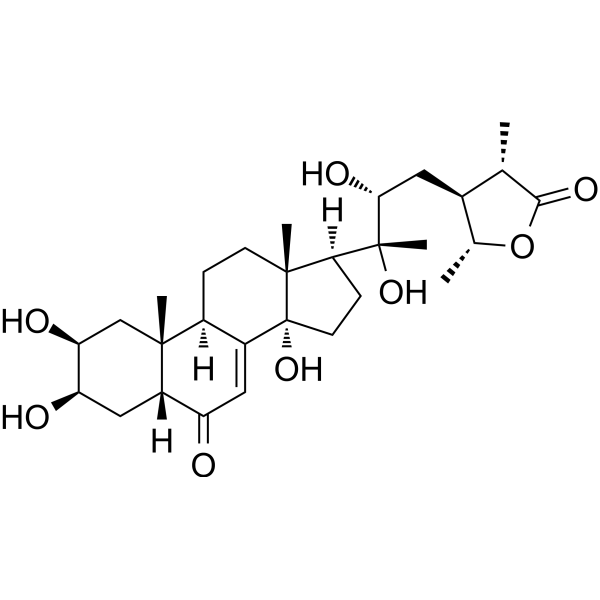
Cyasterone, a natural EGFR inhibitor, mainly isolated from Ajuga decumbens Thunb (Labiatae). Cyasterone manifests anti-proliferation effect by induced apoptosis and cell cycle arrests. Cyasterone may serves as a therapeutic anti-tumor agent against human tumors.
Nur für Forschungszwecke. Wir verkaufen nicht an Patienten.
- Reinheit : 99.58%
- CAS. Nr.: 17086-76-9
- Formel: C29H44O8
- Molecular Weight:520.65
-
Speicherung:
4°C, protect from light
* In solvent : -80°C, 6 months; -20°C, 1 month (protect from light)

To place orders, for customer services and technical support, please contact: MedChemExpress USA
Tel: 609-228-6898 E-mail: [email protected] [email protected]


-
 Biologische Aktivität
-
Chemical Information
-
Lösungsmittel & Löslichkeit
- Protokoll
- Reinheit & Dokumentation
- Verweise
-
 Help & FAQs



Publications Citing Use of MedChemExpress (MCE) Cyasterone
MoreAlle EGFR Isoform-spezifische Produkte anzeigen
More

Biologische Aktivität
Beschreibung
IC50 & Target
IC50: EGFR[1]
In Vitro
Cyasterone exerts cytotoxicity on multiple cell lines: HeLa (IC50=77.24 μg/ml); HepG-2 (IC50=52.03 μg/ml); MCF-7 (IC50=82.07 μg/ml) and MCF-7 (IC50=82.07 μg/ml). And It has none cytotoxicity (IC50>400 μg/ml) on 3 types of carcinoma (HT-29, Caco-2, T47D) and 1 type of normal (NIH 3T3) cell lines[1]. Cyasterone (0-60 μg/ml; 48 hours) causes a significantly decreasing of the cell proliferation. It inhibits cell growth as a dose-dependent manner, exhibits IC50 values of 38.50 μg/ml and 32.96 μg/ml, respectively[1]. Cyasterone (0-60 μg/ml; 24 hours) decreases p-EGFR, p-MEK, and p-mTOR as a dose-dependent manner in A549 cells, it affects the MAPK signaling pathway activities[1].
MedChemExpress (MCE) has not independently confirmed the accuracy of these methods. They are for reference only.
-
Cell Line:A549 cells and MGC823 cells
-
Concentration:0 μg/ml, 20 μg/ml, 40 μg/ml, 60 μg/ml
-
Incubation Time:24 hours
-
Result:Inhibited cell proliferation as a dose-dependent manner.
-
Cell Line:A549 cells
-
Concentration:0 μg/ml, 20 μg/ml, 40 μg/ml, 60 μg/ml
-
Incubation Time:24 hours
-
Result:Surpressed p-EGFR, p-MEK, and p-mTOR expression.
In Vivo
MedChemExpress (MCE) has not independently confirmed the accuracy of these methods. They are for reference only.
-
Animal Model:MGC823 xenograft tumor in BALB/C-nu mice[1]
-
Dosage:5 mg/kg, 10 mg/kg and 15 mg/kg
-
Administration:Intraperitoneal injection
-
Result:Inhibited MGC823 xenograft tumor growth in vivo.


Chemical Information
-
CAS. Nr. 17086-76-9
-
Appearance Solid
-
Molecular Weight 520.65
-
Formel C29H44O8
-
Color White to off-white
-
SMILES
C[C@@]12[C@]([C@](C)([C@H](O)C[C@@H]3[C@@H](C)OC([C@H]3C)=O)O)([H])CC[C@@]1(O)C4=CC([C@]5([H])C[C@@H](O)[C@@H](O)C[C@]5(C)[C@@]4([H])CC2)=O
-
Structure Classification
-
Versand
Room temperature in continental US; may vary elsewhere.
-
Speicherung
4°C, protect from light
* In solvent : -80°C, 6 months; -20°C, 1 month (protect from light)


Publications (4)
-
Journal Impact Factor
-
Most Recent
-
J Ethnopharmacol
The protective mechanism of Tao Hong Si Wu decoction against breast cancer through regulation of EGFR/ERK1/2 signaling. [Abstract]2024 Oct 5:332:118339. PMID: 38777083 -
Curr Med Chem
Cyasterone Improves Mitochondrial Function and Protects against Knee Osteoarthritis by Activating PPARγ. [Abstract]2026 Mar 5. PMID: 41837479 -
PLoS One
hUMSCs-exo@Cyasterone protects the cell model of steroid-induced femur head necrosis by regulating N-glycosylation modification of CTSD-N258A. [Abstract]2026 Apr 3;21(4):e0337562. PMID: 41931502 -
Anticancer Res
Gossypol Inhibits GLI3-dependent SHH Signaling to Selectively Target SPOP-deficient Breast Cancer Cells. [Abstract]2026 May;46(5):2317-2327. PMID: 42049345


Lösungsmittel & Löslichkeit
In Vitro:
DMSO : 100 mg/mL (192.07 mM; Need ultrasonic; Hygroscopic DMSO has a significant impact on the solubility of product, please use newly opened DMSO)
Please refer to the solubility information to select the appropriate solvent. Once prepared, please aliquot and store the solution to prevent product inactivation from repeated freeze-thaw cycles.
Storage method and period of stock solution: -80°C, 6 months; -20°C, 1 month (protect from light). When stored at -80°C, please use it within 6 months. When stored at -20°C, please use it within 1 month.
Please refer to the solubility information to select the appropriate solvent. Once prepared, please aliquot and store the solution to prevent product inactivation from repeated freeze-thaw cycles.
Storage method and period of stock solution: -80°C, 6 months; -20°C, 1 month (protect from light). When stored at -80°C, please use it within 6 months. When stored at -20°C, please use it within 1 month.
Konzentration (Stammlösung) × Volumen (Stammlösung) = Konzentration (Ziellösung) × Volumen (Ziellösung)
In Vivo:
Select the appropriate dissolution method based on your experimental animal and administration route.
- For the following dissolution methods, please ensure to first prepare a clear stock solution using an In Vitro approach and then sequentially add co-solvents:
- To ensure reliable experimental results, the clarified stock solution can be appropriately stored based on storage conditions. As for the working solution for In Vivo experiments, it is recommended to prepare freshly and use it on the same day.
- The percentages shown for the solvents indicate their volumetric ratio in the final prepared solution. If precipitation or phase separation occurs during preparation, heat and/or sonication can be used to aid dissolution.
Add each solvent one by one: 10% DMSO 90% (20% SBE-β-CD in Saline)
Solubility: ≥ 2.5 mg/mL (4.80 mM); Clear solution
This protocol yields a clear solution of ≥ 2.5 mg/mL (saturation unknown).
Taking 1 mL working solution as an example, add 100 μL DMSO stock solution (25.0 mg/mL) to 900 μL 20% SBE-β-CD in Saline, and mix evenly.
Preparation of 20% SBE-β-CD in Saline (4°C, storage for one week): 2 g SBE-β-CD powder is dissolved in 10 mL Saline, completely dissolve until clear.
In Vivo Dissolution Calculator
Please enter the basic information of animal experiments:
-
-
-
-
Recommended: Prepare an additional quantity of animals to account for potential losses during experiments.
Please enter your animal formula composition:
-
%DMSO +
Recommended: Keep the proportion of DMSO in working solution below 2% if your animal is weak.
-
%+
-
+%Tween-80 + +
-
%Saline +
The co-solvents required include: DMSO, . All of co-solvents are available by MedChemExpress (MCE). , Tween 80. All of co-solvents are available by MedChemExpress (MCE).
Working solution concentration: 0.22 mg/mL
Method for preparing stock solution: mg drug dissolved in μL DMSO. Stock solution concentration: mg/mL. * In solvent : -80°C, 6 months; -20°C, 1 month (protect from light)
1. Take μL DMSO stock solution;
2. Add μL .
μL , mix evenly;
3. Then add μL Tween 80, mix evenly;
4. Then add μL
Please ensure that the stock solution in the first step is dissolved to a clear state, and add co-solvents in sequence. You can use ultrasonic heating (ultrasonic cleaner, recommended frequency 20-40 kHz), vortexing, etc. to assist dissolution.


Reinheit & Dokumentation
-
Data Sheet (279 KB)
-
SDS (393 KB)
- English - EN (393 KB)
- Français - FR (393 KB)
- Deutsch - DE (393 KB)
- Norwegian - NO (393 KB)
- Español - ES (393 KB)
- Swedish - SV (393 KB)
- Italian - IT (393 KB)
- Korean - KR (393 KB)
- Portuguese - PT (393 KB)
-
Handling Instructions (2659 KB)
Verweise
[1]. Lu X, et al. Anti-proliferation effects, efficacy of cyasterone in vitro and in vivo and its mechanism. Biomed Pharmacother. 2016 Dec;84:330-339. [Content Brief]
[2]. Lu X,et al. Anti-proliferation effects, efficacy of cyasterone in vitro and in vivo and its mechanism.Biomed Pharmacother. 2016 Dec;84:330-339. [Content Brief]


Complete Stock Solution Preparation Table
Please refer to the solubility information to select the appropriate solvent. Once prepared, please aliquot and store the solution to prevent product inactivation from repeated freeze-thaw cycles.
Storage method and period of stock solution: -80°C, 6 months; -20°C, 1 month (protect from light). When stored at -80°C, please use it within 6 months. When stored at -20°C, please use it within 1 month.
| Optional Solvent | Concentration Solvent Mass | 1 mg | 5 mg | 10 mg | 25 mg |
|---|---|---|---|---|---|
| DMSO | 1 mM | 1.9207 mL | 9.6034 mL | 19.2068 mL | 48.0169 mL |
| 5 mM | 0.3841 mL | 1.9207 mL | 3.8414 mL | 9.6034 mL | |
| 10 mM | 0.1921 mL | 0.9603 mL | 1.9207 mL | 4.8017 mL | |
| 15 mM | 0.1280 mL | 0.6402 mL | 1.2805 mL | 3.2011 mL | |
| 20 mM | 0.0960 mL | 0.4802 mL | 0.9603 mL | 2.4008 mL | |
| 25 mM | 0.0768 mL | 0.3841 mL | 0.7683 mL | 1.9207 mL | |
| 30 mM | 0.0640 mL | 0.3201 mL | 0.6402 mL | 1.6006 mL | |
| 40 mM | 0.0480 mL | 0.2401 mL | 0.4802 mL | 1.2004 mL | |
| 50 mM | 0.0384 mL | 0.1921 mL | 0.3841 mL | 0.9603 mL | |
| 60 mM | 0.0320 mL | 0.1601 mL | 0.3201 mL | 0.8003 mL | |
| 80 mM | 0.0240 mL | 0.1200 mL | 0.2401 mL | 0.6002 mL | |
| 100 mM | 0.0192 mL | 0.0960 mL | 0.1921 mL | 0.4802 mL |


Cyasterone Related Classifications
HY-N0211 Related Classifications
 Powered by Bioz