



Fenoldopam
Based on 4 publication(s) in Google Scholar
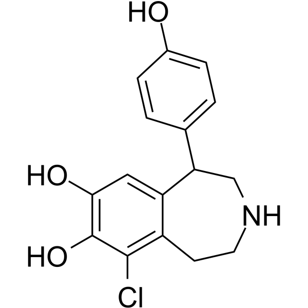
Fenoldopam (SKF-82526) is a selective dopamine D1 receptor agonist. Fenoldopam binds to rat D1B dopamine receptors (Kd = 11 nM) and human D1A receptors (Kd = 17 nM) in COS-7 cell membranes expressing the cloned receptors. Fenoldopam modulates dopamine receptor expression, induces vasorelaxation in vascular tissues, reverses glomerular hyperfiltration in rat models, increases intracellular cAMP levels, promotes DARPP-32 phosphorylation in small cell lung cancer (SCLC) cells, inhibits cytokine secretion, and downregulates T cell activation markers in activated human T cells. Fenoldopam can be used for research on hypertension, acute kidney injury, psoriasis, and small cell lung cancer.
For research use only. We do not sell to patients.
- CAS No.: 67227-56-9
- Formula: C16H16ClNO3
- Molecular Weight:305.76
-
Storage:
Please store the product under the recommended conditions in the Certificate of Analysis.

To place orders, for customer services and technical support, please contact: MedChemExpress USA
Tel: 609-228-6898 E-mail: [email protected] [email protected]


-
 Biological Activity
-
Chemical Information
- Protocols
- Purity & Documentation
- References
-
 Help & FAQs


-
HY-B0410
PramipexoleDopamine Receptor Agonist -
HY-B0735A
Fenoldopam mesylateDopamine DA Receptor Agonist -
HY-14763
CariprazineD3/D2/5-HT Modulator -
HY-12705A
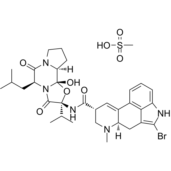
Bromocriptine mesylate -
HY-17355
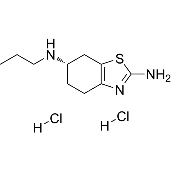
Pramipexole dihydrochlorideDopamine Receptor Agonist -
HY-119486
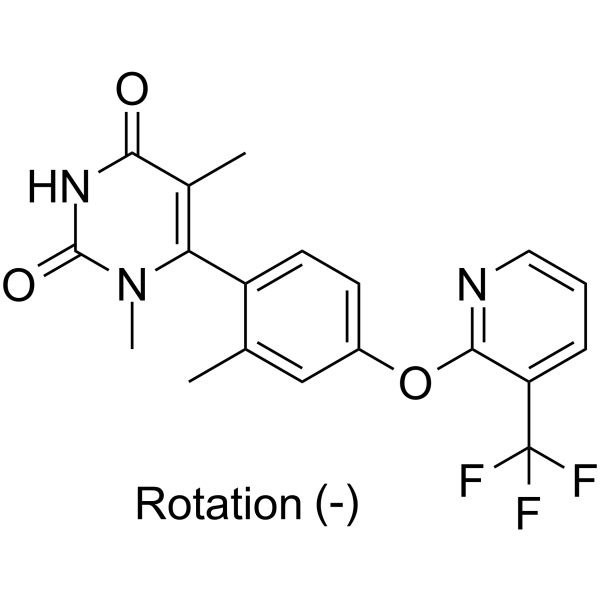
TavapadonDopamine D1/D5 Receptor Agonist -

HY-B0410R
Pramipexole (Standard)Dopamine Receptor Agonist -
HY-14538
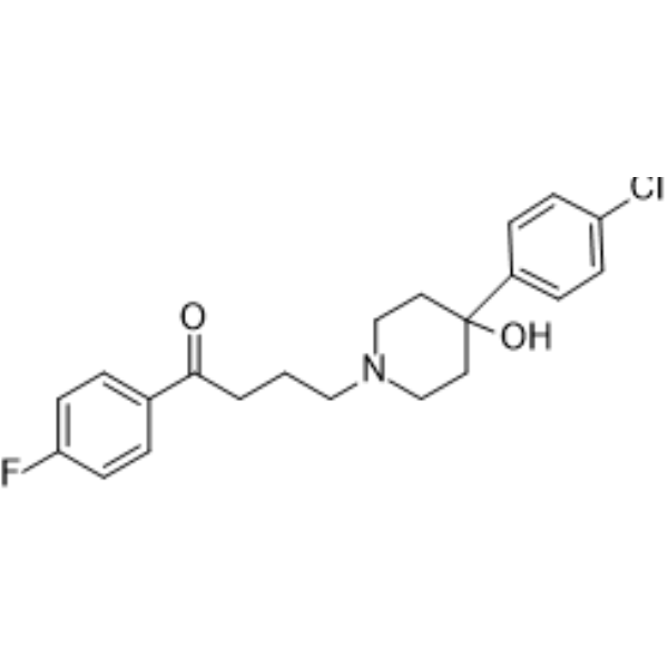
HaloperidolDopamine Receptor Antagonist -
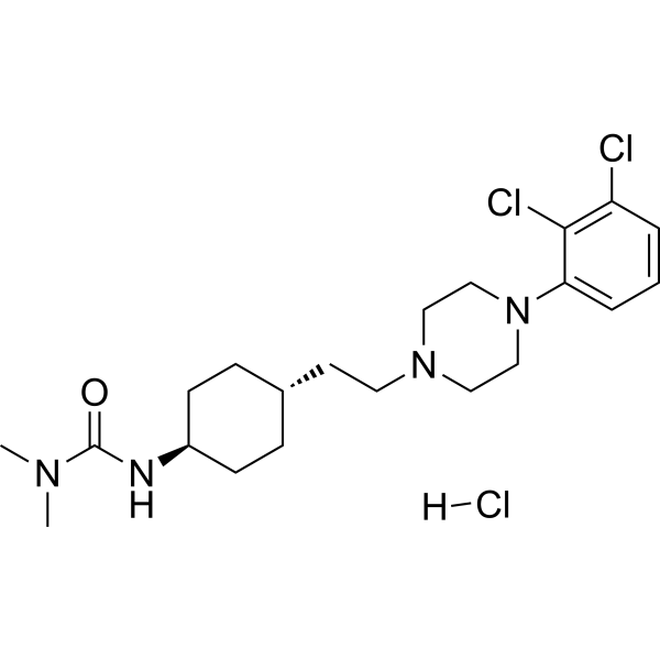
HY-14763A
Cariprazine hydrochlorideDopamine Receptor Modulator -
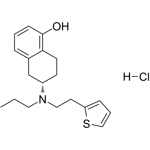
HY-A0007
Rotigotine HydrochlorideDopamine Receptor Agonist -
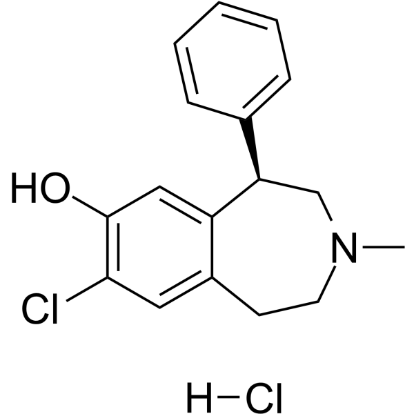
HY-19545A
SCH-23390 hydrochlorideDopamine D1 Receptor Antagonist -

HY-B0735AR
Fenoldopam mesylate (Standard)Reference Standard
Publications Citing Use of MedChemExpress (MCE) Fenoldopam
MoreAll Dopamine Receptor Isoforms
More

Biological Activity
Description
IC50 & Target
[7]|
Human D1 Receptor 17 nM (Kd) |
Rat D1 Receptor 11 nM (Kd) |
Cellular Effect
|
Cell Line

|
Type | Value | Description | References |
|---|---|---|---|---|
| LLC-PK1 | EC50 |
55.5 nM
|
Stimulates cAMP accumulation.
Stimulates cAMP accumulation.
|
2973247 |
In Vitro
Fenoldopam (SKF-82526) (40 min) competes for specific 125I-Sch 23982 binding sites in rat renal cortical membranes with a Ki of 1,975 nM (Tris-HCl buffer) and 1,895 nM (Dulbecco's buffer)[1].
Fenoldopam (1 nM-6 μM; 72 h) does not inhibit the proliferation of CD3/CD28-stimulated Jurkat leukemic T cells, in contrast to its inhibitory effect on normal activated T cells[2].
Fenoldopam (1 μM; 5 h) co-treated Theophylline (HY-B0809) increases intracellular cAMP levels in CD3/CD28-stimulated Jurkat cells[2].
Fenoldopam (1 μM; 10-30 min) increases DARPP-32 phosphorylation in small cell lung cancer (SCLC) H-69 cells and primary SCLC cultures[3].
Fenoldopam (1 μM; 30 180 min) induces cAMP accumulation in SCLC cells and increases BrdU incorporation in SCLC H-69 cells as well as primary SCLC cultures (SCLC H-3)[3].
Fenoldopam (0.1 μM; 48 h) reduces the secretion of TNF-α, IFN-γ, IL-1β, IL-2, IL-4, IL-6, IL-8 and IL-10 by IL-2 activated PBMCs, and downregulates the expression of T cell activation markers CD69, CD28 and intracellular IL-2 in CD3+ T cells from psoriasis patients[4].
Fenoldopam (0.1 μM; 1 h) reduces the chemotactic migration of CD3/CD28-activated normal human T cells towards SDF-1/CXCL12 by 27-55%[4].
Fenoldopam (0.1 μM; 15 sec) induces depolarization of resting and activated normal human T cells[4].
Fenoldopam (10 pM-1 μM; 24 h) increases D1 and D3 receptor protein expression in a concentration- and time-dependent manner in rat aortic smooth muscle A10 cells[6].
Fenoldopam (0.1 μM; 24 h) increases D1/D3 receptor coimmunoprecipitation in A10 cells[6].
Fenoldopam (100 pM-1 mM) induces vasorelaxation in rat mesenteric arterial rings preconstricted with KCl (Emax = 83%, pED50 = 5.0)[6].
Fenoldopam binds to rat D1B dopamine receptors with a Kd of 11 nM and human D1A receptors with a Kd of 17 nM in COS-7 cell membranes expressing the cloned receptors[7].
Fenoldopam (1 nM-100 μM; cumulative) has minimal effect on mouse ductus arteriosus tone, and does not impair O2-or indomethacin-induced ductus constriction[8].
MedChemExpress (MCE) has not independently confirmed the accuracy of these methods. They are for reference only. Further protocols information, click here.
-
Cell Line:Jurkat leukemic T cells, normal activated T cells
-
Concentration:1, 5, 10, 50, 100, 200, 400, 600, 800 nM; 1, 2, 4, 6 μM
-
Incubation Time:72 h
-
Result:Did not inhibit proliferation of CD3/CD28-stimulated Jurkat cells at any concentration tested.
Inhibited proliferation of normal activated T cells with maximum inhibition at 1 μM.
-
Cell Line:SCLC H-69 cells, SCLC primary cultures
-
Concentration:1 μM
-
Incubation Time:10, 30 min
-
Result:Increased DARPP-32 phosphorylation.
-
Cell Line:SCLC H-69 cells, SCLC primary cultures (SCLC-H3)
-
Concentration:1 μM
-
Incubation Time:30, 90, 180 min
-
Result:Induced BrdU incorporation in both cell lines.
-
Cell Line:CD3/CD28-activated normal human T cells
-
Concentration:100 nM
-
Incubation Time:1 h pre-incubation + 4 h migration assay
-
Result:Reduced chemotactic migration towards SDF-1/CXCL12 by 27-55%.
-
Cell Line:A10 cells (rat aortic smooth muscle cells)
-
Concentration:10 pM-1 μM
-
Incubation Time:24 h (concentration-response); 2, 4, 8, 16, 24, 30 h (time-course)
-
Result:Increased D1 and D3 receptor protein expression in a concentration- and time-dependent manner.
In Vivo
Fenoldopam (1, 3, 10 μg/kg; i.v.; 40 min per dose) increases effective renal plasma flow (ERPF) in a dose-dependent manner in conscious rats, with no significant change in GFR, resulting in a decreased filtration fraction[5].
Fenoldopam (1 mg/kg; i.p.; once hourly for 4 doses) does not impair postnatal ductus arteriosus closure in newborn CD-1 mice[8].
MedChemExpress (MCE) has not independently confirmed the accuracy of these methods. They are for reference only.
-
Animal Model:Glycine-induced hyperfiltration study in conscious male SD rats (320-360 g)[5]
-
Dosage:1 μg/kg
-
Administration:i.v.; during glycine infusion
-
Result:Reversed glycine-induced glomerular hyperfiltration and reduced GFR from 1.30 to 1.17 mL/100 g/min.
-
Animal Model:Dose-response study in conscious male SD rats (320-360 g)[5]
-
Dosage:1, 3, 10 μg/kg
-
Administration:i.v.; 40 min per dose
-
Result:Increased effective renal plasma flow (ERPF) in a dose-dependent manner in conscious rats, with no significant change in GFR, resulting in a decreased filtration fraction.
-
Animal Model:Postnatal ductus arteriosus closure study in newborn CD-1 mice[8]
-
Dosage:1 mg/kg
-
Administration:i.p.; once hourly for 4 doses
-
Result:Did not impair postnatal ductus arteriosus closure.
Clinical Trial
| NCT Number | Sponsor | Condition | Start Date |
Phase
|
|---|---|---|---|---|
| NCT01329991 | Plexxikon| | 2011-05 | PHASE1 |


Chemical Information
-
CAS No. 67227-56-9
-
Molecular Weight 305.76
-
Formula C16H16ClNO3
-
SMILES
OC1=C(O)C=C2C(C3=CC=C(O)C=C3)CNCCC2=C1Cl
-
Synonyms
SKF 82526
-
Shipping
Room temperature in continental US; may vary elsewhere.
-
Storage
Please store the product under the recommended conditions in the Certificate of Analysis.


Publications (4)
-
Journal Impact Factor
-
Most Recent
-
Cell
2021 Feb 18;184(4):943-956.e18. PMID: 33571432 -
SLAS Discov
2020 Sep;25(8):895-905. PMID: 32567455 -
Biochem Biophys Res Commun
2022 Jan 15:588:83-89. PMID: 34953210 -
Biomed Pharmacother
Dopamine receptor agonists ameliorate bleomycin-induced pulmonary fibrosis by repressing fibroblast differentiation and proliferation. [Abstract]2021 Jul:139:111500. PMID: 33901873

Protocols
-
Kinase activity and phosphorylation assays
Kinase activity assays measure the ability of kinases to transfer phosphate groups from ATP to specific substrates, while phosphorylation assays detect the presence and levels of phosphorylated proteins. Common methods include radiolabeled ATP incorporation (e. g. ,), ADP release detection via bioluminescence (e. g. ,[3]), enzyme-linked immunosorbent assays (ELISA) for phospho-specific epitopes (e. g. ,[6]), and microtiter-based formats for high-throughput screening (e. g. ,[8]). The ADP-Glo assay quantifies kinase activity by measuring ADP produced during phosphorylation using a luciferase-based system. Radiometric assays involve autoradiography or scintillation counting after incorporation of 32P-labeled ATP into substrate proteins. ELISA-based approaches rely on phospho-specific antibodies to detect activated kinases in cell lysates or purified samples.
-
RNA extraction experimental
By lysing cells, releasing RNA, and removing impurities such as proteins and DNA, high-purity RNA products are finally obtained. The commonly used traditional method is the guanidine isothiocyanate/phenol/chloroform method (Trizol), which is suitable for a variety of animal materials including animal tissues, microorganisms, cultured cells, etc., and most plant materials.
-
Western Blot
Western blotting (WB) is a commonly used experimental method in molecular biology, biochemistry, and immunogenetics for identifying and quantifying target proteins. It combines gel electrophoresis with immunoassay, enabling researchers to analyze protein expression, post-translational modifications, and molecular weight.
-
Nephrotoxicity Study
This protocol assesses nephrotoxicity by combining functional kidney injury readouts, urinary/tissue injury biomarkers, and renal histopathology. Serum creatinine and BUN reflect impaired kidney function, while KIM-1, NGAL, clusterin, osteopontin, IL-18, cystatin C, nephrin, Oat5, urinary protein, glucose, and alkaline phosphatase have been used to detect tubular injury in cisplatin-, gentamicin-, and acetaminophen-induced nephrotoxicity models.
-
Imiquimod-Induced Psoriasiform Dermatitis
Imiquimod (IMQ)-induced psoriasiform dermatitis is a widely used murine model in which topical application of IMQ, a Toll-like receptor 7 (TLR7) agonist, triggers innate immune activation in the skin and induces a psoriasis-like inflammatory cascade characterized by epidermal hyperplasia, immune cell infiltration, and cytokine production dominated by the IL-23/IL-17 axis. This inflammatory response is mediated through activation of dendritic cells and downstream induction of IL-23, IL-17A, IL-22, and related pro-inflammatory mediators, recapitulating key features of human plaque psoriasis and enabling mechanistic and therapeutic studies. The model is commonly induced using Aldara (5% IMQ cream) applied topically to murine skin, resulting in rapid onset of erythema, scaling, and thickening that can be quantified as disease severity indices and validated histologically.
-
Research Protocol for Cardiovascular Diseases
Cardiovascular disease can be modeled as maladaptive cardiac remodeling, where ischemic injury or pressure overload activates inflammatory signaling, fibroblast activation, extracellular-matrix deposition, cardiomyocyte hypertrophy, vascular remodeling, and progressive ventricular dysfunction. The TGF-β/SMAD axis is a central profibrotic pathway after myocardial injury and pressure overload, while innate immune and cytokine pathways regulate leukocyte recruitment, scar formation, and adverse remodeling. Key unresolved questions include which inflammatory signals are reparative versus harmful, when fibrosis is protective versus maladaptive, and whether pathway inhibition improves function without weakening necessary infarct healing or compensatory remodeling.
-
Protocol for Kinase activity and phosphorylation assays
Kinase activity assays measure transfer of phosphate from ATP to a protein or peptide substrate, generating phosphorylated substrate, ADP, or incorporated radiolabeled phosphate as the readout; phosphorylation assays measure site-specific phosphorylation in cells or tissues as a proxy for kinase-pathway activation, inhibition, or substrate regulation. Phosphorylation can be detected by phospho-specific Western blot, immunoprecipitation kinase assay, phospho-immunofluorescence, phospho-flow cytometry, luminescent ADP detection, radiolabeled ATP incorporation, or reporter-based pathway assays, and these readouts can be applied to cancer cells, primary neurons, mouse tumors, organoids, inflammatory macrophages, ferroptosis studies, and mitophagy studies when the kinase target is biologically relevant.
-
Research Protocol for Inflammation-related Diseases
The NLRP3 inflammasome is a cytosolic innate immune signaling platform that integrates priming signals and danger-signal activation to promote caspase-1 activation, maturation of IL-1β and IL-18, and gasdermin D-mediated pyroptotic cell death. The core experimental logic is to determine whether inflammatory disease phenotypes are driven by increased NLRP3 expression, ASC-containing inflammasome assembly, caspase-1 cleavage, GSDMD cleavage, and extracellular release of IL-1β/IL-18 rather than by nonspecific cell injury alone. The pathway is strongly linked to inflammation-related disease phenotypes because monosodium urate crystals activate NALP3/NLRP3 inflammasome signaling in gout-like crystal inflammation, cholesterol crystals activate NLRP3 inflammasomes in atherogenesis models, and DSS-induced intestinal inflammation has been reported to involve NLRP3 inflammasome activity. However, experimental colitis studies also show context-dependent protective effects of NLRP3 inflammasome co
-
Research Protocol for Cancer Immunology
Cancer immunology studies how the immune system recognizes, suppresses, edits, or fails to eliminate malignant cells through tumor antigen release, antigen presentation, T-cell priming, immune trafficking, tumor-cell killing, and feedback inhibition in the tumor microenvironment. The cancer-immunity cycle links tumor antigenicity, dendritic-cell priming, CD8+ T-cell infiltration, cytotoxic function, and immune-checkpoint regulation to tumor rejection or immune escape. Immune-checkpoint pathways such as PD-1/PD-L1 and CTLA-4 suppress antitumor T-cell activity and can be therapeutically blocked, but many tumors remain resistant because of poor antigen presentation, weak T-cell infiltration, suppressive myeloid cells, regulatory T cells, and tumor-intrinsic immune-exclusion programs. Unresolved questions include which immune-cell states predict response, how tumor-intrinsic pathways exclude immune cells, how myeloid suppression limits checkpoint blockade, and which combination strategies


Purity & Documentation
References
[1]. Felder RA, et al. Dopamine1 receptors in rat kidneys identified with 125I-Sch 23982. Am J Physiol. 1988 Nov;255(5 Pt 2):F970-6. [Content Brief]
[2]. Basu B, et al. D1 and D2 dopamine receptor-mediated inhibition of activated normal T cell proliferation is lost in jurkat T leukemic cells. J Biol Chem. 2010 Aug 27;285(35):27026-27032. [Content Brief]
[3]. Cherubini E, et al. Genetic and Functional Analysis of Polymorphisms in the Human Dopamine Receptor and Transporter Genes in Small Cell Lung Cancer. J Cell Physiol. 2016 Feb;231(2):345-56. [Content Brief]
[4]. Keren A, et al. Instantaneous depolarization of T cells via dopamine receptors, and inhibition of activated T cells of Psoriasis patients and inflamed human skin, by D1-like receptor agonist: Fenoldopam. Immunology. 2019 Nov;158(3):171-193. [Content Brief]
[5]. Gillies MA, et al. Fenoldopam to prevent acute kidney injury after major surgery-a systematic review and meta-analysis. Crit Care. 2015 Dec 25;19:449. [Content Brief]
[6]. Zeng C, et al. Dopamine D1 receptor augmentation of D3 receptor action in rat aortic or mesenteric vascular smooth muscles. Hypertension. 2004 Mar;43(3):673-9. [Content Brief]
[7]. Tiberi M, et al. Cloning, molecular characterization, and chromosomal assignment of a gene encoding a second D1 dopamine receptor subtype: differential expression pattern in rat brain compared with the D1A receptor. Proc Natl Acad Sci U S A. 1991 Sep 1;88(17):7491-5. [Content Brief]
[8]. Crockett SL, et al. Role of dopamine and selective dopamine receptor agonists on mouse ductus arteriosus tone and responsiveness. Pediatr Res. 2020 May;87(6):991-997. [Content Brief]

Calculators
Concentration (start) × Volume (start) = Concentration (final) × Volume (final)


Fenoldopam Related Classifications
HY-B0735 Related Classifications
 Powered by Bioz