



Wright's stain

Based on 1 Customer Validation
Wright's stain is a composite cell stain that mainly binds to intracellular nucleic acids, proteins and other components through thiazine dyes (such as methylene blue) and eosin. Wright's stain is pH-dependent (optimal pH 6.4-6.7) and achieves cell morphology resolution by differentially staining the cytoplasm and nucleus. Under alkaline conditions, thiazine dyes bind to nucleic acids to form purple, and acidic eosin binds to cytoplasmic proteins to form red, which can form contrasting cell morphological features. Wright's stain can clearly display the fine structures of blood cells and bone marrow cells (such as nuclear chromatin and granules) and quickly evaluate cell morphological abnormalities.
For research use only. We do not sell to patients.
- Purity: 95%
- CAS No.: 68988-92-1
-
Storage:
4°C, protect from light
* In solvent : -80°C, 6 months; -20°C, 1 month (protect from light)


To place orders, for customer services and technical support, please contact: MedChemExpress USA
Tel: 609-228-6898 E-mail: [email protected] [email protected]


-
 Biological Activity
-
Chemical Information
- Purity & Documentation
- References
-
 Help & FAQs


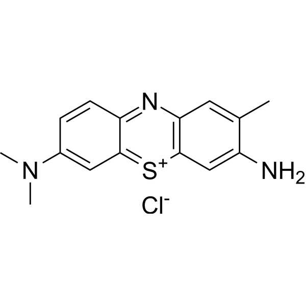
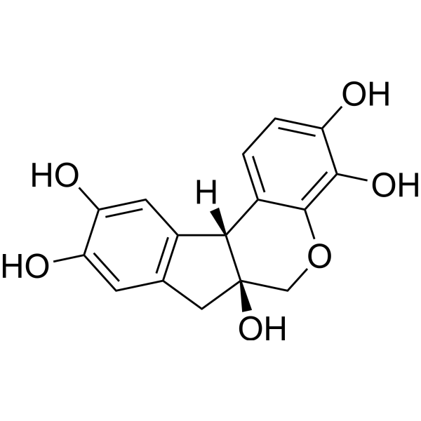
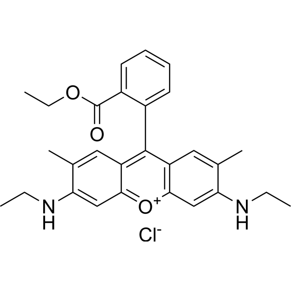
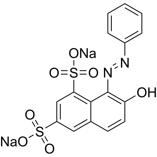
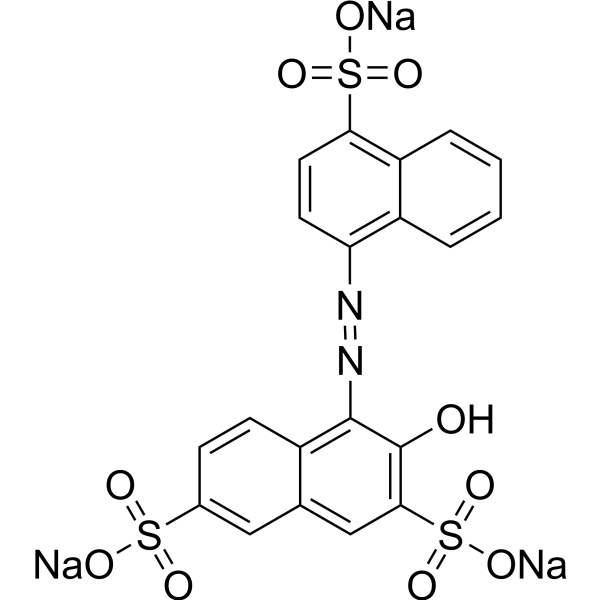
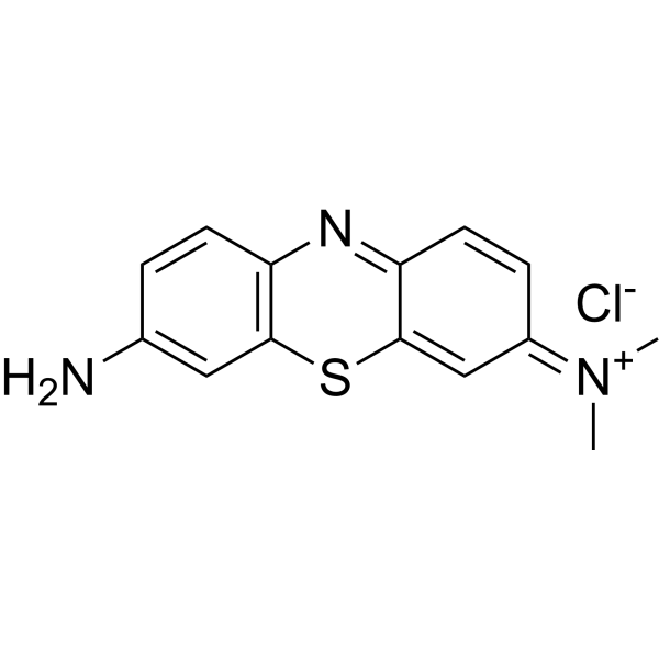
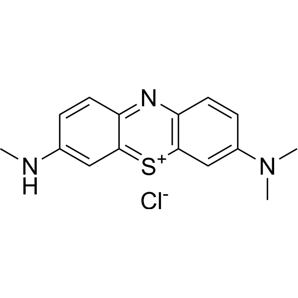
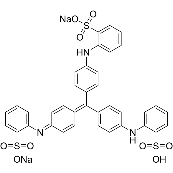


Biological Activity
1. Automated staining of bone marrow and peripheral blood smears: Wright's stain (12 min for peripheral blood smears, 20 min for bone marrow smears) was performed in the Hematrak instrument through a two-stage staining method (first fixation with Wright's solution, then staining with Wright's-buffer-Eosin mixture), which made the red blood cells appear bright orange-red and the white blood cell nuclear chromatin clear, significantly improving the recognition of cell morphology, and with small batch-to-batch differences[1].
2. Combined immunofluorescence detection of leukemia cells: Wright's stain (conventional concentration; 40 min) was combined with upconversion nanoparticle immunolabeling to achieve simultaneous observation of cell morphology (cytoplasm/nuclear structure) and immunofluorescence signals in DLBCL cells and clinical samples, assisting in the identification of leukemia cell phenotypes, with a detection limit as low as 1.54 cell/μL[2].
Wright's stain staining method
Reagents:
(1) 6.3 g Wright's stain dissolved in 3.8 L acetone-free methanol, aged for 1 month.
(2) Phosphate buffer (pH 6.5): Mix 250 mL 0.067 M?Na2HPO4, 250 mL 0.06 M?KH2PO4, and distilled water to 3 L.
(3) Eosin Y solution: 12.5 g Eosin Y, 575 mL distilled water, 1625 mL 95% methanol.
Procedure Fixation and Primary Staining:
Immerse blood/bone marrow smears in Wright's stain for 3 minutes to fix cells and initiate staining.
Buffer-mediated staining: Prepare a 1:5.25 mixture of Wright's stain and phosphate buffer. Stain peripheral blood smears for 5-8 minutes; bone marrow smears for 10-15 minutes to provide nuclear and cytoplasmic contrast.
Secondary Eosin Y stain: Prepare a mixture of Eosin Y solution, Wright's stain, and buffer (1:4:28 ratio). Stain smears for 1-2 minutes to enhance red blood cell staining (bright orange-red) while preserving white blood cell nuclear detail.
Rinse and dry: Wash smears in running distilled water or pH 6.3 buffer for 2 minutes. Air dry completely before microscopic observation.
Key parameters Optimal pH: 6.5-7.15, which allows for balanced red and white blood cell staining.
Total time: approximately 12 minutes for peripheral blood smears; approximately 20 minutes for bone marrow smears.
MedChemExpress (MCE) has not independently confirmed the accuracy of these methods. They are for reference only.


Chemical Information
-
CAS No. 68988-92-1
-
Appearance Solid
-
Color Dark green to black
-
SMILES
[Wright's stain]
-
Shipping
Room temperature in continental US; may vary elsewhere.
-
Storage
4°C, protect from light
* In solvent : -80°C, 6 months; -20°C, 1 month (protect from light)


Purity & Documentation
-
Data Sheet (270 KB)
-
SDS (393 KB)
- English - EN (393 KB)
- Français - FR (393 KB)
- Deutsch - DE (393 KB)
- Norwegian - NO (393 KB)
- Español - ES (393 KB)
- Swedish - SV (393 KB)
- Italian - IT (393 KB)
- Korean - KR (393 KB)
- Portuguese - PT (393 KB)
-
Handling Instructions (2659 KB)
References
[1]. N Kutaish. Automated staining of bone marrow and peripheral blood by a modified Wright's technic. Am J Clin Pathol. 1982 Mar;77(3):319-20. [Content Brief]
[2]. Chen L, et al. A Novel and Rapid Smear Cytomorphology Detection Strategy Based on Upconversion Nanoparticles Immunolabeling Integrated with Wright's Staining for Accurate Diagnosis of Leukemia. Int J Nanomedicine. 2023 Sep 13;18:5213-5224. [Content Brief]

Calculators
Concentration (start) × Volume (start) = Concentration (final) × Volume (final)


Wright's stain Related Classifications
HY-W088068 Related Classifications
 Powered by Bioz