


MitoTEMPO hydrate
Based on 179 publication(s) in Google Scholar
MitoTEMPO hydrate is a mitochondria-targeted antioxidant. MitoTEMPO hydrate induces mitophagy by activating the PINK1/Parkin pathway, inhibits NLRP3 inflammasome activation, restores mitochondrial membrane potential, and improves renal function and podocyte injury. MitoTEMPO hydrate regulates Ca2+ homeostasis, inhibits Bnip3 overexpression, shortens action potential duration, and exerts antiarrhythmic effects. MitoTEMPO hydrate reverses premature senescence, reduces trabecular bone loss, and decreases cell apoptosis. MitoTEMPO hydrate can be used in studies of chronic kidney disease, age-related cardiac dysfunction, postmenopausal osteoporosis, and ischemic stroke.
For research use only. We do not sell to patients.
- Purity: 98.03%
- CAS No.: 1569257-94-8
- Formula: C29H37ClN2O3P
- Molecular Weight:528.04
-
Storage:
-20°C, sealed storage, away from moisture
* In solvent : -80°C, 6 months; -20°C, 1 month (sealed storage, away from moisture)

To place orders, for customer services and technical support, please contact: MedChemExpress USA
Tel: 609-228-6898 E-mail: [email protected] [email protected]


-
 Biological Activity
-
Chemical Information
- Purity & Documentation
- References
-
 Help & FAQs
-


Apoptosis Compound Library
HY-L003
-
Immunology/Inflammation Compound Library
HY-L007
-
Membrane Transporter/Ion Channel Compound Library
HY-L011
-
Metabolism/Protease Compound Library
HY-L012
-
Neuronal Signaling Compound Library
HY-L013
-
NF-κB Signaling Compound Library
HY-L014
-
Stem Cell Signaling Compound Library
HY-L017
-
Autophagy Compound Library
HY-L029
-
Anti-Aging Compound Library
HY-L034
-
Antioxidant Compound Library
HY-L037
-
Oxygen Sensing Compound Library
HY-L045
-
Anti-Cardiovascular Disease Compound Library
HY-L046
-
Endocrinology Compound Library
HY-L047
-
Pyroptosis Compound Library
HY-L059
-
Glutamine Metabolism Compound Library
HY-L064
-
Anti-Cancer Metabolism Compound Library
HY-L083
-
Mitochondria-Targeted Compound Library
HY-L089
-
Glucose Metabolism Compound Library
HY-L092
-
Antidepressant Compound Library
HY-L108
-
Cuproptosis Compound Library
HY-L133
-
Mitochondrial Protection Compound Library
HY-L144
-
Metabolic Enzyme Compound Library
HY-L146
-
Membrane Protein-targeted Compound Library
HY-L149
-
Membrane Receptor-targeted Compound Library
HY-L150
-
Mitochondrial Toxicity Compound Library
HY-L155
-
Autoimmune Disease Compound Library
HY-L156
-
Cell Death Library
HY-L162
-
Ion Channel Compound Library
HY-L166
-
Inflammasomes related Compound Library
HY-L175
-
Multi-Target Compound Library
HY-L176
-
Radioprotector Library
HY-L178
-
Mitophagy Compound Library
HY-L180
-
Bioactive Compound Library Max
HY-L181
-
MCE Bioactive Compound Library
HY-L001V
-
Bioactive Compound Library
HY-L001
-
Anti-Fibrosis Compound Library
HY-L185
-
Non-Alcoholic Fatty Liver Disease (NAFLD) Compound Library
HY-L199
-
High-Throughput Bioactive Compound Library
HY-L205
-
Pattern Recognition Receptors Library
HY-L237


Publications Citing Use of MedChemExpress (MCE) MitoTEMPO hydrate
More- Science. 2025 Mar 7;387(6738):eadq2509. [Abstract]
- Immunity. 2025 Apr 8;58(4):811-825.e7. [Abstract]
- Immunity. 2024 Jun 19:S1074-7613(24)00305-4. [Abstract]
- Nat Microbiol. 2025 Oct;10(10):2521-2536. [Abstract]
- ACS Nano. 2025 Mar 25;19(11):11029-11048. [Abstract]
- Nat Commun. 2026 Jun 8. [Abstract]
- Nat Commun. 2026 Jan 31;17(1):2227. [Abstract]
- Nat Commun. 2025 Aug 26;16(1):7954. [Abstract]
- Nat Commun. 2023 Feb 16;14(1):872. [Abstract]
- Cell Death Differ. 2024 Nov;31(11):1519-1533. [Abstract]
- Acta Pharm Sin B. 2022 Feb;12(2):759-773. [Abstract]
- J Extracell Vesicles. 2026 Apr;15(4):e70277. [Abstract]
- Autophagy. 2026 Jun 16:1-20. [Abstract]
- Autophagy. 2024 Apr;20(4):752-768. [Abstract]
- Adv Sci (Weinh). 2026 Apr;13(19):e16090. [Abstract]
- Adv Sci (Weinh). 2025 Sep 26:e05486. [Abstract]
- Adv Sci (Weinh). 2025 Sep 15:e07283. [Abstract]
- Adv Sci (Weinh). 2023 Mar;10(9):e2207084. [Abstract]
- Nat Plants. 2026 Mar;12(3):635-652. [Abstract]
- Leukemia. 2023 Apr;37(4):765-775. [Abstract]
- Theranostics. 2025 Jan 20;15(6):2360-2374. [Abstract]
- Biomaterials. 2025 Sep:320:123259. [Abstract]
- J Exp Clin Cancer Res. 2025 Jul 4;44(1):193. [Abstract]
- J Nanobiotechnology. 2026 Apr 2;24(1):431. [Abstract]
- Sci Adv. 2025 Jul 18;11(29):eadw5228. [Abstract]
- J Biomed Sci. 2023 Nov 7;30(1):91. [Abstract]
- Redox Biol. 2026 Jun 11:95:104255. [Abstract]
- Redox Biol. 2026 Jun:93:104179. [Abstract]
- Redox Biol. 2025 Sep:85:103750. [Abstract]
- Redox Biol. 2024 Nov 20:78:103439. [Abstract]
- Redox Biol. 2023 Sep:65:102828. [Abstract]
- Redox Biol. 2023 Jun:62:102702. [Abstract]
- Redox Biol. 2023 Feb:59:102587. [Abstract]
- Redox Biol. 2020 Jul;34:101559. [Abstract]
- Environ Sci Technol. 2025 Aug 5;59(30):15705-15719. [Abstract]
- J Hazard Mater. 2024 Jun 7:475:134854. [Abstract]
- J Hazard Mater. 2024 May 15:470:134142. [Abstract]
- J Hazard Mater. 2024 Apr 5:467:133719. [Abstract]
- J Hazard Mater. 2023 Sep 5:457:131750. [Abstract]
- J Hazard Mater. 2022 Feb 15;424(Pt A):127268. [Abstract]
- J Immunother Cancer. 2024 Nov 24;12(11):e009805. [Abstract]
- Mol Biomed. 2025 Oct 2;6(1):75. [Abstract]
- Int J Biol Sci. 2024 Aug 19;20(11):4476-4495. [Abstract]
- Int J Biol Sci. 2024 Apr 29;20(7):2658-2685. [Abstract]
- Int J Biol Sci. 2022 Mar 6;18(6):2261-2276. [Abstract]
- Environ Int. 2023 Aug:178:108138. [Abstract]
- Cell Death Dis. 2026 Mar 27;17(1):413. [Abstract]
- Cell Death Dis. 2025 Jul 1;16(1):482. [Abstract]
- Cell Death Dis. 2020 May 5;11(5):319. [Abstract]
- Genes Dis. 2025 Sep 23.
- Cell Commun Signal. 2023 May 25;21(1):123. [Abstract]
- Dev Cell. 2025 Apr 18:S1534-5807(25)00206-0. [Abstract]
- Int J Biol Macromol. 2026 Apr:355:151129. [Abstract]
- Int J Biol Macromol. 2025 Dec;334(Pt 1):149021. [Abstract]
- Acta Pharmacol Sin. 2025 Jul;46(7):1974-1989. [Abstract]
- Acta Pharmacol Sin. 2024 Aug;45(8):1660-1672. [Abstract]
- Phytomedicine. 2024 Nov 14:136:156260. [Abstract]
- Phytomedicine. 2024 Dec:135:156139. [Abstract]
- Phytomedicine. 2024 Jul:129:155570. [Abstract]
- Free Radic Biol Med. 2026 Jul 8:254:690-700. [Abstract]
- Free Radic Biol Med. 2026 May:248:255-271. [Abstract]
- Free Radic Biol Med. 2025 Jun 14:237:585-599. [Abstract]
- Free Radic Biol Med. 2025 Apr 5:S0891-5849(25)00215-1. [Abstract]
- Free Radic Biol Med. 2025 Mar 28:S0891-5849(25)00192-3. [Abstract]
- Free Radic Biol Med. 2025 Feb 16:228:183-196. [Abstract]
- Free Radic Biol Med. 2024 Nov 5:225:856-870. [Abstract]
- Free Radic Biol Med. 2024 Jul 9:S0891-5849(24)00552-5. [Abstract]
- Free Radic Biol Med. 2024 Aug 1:220:179-191. [Abstract]
- Free Radic Biol Med. 2024 Feb 20:212:493-504. [Abstract]
- Sci Total Environ. 2023 Dec 20:905:166890. [Abstract]
- Basic Res Cardiol. 2022 Aug 23;117(1):40. [Abstract]
- J Orthop Translat. 2026 Jan 6.
- J Transl Med. 2025 Nov 10;23(1):1254. [Abstract]
- Biomed Pharmacother. 2022 Sep:153:113280. [Abstract]
- Redox Rep. 2026 Dec;31(1):2632434. [Abstract]
- Redox Rep. 2025 Dec;30(1):2550412. [Abstract]
- Redox Rep. 2024 Dec;29(1):2377870. [Abstract]
- Oncogene. 2025 Sep 16:10.1038/s41388-025-03571-1. [Abstract]
- Environ Pollut. 2024 May 15:349:123874. [Abstract]
- Aging Cell. 2026 Jun;25(6):e70573. [Abstract]
- Cell Death Discov. 2023 Nov 18;9(1):419. [Abstract]
- Cell Rep. 2026 Feb 18;45(3):116978. [Abstract]
- Clin Transl Med. 2025 Jun;15(6):e70385. [Abstract]
- Clin Transl Med. 2025 Apr;15(4):e70289. [Abstract]
- Clin Transl Med. 2024 Apr;14(4):e1653. [Abstract]
- Antioxidants (Basel). 2022 Jul 26;11(8):1455. [Abstract]
- Phytother Res. 2025 Feb;39(2):581-592. [Abstract]
- J Agric Food Chem. 2026 Apr 29;74(16):13213-13229. [Abstract]
- Cell Mol Life Sci. 2025 Jun 5;82(1):226. [Abstract]
- J Agric Food Chem. 2025 Apr 10. [Abstract]
- J Agric Food Chem. 2023 Nov 1;71(43):16310-16322. [Abstract]
- J Agric Food Chem. 2023 Aug 30;71(34):12645-12656. [Abstract]
- Ecotoxicol Environ Saf. 2026 Jan 15:310:119774. [Abstract]
- Ecotoxicol Environ Saf. 2025 Apr 3:295:118139. [Abstract]
- Ecotoxicol Environ Saf. 2024 Mar 15:273:116150. [Abstract]
- Int J Mol Med. 2025 Jun;55(6):96. [Abstract]
- Biochem Pharmacol. 2025 Apr 4:116932. [Abstract]
- Cell Prolif. 2023 Sep;56(9):e13442. [Abstract]
- NanoImpact. 2025 Jun 15:39:100572. [Abstract]
- J Ethnopharmacol. 2025 Nov 28:358:120957. [Abstract]
- Mar Drugs. 2025 Mar 12;23(3):123. [Abstract]
- Chem Biol Interact. 2024 Dec 31:111368. [Abstract]
- Chem Biol Interact. 2025 Jan 5:405:111269. [Abstract]
- J Ethnopharmacol. 2024 Jun 11:118455. [Abstract]
- Life Sci. 2026 Mar 1:388:124199. [Abstract]
- Life Sci. 2025 May 30:123780. [Abstract]
- PLoS Pathog. 2024 Nov 5;20(11):e1012614. [Abstract]
- Int J Mol Sci. 2023 Mar 27;24(7):6309. [Abstract]
- Int Immunopharmacol. 2026 May 14:182:116841. [Abstract]
- Int Immunopharmacol. 2026 Jan 1;168(Pt 1):115862. [Abstract]
- Eur J Pharmacol. 2025 Oct 15:1005:178118. [Abstract]
- Int Immunopharmacol. 2025 Aug 11:164:115325. [Abstract]
- Int Immunopharmacol. 2025 May 30:160:114984. [Abstract]
- Int Immunopharmacol. 2024 Oct 29;143(Pt 2):113504. [Abstract]
- Eur J Pharmacol. 2024 Apr 15:969:176459. [Abstract]
- Arthritis Res Ther. 2025 Dec 15;27(1):226. [Abstract]
- Hepatol Commun. 2024 Mar 18;8(4):e0399. [Abstract]
- Arthritis Res Ther. 2023 Jul 19;25(1):121. [Abstract]
- Molecules. 2023 Jun 14;28(12):4770. [Abstract]
- Mol Oncol. 2025 Dec 2. [Abstract]
- Cancers (Basel). 2025 Dec 27;18(1):92. [Abstract]
- FASEB J. 2026 May 31;40(10):e71812. [Abstract]
- J Cell Mol Med. 2025 Jul;29(13):e70613. [Abstract]
- Biochim Biophys Acta Mol Basis Dis. 2025 Apr 26;1871(6):167874. [Abstract]
- Biochim Biophys Acta Mol Basis Dis. 2025 Apr 23;1871(6):167866. [Abstract]
- Biochim Biophys Acta Mol Basis Dis. 2023 Aug;1869(6):166740. [Abstract]
- FASEB J. 2023 Jun;37(6):e22954. [Abstract]
- Biochim Biophys Acta Mol Basis Dis. 2023 Mar;1869(3):166613. [Abstract]
- FASEB J. 2022 Aug;36(8):e22475. [Abstract]
- J Inflamm Res. 2025 Aug 14:18:11095-11108. [Abstract]
- Cell Calcium. 2025 Aug 26:132:103071. [Abstract]
- Diabetol Metab Syndr. 2025 Jul 16;17(1):267. [Abstract]
- Aquaculture. 2025 Feb 15.
- Fish Shellfish Immunol. 2024 Jan:144:109261. [Abstract]
- Aging (Albany NY). 2020 Jun 11;12(11):11116-11138. [Abstract]
- Biochim Biophys Acta Mol Cell Res. 2025 Dec;1872(8):120059. [Abstract]
- Cell Signal. 2025 Apr 22:111829. [Abstract]
- Cell Signal. 2022 May:93:110304. [Abstract]
- Biol Trace Elem Res. 2026 Mar 27. [Abstract]
- J Proteome Res. 2025 Aug 1;24(8):4098-4113. [Abstract]
- Biol Trace Elem Res. 2024 Sep;202(9):4180-4190. [Abstract]
- Front Med. 2022 Jul 22:9:887062. [Abstract]
- Mol Hum Reprod. 2020 Oct 1;26(10):773-783. [Abstract]
- Front Oncol. 2022 May 18:12:874900. [Abstract]
- OncoTargets and Therapy Dovepress. 2020 May 18;15:1093-1101. [Abstract]
- Arch Biochem Biophys. 2026 Jun:780:110790. [Abstract]
- Arch Biochem Biophys. 2024 Jul 17:110102. [Abstract]
- J Diabetes Investig. 2023 Jan;14(1):28-36. [Abstract]
- FEBS Lett. 2021 Oct;595(19):2447-2462. [Abstract]
- J Clin Lab Anal. 2026 Mar;40(5):e70172. [Abstract]
- Oral Dis. 2025 Feb;31(2):577-588. [Abstract]
- Mol Pain. 2025 Aug 28:17448069251377633. [Abstract]
- Virus Res. 2023 Dec:338:199238. [Abstract]
- PLoS One. 2020 Oct 1;15(10):e0239659. [Abstract]
- Tissue Cell. 2025 Sep 18:98:103141. [Abstract]
- Placenta. 2023 Nov:143:1-11. [Abstract]
- Vet Sci. 2024 Dec 12;11(12):643. [Abstract]
- Biomol Biomed. 2025 Oct 29. [Abstract]
- Biochem Biophys Rep. 2025 Jan 9:41:101909. [Abstract]
- Biochem Biophys Res Commun. 2023 Nov 5:680:184-193. [Abstract]
- Arch Oral Biol. 2023 Mar:147:105632. [Abstract]
- Dev Neurosci. 2022;44(4-5):309-319. [Abstract]
- Biol Pharm Bull. 2025;48(11):1741-1752. [Abstract]
- Biosci Biotechnol Biochem. 2024 May 8:zbae058. [Abstract]
- bioRxiv. 2026 May 29:2026.05.26.727520. [Abstract]
- Res Sq. 2026 Apr 21.
- bioRxiv. 2025 Nov 11.
- SSRN. 2025 Sep 17.
- Keimyung University. 2025.
- SSRN. 2025 Mar 20.
- Res Sq. 2024 Sep 22.
- Res Sq. 2024 Jun 09.
- Clin Complement Med Pharmacol. 2024 May 21.
- Research Square Preprint. 2024 Mar 19.
- Research Square Preprint. 2024 Mar 19.
- Authorea. 2023 Aug 7.
- Oxid Med Cell Longev. 2023 Feb 10:2023:6726654. [Abstract]
- Oxid Med Cell Longev. 2023 Feb 17:2023:1708251. [Abstract]
- Research Square Preprint. 2022 May.
 Customer Validation & Images

-
IF
-
WB
-
WB
All Calcium Channel Isoforms
More

Biological Activity
MitoTEMPO (200 μM; 24 h) hydrate inhibits the activation of the NLRP3 inflammasome and protects human podocytes (HPC) from TNF-α-induced injury[1].
MitoTEMPO (200 μM; 24 h) hydrate improves mitochondrial function and induces PINK1/Parkin pathway-mediated mitophagy in TNF-α-injured human podocytes (HPC)[1].
MitoTEMPO (200 μM; 24 h) hydrate inhibits the activation of the NLRP3 inflammasome in TNF-α-injured human podocytes (HPC) in a Parkin-dependent manner[1].
MitoTEMPO (0.1-10 μM; 2 days) hydrate significantly reduces mitochondrial superoxide levels in bone marrow mesenchymal stem cells (BMSCs) of rats in the sham-operated group, short-term ovariectomized (ST-OVX) group, and long-term ovariectomized (LT-OVX) group, with the most prominent effect on BMSCs in the LT-OVX group at the concentration of 1 μM[3].
MitoTEMPO (1 μM; 7 days) hydrate upregulates the activity of alkaline phosphatase (ALP), an early osteogenic marker, in bone marrow mesenchymal stem cells (BMSCs) of rats with long-term ovariectomy (LT-OVX), and reverses the activity reduction caused by estrogen deficiency[3].
MitoTEMPO (1 μM; 2 days) hydrate alleviates premature senescence of bone marrow mesenchymal stem cells (BMSCs) in ovariectomized (LT-OVX) rats by reducing the expression of senescence markers, decreasing DNA damage, and ameliorating cell proliferation impairment[3].
MitoTEMPO (1 μM; 2 days) hydrate restores lysosomal acidity, reduces mature CTSB levels, and alleviates lysosomal dysfunction in bone marrow mesenchymal stem cells (BMSCs) from ovariectomized (LT-OVX) rats induced by estrogen deficiency[3].
MitoTEMPO (1 μM; 2 days) hydrate enhances mitophagy in bone marrow mesenchymal stem cells (BMSCs) from ovariectomized (LT-OVX) rats, which is evidenced by increased colocalization of mitochondrial markers and lysosomal markers[3].
MitoTEMPO (1 μM; 2 days) hydrate inhibits the activation of mitochondrial unfolded protein response (UPRmt) in bone marrow mesenchymal stem cells (BMSCs) of ovariectomized (LT-OVX) rats induced by estrogen deficiency by reducing the expression of HSP60 and CLPP proteins[3].
MitoTEMPO (1 μM; 2 days) hydrate restores mitochondrial membrane potential in bone marrow mesenchymal stem cells (BMSCs) from ovariectomized (LT-OVX) rats and alleviates estrogen deficiency-induced mitochondrial dysfunction[3].
MitoTEMPO (1 μM; 2 days) hydrate improves mitochondrial respiratory function, including basal respiration, ATP-coupled respiration, and maximal respiration, in bone marrow mesenchymal stem cells (BMSCs) from ovariectomized (LT-OVX) rats[3].
MedChemExpress (MCE) has not independently confirmed the accuracy of these methods. They are for reference only.
-
Cell Line:human podocyte cells (HPC)
-
Concentration:200 μM (Target Reagent); 100 nM (Parkin siRNA)
-
Incubation Time:24 h (co-treated with TNF-α); 24 h (Parkin siRNA transfection prior to TNF-α and Target Reagent treatment)
-
Result:Lost the ability to significantly reduce the levels of NLRP3, cleaved caspase-1, or mature IL-1β in Parkin-silenced HPC, reversing the inhibitory effect observed in non-silenced cells.
MitoTEMPO (0.6 mg/kg; i.p.; once daily for 4 weeks) hydrate alleviates trabecular bone loss and reduces the expression of senescence and mitochondrial stress markers in ovariectomized rats with osteoporosis[3].
MitoTEMPO (0.7 mg/kg/day; i.p.; once daily for 14 consecutive days) hydrate exerts protective effects against ischemia-reperfusion-induced cardiac and neurological dysfunction in male Wistar albino rats, as evidenced by the normalization of hemodynamic, electrocardiographic, biochemical and histological parameters compared with the IR group[4].
MedChemExpress (MCE) has not independently confirmed the accuracy of these methods. They are for reference only.
-
Animal Model:Sprague-Dawley (male, 6-8 weeks old, 180-220 g, CKD induced by subcutaneous injection of 1 mg C-BSA emulsion followed by tail vein injection of 0.5 mg C-BSA every other day for 21 days)[1]
-
Dosage:0.7 mg/kg
-
Administration:i.p.; daily; 7 days
-
Result:Significantly reduced 24-hour urinary protein levels at 35 days post-modeling.
Decreased serum creatinine (SCR) and blood urea nitrogen (BUN) levels compared to CKD model rats.
Reduced mean density of podocyte injury marker desmin by ~55% compared to CKD model rats.
Increased mean density of slit diaphragm protein podocin compared to CKD model rats.
Suppressed podocyte foot process fusion and reduced thickened glomerular basement membrane (GBM) thickness compared to CKD model rats.
Reduced colocalization of NLRP3 and ASC in glomeruli compared to CKD model rats.
Decreased protein levels of NLRP3, cleaved caspase-1, and mature IL-1β compared to CKD model rats.
Lowered mRNA levels of IL-1β and TNF-α compared to CKD model rats .
Increased LC3 II/I ratio, PINK1, and Parkin protein levels compared to CKD model rats.
Decreased p62 protein levels compared to CKD model rats.
Enhanced colocalization of LC3 and mitochondrial marker COX IV compared to CKD model rats.
-
Animal Model:Sprague-Dawley (female, 10 weeks old at procurement, average weight 220 g; bilateral ovariectomy-induced osteoporosis model)[3]
-
Dosage:0.6 mg/kg
-
Administration:i.p.; daily; 4 weeks
-
Result:Significantly inhibited trabecular bone loss in OVX rats.
Improved trabecular bone thickness and density in treated OVX rats compared to vehicle-treated OVX rats.
Restored bone mineralization and deposition capabilities in treated OVX rats.
Reduced the expression of senescence-related marker p53 in the trabecular bone region of the proximal tibia.
Significantly decreased the expression of HSP60 and CLPP, markers of mitochondrial unfolded protein response, in OVX rats.
-
Animal Model:Wistar Albino (17-week-old male, initial body weight 250-304 g, middle cerebral artery occlusion followed by 3 days of reperfusion)[4]
-
Dosage:0.7 mg/kg/day
-
Administration:i.p.; daily; 14 days
-
Result:Increased final body weight to 302.57 g, compared to 285.75 g for the distilled water group.
Reduced heart weight/body weight ratio to 3.01 mg/g, which was statistically significantly lower than the ischemia-reperfusion (IR) group's 3.39 mg/g.
Normalized volume of electrically participating tissue, left ventricular ejection time, and heart rate to levels not significantly different from the sham group, and statistically significantly different from the IR group.
Normalized P-R interval, QTc, T-wave time, T-wave repolarization time, and R-R interval to levels not significantly different from the sham group, and statistically significantly different from the IR group.
Increased blood serum total antioxidant status (TAS) compared to the IR group; decreased total oxidant status (TOS) and oxidative stress index (OSI) compared to the IR group.
Increased heart left ventricle cyclic adenosine monophosphate (cAMP) and inositol triphosphate (IP3) levels to values close to the sham group and statistically significantly higher than the IR group.
Decreased brain right cerebral hemisphere catalase levels compared to the IR group.
Improved heart left ventricle tissue morphology to show mostly normal cardiac muscle cells with minimal myofibril degeneration, compared to widespread abnormalities in the IR group.
Improved brain right cerebral hemisphere tissue morphology to show mostly normal neurons and glial cells with fewer degenerative changes than the IR group.


Chemical Information
-
CAS No. 1569257-94-8
-
Appearance Solid
-
Molecular Weight 528.04
-
Formula C29H37ClN2O3P
-
Color Orange to red
-
SMILES
[O]N1C(C)(C)CC(NC(C[P+](C2=CC=CC=C2)(C3=CC=CC=C3)C4=CC=CC=C4)=O)CC1(C)C.[Cl-].O
-
Shipping
Room temperature in continental US; may vary elsewhere.
-
Storage
-20°C, sealed storage, away from moisture
* In solvent : -80°C, 6 months; -20°C, 1 month (sealed storage, away from moisture)


Publications (179)
-
Journal Impact Factor
-
Most Recent
-
Science
2025 Mar 7;387(6738):eadq2509. PMID: 40048515 -
Immunity
Mitochondrial DNA released by senescent tumor cells enhances PMN-MDSC-driven immunosuppression through the cGAS-STING pathway. [Abstract]2025 Apr 8;58(4):811-825.e7. PMID: 40203808 -
Immunity
A metabolic switch orchestrated by IL-18 and the cyclic dinucleotide cGAMP programs intestinal tolerance. [Abstract]2024 Jun 19:S1074-7613(24)00305-4. PMID: 38906145 -
Nat Microbiol
Oxaloacetate sensing promotes innate immune antiviral defence against influenza virus infection. [Abstract]2025 Oct;10(10):2521-2536. PMID: 40983701 -
ACS Nano
Adverse Outcome Pathway-Based Strategies to Mitigate Ag2Se Quantum Dot-Induced Neurotoxicity. [Abstract]2025 Mar 25;19(11):11029-11048. PMID: 40063898 -
Nat Commun
Mycobacterium tuberculosis IDH-PPARγ interaction suppresses GPX4 to drive macrophage ferroptosis and sustain persistent infection. [Abstract]2026 Jun 8. PMID: 42259813 -
Nat Commun
Microbiota-induced EI24 improves homeostasis but impedes function of alveolar macrophages via metabolic regulation. [Abstract]2026 Jan 31;17(1):2227. PMID: 41620436 -
Nat Commun
Helicobacter hepaticus promotes hepatic steatosis through CdtB-induced mitochondrial stress and lipid metabolism reprogramming. [Abstract]2025 Aug 26;16(1):7954. PMID: 40858606 -
Nat Commun
Oxidized mitochondrial DNA induces gasdermin D oligomerization in systemic lupus erythematosus. [Abstract]2023 Feb 16;14(1):872. PMID: 36797275 -
Cell Death Differ
Vps34 sustains Treg cell survival and function via regulating intracellular redox homeostasis. [Abstract]2024 Nov;31(11):1519-1533. PMID: 39117783 -
Acta Pharm Sin B
Targeting glutamine utilization to block metabolic adaptation of tumor cells under the stress of carboxyamidotriazole-induced nutrients unavailability. [Abstract]2022 Feb;12(2):759-773. PMID: 35256945 -
J Extracell Vesicles
Chemoradiotherapy-Integrated Tumor Cell-Derived Microparticles Mediate Tumor Eradication in Malignant Pleural Effusion. [Abstract]2026 Apr;15(4):e70277. PMID: 41944389 -
Autophagy
Porcine reproductive and respiratory syndrome virus hijacks the non-canonical enzymatic function of PHGDH to arrest autophagic flux for viral replication. [Abstract]2026 Jun 16:1-20. PMID: 42260976 -
Autophagy
2024 Apr;20(4):752-768. PMID: 37798944 -
Adv Sci (Weinh)
Connexin43 Deficiency Leads to Ventricular Arrhythmias by Reprogramming Proline Metabolism. [Abstract]2026 Apr;13(19):e16090. PMID: 41618855 -
Adv Sci (Weinh)
2025 Sep 26:e05486. PMID: 41013886 -
Adv Sci (Weinh)
Redox Cascade in Chicken Skeletal Muscle: SELENOT Suppression in Selenium Deficiency Triggers Disulfidptosis via mtROS-NADPH Dysregulation. [Abstract]2025 Sep 15:e07283. PMID: 40953299 -
Adv Sci (Weinh)
Extracellularly Detectable Electrochemical Signals of Living Cells Originate from Metabolic Reactions. [Abstract]2023 Mar;10(9):e2207084. PMID: 36737855 -
Nat Plants
2026 Mar;12(3):635-652. PMID: 41644634 -
Leukemia
2023 Apr;37(4):765-775. PMID: 36739349
MitoTEMPO hydrate purchased from MedChemExpress. Usage Cited in: Leukemia. 2023 Apr;37(4):765-775. [Abstract]
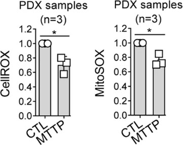
Mito-TEMPO (MTTP; 50 nM; 48 h) efficiently reduces total and mitochondrial ROS content in PDX AML cells. CellROX dye, left panel, MitoSOX dye, right panel.
-
Theranostics
Inhibition of macrophage inflammasome assembly and pyroptosis with GC-1 ameliorates acute lung injury. [Abstract]2025 Jan 20;15(6):2360-2374. PMID: 39990234 -
Biomaterials
Selective and iron-independent ferroptosis in cancer cells induced by manipulation of mitochondrial fatty acid oxidation. [Abstract]2025 Sep:320:123259. PMID: 40112511 -
J Exp Clin Cancer Res
M2 macrophage-secreted KYNU promotes stemness remodeling and malignant behavior in endometrial cancer via the SOD2-mtROS-ERO1α-UPRER axis. [Abstract]2025 Jul 4;44(1):193. PMID: 40616124 -
J Nanobiotechnology
Ag2Se quantum dots neurotoxicity: mitochondrial stress drives ferroptosis-dependent microglial inflammation. [Abstract]2026 Apr 2;24(1):431. PMID: 41928263 -
Sci Adv
2025 Jul 18;11(29):eadw5228. PMID: 40668928 -
J Biomed Sci
Spatiotemporal roles of AMPK in PARP-1- and autophagy-dependent retinal pigment epithelial cell death caused by UVA. [Abstract]2023 Nov 7;30(1):91. PMID: 37936170 -
Redox Biol
TRPV4 drives macrophage pyroptosis via mitochondrial dysfunction and mtROS-dependent NLRP3 inflammasome activation in acute lung injury. [Abstract]2026 Jun 11:95:104255. PMID: 42314332 -
Redox Biol
Redox-triggered USP18 confers cisplatin resistance in ovarian cancer by selectively activating a non-canonical FSP1-dependent ferroptosis escape pathway. [Abstract]2026 Jun:93:104179. PMID: 42030646 -
Redox Biol
EMAP-II from macrophage-derived extracellular vesicles drives neutrophil extracellular traps formation via PI3K/AKT/mtROS in lung ischemia/reperfusion injury. [Abstract]2025 Sep:85:103750. PMID: 40616949 -
Redox Biol
Podocyte SIRPα reduction in diabetic nephropathy aggravates podocyte injury by promoting pyruvate kinase M2 nuclear translocation. [Abstract]2024 Nov 20:78:103439. PMID: 39586122 -
Redox Biol
Epigenetic modulation of Drp1-mediated mitochondrial fission by inhibition of S-adenosylhomocysteine hydrolase promotes vascular senescence and atherosclerosis. [Abstract]2023 Sep:65:102828. PMID: 37517319 -
Redox Biol
BNP protects against diabetic cardiomyopathy by promoting Opa1-mediated mitochondrial fusion via activating the PKG-STAT3 pathway. [Abstract]2023 Jun:62:102702. PMID: 37116257 -
Redox Biol
PM2.5 increases susceptibility to acute exacerbation of COPD via NOX4/Nrf2 redox imbalance-mediated mitophagy. [Abstract]2023 Feb:59:102587. PMID: 36608590 -
Redox Biol
Baicalin combats glutamate excitotoxicity via protecting glutamine synthetase from ROS-induced 20S proteasomal degradation. [Abstract]2020 Jul;34:101559. PMID: 32473460
MitoTEMPO hydrate purchased from MedChemExpress. Usage Cited in: Redox Biol. 2020 Jul;34:101559. [Abstract]
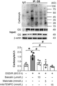
GS carbonylation in astrocytes under OGD/R. Mito-TEMPO decreases GS carbonylation in astrocytes.
-
Environ Sci Technol
Chronic Dietary Exposure to Environmental Levels of Glyphosate Increases the Risk of Reproductive Dysfunction in Male Mice. [Abstract]2025 Aug 5;59(30):15705-15719. PMID: 40698942 -
J Hazard Mater
Potential mechanisms of aortic medial degeneration promoted by co-exposure to microplastics and lead. [Abstract]2024 Jun 7:475:134854. PMID: 38889468 -
J Hazard Mater
Environmental cadmium inhibits testicular testosterone synthesis via Parkin-dependent MFN1 degradation. [Abstract]2024 May 15:470:134142. PMID: 38555669 -
J Hazard Mater
Sodium sulfite triggered hepatic apoptosis, necroptosis, and pyroptosis by inducing mitochondrial damage in mice and AML-12 cells. [Abstract]2024 Apr 5:467:133719. PMID: 38335615 -
J Hazard Mater
MDM2 upregulation induces mitophagy deficiency via Mic60 ubiquitination in fetal microglial inflammation and consequently neuronal DNA damage caused by exposure to ZnO-NPs during pregnancy. [Abstract]2023 Sep 5:457:131750. PMID: 37315416 -
J Hazard Mater
Gestational exposure to environmental cadmium induces placental apoptosis and fetal growth restriction via Parkin-modulated MCL-1 degradation. [Abstract]2022 Feb 15;424(Pt A):127268. PMID: 34583167 -
J Immunother Cancer
Propafenone facilitates mitochondrial-associated ferroptosis and synergizes with immunotherapy in melanoma. [Abstract]2024 Nov 24;12(11):e009805. PMID: 39581704 -
Mol Biomed
Iron accumulation in hypothalamus promotes age-dependent obesity and metabolic dysfunction of male mice. [Abstract]2025 Oct 2;6(1):75. PMID: 41037185 -
Int J Biol Sci
Chronic Stress-induced Serotonin Impairs Intestinal Epithelial Cell Mitochondrial Biogenesis via the AMPK-PGC-1α Axis. [Abstract]2024 Aug 19;20(11):4476-4495. PMID: 39247815 -
Int J Biol Sci
Mitochondrial Dysfunction by FADDosome Promotes Gastric Mucosal Injury in Portal Hypertensive Gastropathy. [Abstract]2024 Apr 29;20(7):2658-2685. PMID: 38725851 -
Int J Biol Sci
Yap is essential for uterine decidualization through Rrm2/GSH/ROS pathway in response to Bmp2. [Abstract]2022 Mar 6;18(6):2261-2276. PMID: 35414789 -
Environ Int
PPARα/ACOX1 as a novel target for hepatic lipid metabolism disorders induced by per- and polyfluoroalkyl substances: An integrated approach. [Abstract]2023 Aug:178:108138. PMID: 37572494 -
Cell Death Dis
2026 Mar 27;17(1):413. PMID: 41896546 -
Cell Death Dis
Exploiting mitochondrial dysfunction to overcome BRAF inhibitor resistance in advanced melanoma: the role of disulfiram as a copper ionophore. [Abstract]2025 Jul 1;16(1):482. PMID: 40592836 -
Cell Death Dis
Nestin protects podocyte from injury in lupus nephritis by mitophagy and oxidative stress. [Abstract]2020 May 5;11(5):319. PMID: 32371936
MitoTEMPO hydrate purchased from MedChemExpress. Usage Cited in: Cell Death Dis. 2020 May 5;11(5):319. [Abstract]
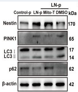
Western blot assay shows the expression of nestin, PINK1, LC3 and p62 in the MPCs, which are pretreated with Mito-TEMPO and exposed to lupus nephritis plasma for 24 h.
-
-
Cell Commun Signal
Tricyclic antidepressants induce liver inflammation by targeting NLRP3 inflammasome activation. [Abstract]2023 May 25;21(1):123. PMID: 37231437 -
Dev Cell
2025 Apr 18:S1534-5807(25)00206-0. PMID: 40280135 -
Int J Biol Macromol
Targeting OXCT1 with the methyl donor S-adenosylmethionine as a therapeutic strategy for cerebral cavernous malformations. [Abstract]2026 Apr:355:151129. PMID: 41865434 -
Int J Biol Macromol
Lipocalin 2 deficiency attenuates NLRP3 inflammasome activation through glycolysis impairment and MGST1-mediated mitochondrial ROS reduction. [Abstract]2025 Dec;334(Pt 1):149021. PMID: 41260431 -
Acta Pharmacol Sin
YY1/HIF-1α/mROS positive-feedback loop exacerbates glomerular mesangial cell proliferation in mouse early diabetic kidney disease. [Abstract]2025 Jul;46(7):1974-1989. PMID: 40038466 -
Acta Pharmacol Sin
Neutrophil extracellular traps promote acetaminophen-induced acute liver injury in mice via AIM2. [Abstract]2024 Aug;45(8):1660-1672. PMID: 38589685 -
Phytomedicine
Salvianolic acid B alleviated myocardial ischemia-reperfusion injury via modulating SIRT3-mediated crosstalk between mitochondrial ROS and NLRP3. [Abstract]2024 Nov 14:136:156260. PMID: 39579610 -
Phytomedicine
Shikonin induces ferroptosis in osteosarcomas through the mitochondrial ROS-regulated HIF-1α/HO-1 axis. [Abstract]2024 Dec:135:156139. PMID: 39423479 -
Phytomedicine
Baicalin attenuates neuronal damage associated with SDH activation and PDK2-PDH axis dysfunction in early reperfusion. [Abstract]2024 Jul:129:155570. PMID: 38579645 -
Free Radic Biol Med
Polystyrene nanoplastics exacerbated the toxicity of cadmium: from the perspective of the placenta-uterus micro-environment. [Abstract]2026 Jul 8:254:690-700. PMID: 42419651 -
Free Radic Biol Med
Lysophosphatidylcholine promotes pulmonary fibrosis following lung injury by facilitating alveolar type 2 cell senescence via Mfsd2a-dependent, Drp1-mediated mitochondrial fission. [Abstract]2026 May:248:255-271. PMID: 41740689 -
Free Radic Biol Med
Alexidine dihydrochloride enhances the sensitivity of human hepatocellular carcinoma to disulfidptosis via ATF4-DDIT3 activation. [Abstract]2025 Jun 14:237:585-599. PMID: 40518125 -
Free Radic Biol Med
Endoplasmic reticulum stress-autophagy axis is involved in copper-induced ovarian ferroptosis. [Abstract]2025 Apr 5:S0891-5849(25)00215-1. PMID: 40194638 -
Free Radic Biol Med
STING immune activation of microglia aggravating neurovascular unit damage in diabetic retinopathy. [Abstract]2025 Mar 28:S0891-5849(25)00192-3. PMID: 40158743 -
Free Radic Biol Med
Lipotoxicity-induced upregulation of FIS1 exacerbates mitochondrial fragmentation and promotes NLRP3-dependent pyroptosis in diabetic cardiomyopathy. [Abstract]2025 Feb 16:228:183-196. PMID: 39734056 -
Free Radic Biol Med
Targeting DUSP26 to drive cardiac mitochondrial dynamics via FAK-ERK signaling in diabetic cardiomyopathy. [Abstract]2024 Nov 5:225:856-870. PMID: 39510451 -
Free Radic Biol Med
2024 Jul 9:S0891-5849(24)00552-5. PMID: 38992393 -
Free Radic Biol Med
Atorvastatin combined with imipenem alleviates lung injury in sepsis by inhibiting neutrophil extracellular trap formation via the ERK/NOX2 signaling pathway. [Abstract]2024 Aug 1:220:179-191. PMID: 38704053 -
Free Radic Biol Med
2024 Feb 20:212:493-504. PMID: 38184120 -
Sci Total Environ
New insights into brain injury in chickens induced by bisphenol A and selenium deficiency-Mitochondrial reactive oxygen species and mitophagy-apoptosis crosstalk homeostasis. [Abstract]2023 Dec 20:905:166890. PMID: 37683847 -
Basic Res Cardiol
Mitochondrial calpain-1 activates NLRP3 inflammasome by cleaving ATP5A1 and inducing mitochondrial ROS in CVB3-induced myocarditis. [Abstract]2022 Aug 23;117(1):40. PMID: 35997820 -
-
J Transl Med
Peroxynitrite regulates ER stress-mediated Ca2+ flux to mitochondria characterizing cardiac microvascular ischemia-reperfusion injury associated with hyperhomocysteinemia. [Abstract]2025 Nov 10;23(1):1254. PMID: 41214689 -
Biomed Pharmacother
2022 Sep:153:113280. PMID: 35724508 -
Redox Rep
Prophylactic quercetin administration attenuates pulmonary fibrosis via ferroptosis-resistant priming of alveolar epithelial cells. [Abstract]2026 Dec;31(1):2632434. PMID: 41697789 -
Redox Rep
FASN regulates CSE-induced apoptosis, oxidative stress and mitochondrial damage in type 2 alveolar epithelial cells by regulating NRF2 expression and nuclear translocation. [Abstract]2025 Dec;30(1):2550412. PMID: 41025365 -
Redox Rep
CISD2 regulates oxidative stress and mitophagy to maintain the balance of the follicular microenvironment in PCOS. [Abstract]2024 Dec;29(1):2377870. PMID: 39010730 -
Oncogene
2025 Sep 16:10.1038/s41388-025-03571-1. PMID: 40957950 -
Environ Pollut
Nano-sized microplastics exposure induces skin cell senescence via triggering the mitochondrial localization of GSDMD. [Abstract]2024 May 15:349:123874. PMID: 38552769 -
Aging Cell
Oxidative Stress and Diminished Mitochondrial Proteostatic Reserve Are Linked to Enhanced mtUPR Initiation in Aged Mouse Muscle. [Abstract]2026 Jun;25(6):e70573. PMID: 42243621 -
Cell Death Discov
Iron derived from NCOA4-mediated ferritinophagy causes cellular senescence via the cGAS-STING pathway. [Abstract]2023 Nov 18;9(1):419. PMID: 37980349 -
Cell Rep
2026 Feb 18;45(3):116978. PMID: 41719124 -
Clin Transl Med
OXA1L deficiency causes mitochondrial myopathy via reactive oxygen species regulated nuclear factor kappa B signalling pathway. [Abstract]2025 Jun;15(6):e70385. PMID: 40551575 -
Clin Transl Med
ATG16L1 restrains macrophage NLRP3 activation and alveolar epithelial cell injury during septic lung injury. [Abstract]2025 Apr;15(4):e70289. PMID: 40211890 -
Clin Transl Med
Mitochondrial dysfunction induced by HIF-1α under hypoxia contributes to the development of gastric mucosal lesions. [Abstract]2024 Apr;14(4):e1653. PMID: 38616702 -
Antioxidants (Basel)
Muscone and (+)-Borneol Cooperatively Strengthen CREB Induction of Claudin 5 in IL-1 β-Induced Endothelium Injury. [Abstract]2022 Jul 26;11(8):1455. PMID: 35892657 -
Phytother Res
Platycodin D2 Mediates Incomplete Autophagy and Ferroptosis in Breast Cancer Cells by Regulating Mitochondrial ROS. [Abstract]2025 Feb;39(2):581-592. PMID: 39581858 -
J Agric Food Chem
Polystyrene Microplastics Induced Hepatocytes Pyroptosis, Apoptosis and Ferroptosis via GSDMD-N-Mediated Mitochondrial Damage. [Abstract]2026 Apr 29;74(16):13213-13229. PMID: 41980172 -
Cell Mol Life Sci
Mitochondrial oxidative stress inhibited Sirt3/Foxo3/PPARα pathway and aggravated copper and zinc co-deficiency-induced hepatic lipotoxicity in a fish model. [Abstract]2025 Jun 5;82(1):226. PMID: 40471448 -
J Agric Food Chem
Apigenin-Mediated ESCRT-III Activation and Mitophagy Alleviate LPS-Induced Necroptosis in Renal Cells. [Abstract]2025 Apr 10. PMID: 40211127 -
J Agric Food Chem
Sodium Sulfite-Triggered Hepatocyte Ferroptosis via mtROS/Lysosomal Membrane Permeabilization-Mediated Lysosome Iron Efflux. [Abstract]2023 Nov 1;71(43):16310-16322. PMID: 37871339 -
J Agric Food Chem
Free Fatty Acids Induce Apoptosis of Mammary Epithelial Cells of Ketotic Dairy Cows via the Mito-ROS/NLRP3 Signaling Pathway. [Abstract]2023 Aug 30;71(34):12645-12656. PMID: 37585786 -
Ecotoxicol Environ Saf
Indoleamine 2,3-dioxygenase 1 mediated macrophage oxidative phosphorylation impairment drives pro-inflammatory M1 polarization aggravates acetaminophen-induced acute liver injury. [Abstract]2026 Jan 15:310:119774. PMID: 41576503 -
Ecotoxicol Environ Saf
Arsenic exposure provoked prostatic PANoptosis by inducing mitochondrial dysfunction in mice and WPMY-1 cells. [Abstract]2025 Apr 3:295:118139. PMID: 40185034 -
Ecotoxicol Environ Saf
2024 Mar 15:273:116150. PMID: 38430579 -
Int J Mol Med
Honokiol ameliorates pyroptosis in intestinal ischemia‑reperfusion injury by regulating the SIRT3‑mediated NLRP3 inflammasome. [Abstract]2025 Jun;55(6):96. PMID: 40280115 -
Biochem Pharmacol
Scutellarein ameliorates pulmonary arterial hypertension via sirtuin 1 mediated deacetylation of nicotinamide nucleotide transhydrogenase. [Abstract]2025 Apr 4:116932. PMID: 40189160 -
Cell Prolif
2023 Sep;56(9):e13442. PMID: 37086012 -
NanoImpact
CdTe quantum dots induce apoptosis in RSC96 cells by disrupting calcium homeostasis and triggering subcellular structural dysfunction. [Abstract]2025 Jun 15:39:100572. PMID: 40527434 -
J Ethnopharmacol
Duhuo Jisheng decoction alleviates intervertebral disc degeneration by inhibiting nucleus pulposus cell pyroptosis via gallic acid-mediated downregulation of HIF-1α. [Abstract]2025 Nov 28:358:120957. PMID: 41317810 -
Mar Drugs
Fucoxanthin from Laminaria japonica Targeting PANoptosis and Ferroptosis Pathways: Insights into Its Therapeutic Potential Against Ovarian Cancer. [Abstract]2025 Mar 12;23(3):123. PMID: 40137309 -
Chem Biol Interact
Integrated network pharmacology, bioinformatics, and experiment analysis to decipher the molecular mechanism of Salidroside on Gastric cancer via targeting NCOA4-mediated ferritinophagy. [Abstract]2024 Dec 31:111368. PMID: 39746501 -
Chem Biol Interact
Aristolochic acid I induced mitochondrial Ca2+ accumulation triggers the production of MitoROS and activates Src/FAK pathway in hepatocellular carcinoma cells. [Abstract]2025 Jan 5:405:111269. PMID: 39426658 -
J Ethnopharmacol
Octahydroindolizine alkaloid Homocrepidine A from Dendrobium crepidatum attenuate P. acnes-induced inflammatory in vitro and in vivo. [Abstract]2024 Jun 11:118455. PMID: 38871011 -
Life Sci
Sinomenine ameliorates myocardial ischemia/reperfusion injury by inhibiting mitochondrial oxidative stress-mediated PANoptosis and ferroptosis via α7nAChR. [Abstract]2026 Mar 1:388:124199. PMID: 41520699 -
Life Sci
METTL3-mediated m6A modification of FUNDC1/IP3R2 pathway facilitates cardiac hypertrophy in obesity hypertension. [Abstract]2025 May 30:123780. PMID: 40451326 -
PLoS Pathog
2024 Nov 5;20(11):e1012614. PMID: 39499730 -
Int J Mol Sci
Hyperglycemia Aggravates Periodontitis via Autophagy Impairment and ROS-Inflammasome-Mediated Macrophage Pyroptosis. [Abstract]2023 Mar 27;24(7):6309. PMID: 37047282 -
Int Immunopharmacol
Albiflorin contributes to Xuebijing-mediated protection against sepsis-associated acute kidney injury by modulating the succinate-PFKFB3 immunometabolic axis. [Abstract]2026 May 14:182:116841. PMID: 42134292 -
Int Immunopharmacol
Inhibition of neutrophil infiltration and NETosis ameliorates sepsis-induced cardiac injury and reduces myocardial inflammation and apoptosis. [Abstract]2026 Jan 1;168(Pt 1):115862. PMID: 41248570 -
Eur J Pharmacol
MPTP controls the release of mtDNA and induces endothelial cell PANoptosis in trichloroethylene-induced immune kidney injury. [Abstract]2025 Oct 15:1005:178118. PMID: 40912515 -
Int Immunopharmacol
ACE2/Ang(1-7)/MasR axis exerts protective effects on lung ischemia/reperfusion injury via NF-κB-dependent mitochondrial adaptation and epithelial cell pyroptosis. [Abstract]2025 Aug 11:164:115325. PMID: 40795499 -
Int Immunopharmacol
Mitophagy-mtROS axis contributes to anti-tuberculosis-induced liver injury through activation of the cGAS-STING pathway in rat hepatocytes. [Abstract]2025 May 30:160:114984. PMID: 40449272 -
Int Immunopharmacol
NOX2 deficiency promotes GSDME-related pyroptosis by reducing AMPK activation in neutrophils. [Abstract]2024 Oct 29;143(Pt 2):113504. PMID: 39476568 -
Eur J Pharmacol
Tetrandrine alleviates pulmonary fibrosis by inhibiting alveolar epithelial cell senescence through PINK1/Parkin-mediated mitophagy. [Abstract]2024 Apr 15:969:176459. PMID: 38438063 -
Arthritis Res Ther
Bone marrow mesenchymal stem cell-derived exosomes improve pyroptosis and mitochondrial integrity through miR-515-5p-mediated TLR4/NLRP3/GSDMD axis in rheumatoid arthritis. [Abstract]2025 Dec 15;27(1):226. PMID: 41398286 -
Hepatol Commun
ER stress promotes mitochondrial calcium overload and activates the ROS/NLRP3 axis to mediate fatty liver ischemic injury. [Abstract]2024 Mar 18;8(4):e0399. PMID: 38497930 -
Arthritis Res Ther
Sirt3 improves monosodium urate crystal-induced inflammation by suppressing Acod1 expression. [Abstract]2023 Jul 19;25(1):121. PMID: 37468929 -
Molecules
Attenuation of Sepsis-Induced Acute Kidney Injury by Exogenous H2S via Inhibition of Ferroptosis. [Abstract]2023 Jun 14;28(12):4770. PMID: 37375325 -
Mol Oncol
Exploiting metabolic adaptations to overcome dabrafenib treatment resistance in melanoma cells. [Abstract]2025 Dec 2. PMID: 41332139 -
Cancers (Basel)
Synthesis and Biological Evaluation of a Caffeic Acid Phenethyl Ester Derivatives as Anti-Hepatocellular Carcinoma Agents via Inhibition of Mitochondrial Respiration and Disruption of Cellular Metabolism. [Abstract]2025 Dec 27;18(1):92. PMID: 41514605 -
FASEB J
Atf4a Regulates Mitochondrial Homeostasis Through Parkin-Mediated Mitophagy to Enhance Hypoxia Tolerance in Zebrafish (Danio rerio). [Abstract]2026 May 31;40(10):e71812. PMID: 42118049 -
J Cell Mol Med
Dexamethasone Promotes Autophagy Dependent Ferroptosis of Placental Trophoblast Cells Through GRα. [Abstract]2025 Jul;29(13):e70613. PMID: 40624893 -
Biochim Biophys Acta Mol Basis Dis
PGC-1α role in rescuing ferroptosis in cerebral ischemia/reperfusion injury through promoting mitochondrial biogenesis and UCP2 expression. [Abstract]2025 Apr 26;1871(6):167874. PMID: 40294850 -
Biochim Biophys Acta Mol Basis Dis
2025 Apr 23;1871(6):167866. PMID: 40280203 -
Biochim Biophys Acta Mol Basis Dis
Phenethyl isothiocyanate induces oxidative cell death in osteosarcoma cells with regulation on mitochondrial network, function and metabolism. [Abstract]2023 Aug;1869(6):166740. PMID: 37142133 -
FASEB J
Artesunate protects against ocular fibrosis by suppressing fibroblast activation and inducing mitochondria-dependent ferroptosis. [Abstract]2023 Jun;37(6):e22954. PMID: 37159329 -
Biochim Biophys Acta Mol Basis Dis
Necroptosis signaling and NLRP3 inflammasome cross-talking in epithelium facilitate Pseudomonas aeruginosa mediated lung injury. [Abstract]2023 Mar;1869(3):166613. PMID: 36470578 -
FASEB J
Extracellular HSP90α promotes cellular senescence by modulating TGF-β signaling in pulmonary fibrosis. [Abstract]2022 Aug;36(8):e22475. PMID: 35899478 -
J Inflamm Res
ALDH2 Ameliorates Acute Gouty Arthritis Through Inhibiting NLRP3 Inflammasome and Pyroptosis by Nrf2/ROS Pathway. [Abstract]2025 Aug 14:18:11095-11108. PMID: 40831518 -
Cell Calcium
Silencing CALB1 enhances prostate cancer radiosensitivity via calcium-mediated mitochondrial dysfunction and cellular senescence. [Abstract]2025 Aug 26:132:103071. PMID: 40902499 -
Diabetol Metab Syndr
UCP2 inhibition exaggerates diabetic cardiomyopathy by facilitating the activation of NLRP3 and pyroptosis. [Abstract]2025 Jul 16;17(1):267. PMID: 40671041 -
-
Fish Shellfish Immunol
Grass carp Il-2 promotes neutrophil extracellular traps formation via inducing ROS production and autophagy in vitro. [Abstract]2024 Jan:144:109261. PMID: 38040137 -
Aging (Albany NY)
Deoxyelephantopin induces apoptosis via oxidative stress and enhances gemcitabine sensitivity in vitro and in vivo through targeting the NF-κB signaling pathway in pancreatic cancer. [Abstract]2020 Jun 11;12(11):11116-11138. PMID: 32526702 -
Biochim Biophys Acta Mol Cell Res
SYK overexpression enhances microtubule instability in an MDA-MB-231-derived paclitaxel-resistant cell line. [Abstract]2025 Dec;1872(8):120059. PMID: 40930321 -
Cell Signal
Deficiency of FUN14 domain-containing 1 enhances the migration and invasion of fibroblast-like synoviocytes in rheumatoid arthritis through mitochondrial dysregulation. [Abstract]2025 Apr 22:111829. PMID: 40274085 -
Cell Signal
ATRA-mediated-crosstalk between stellate cells and Kupffer cells inhibits autophagy and promotes NLRP3 activation in acute liver injury. [Abstract]2022 May:93:110304. PMID: 35278669 -
Biol Trace Elem Res
Realgar Transforming Solution as a Novel Arsenic Agent Triggers PINK1/Parkin-Dependent Mitophagy and Apoptosis in the Molm-13 Acute Myeloid Leukemia Cell Line. [Abstract]2026 Mar 27. PMID: 41896471 -
J Proteome Res
Mitochondria-Derived Reactive Oxygen Species Regulation of Tardigrade Osmobiosis Revealed by Proteomics of Hypsibius exemplaris. [Abstract]2025 Aug 1;24(8):4098-4113. PMID: 40623961 -
Biol Trace Elem Res
2024 Sep;202(9):4180-4190. PMID: 38102534 -
Front Med
Mitochondria-Related Ferroptosis Drives Cognitive Deficits in Neonatal Mice Following Sevoflurane Administration. [Abstract]2022 Jul 22:9:887062. PMID: 35935755 -
Mol Hum Reprod
Ionomycin-induced mouse oocyte activation can disrupt preimplantation embryo development through increased reactive oxygen species reaction and DNA damage. [Abstract]2020 Oct 1;26(10):773-783. PMID: 32697831 -
Front Oncol
Solasonine Causes Redox Imbalance and Mitochondrial Oxidative Stress of Ferroptosis in Lung Adenocarcinoma. [Abstract]2022 May 18:12:874900. PMID: 35664792 -
OncoTargets and Therapy Dovepress
2020 May 18;15:1093-1101. PMID: 32546997 -
Arch Biochem Biophys
Selective mitochondrial damage and dysfunction in cholesterol-exposed neuronal cells: Role of mitochondrial lipid peroxidation. [Abstract]2026 Jun:780:110790. PMID: 41825807 -
Arch Biochem Biophys
GSDMB involvement in the pathogenesis of abdominal aortic aneurysm through regulation of macrophage non-canonical pyroptosis. [Abstract]2024 Jul 17:110102. PMID: 39029644 -
J Diabetes Investig
Increased oxidative stress caused by impaired mitophagy aggravated liver ischemia and reperfusion injury in diabetic mice. [Abstract]2023 Jan;14(1):28-36. PMID: 36345578 -
FEBS Lett
2021 Oct;595(19):2447-2462. PMID: 34387860 -
J Clin Lab Anal
Ginsenoside Rb1 Targets the HRD1-STING Axis to Mitigate Cholesterol-Induced VSMC Senescence. [Abstract]2026 Mar;40(5):e70172. PMID: 41626813 -
Oral Dis
2025 Feb;31(2):577-588. PMID: 39054859 -
Mol Pain
2025 Aug 28:17448069251377633. PMID: 40878369 -
Virus Res
Rhein suppresses African swine fever virus replication in vitro via activating the caspase-dependent mitochondrial apoptosis pathway. [Abstract]2023 Dec:338:199238. PMID: 37827302 -
PLoS One
Estrogen protects against liver damage in sepsis through inhibiting oxidative stress mediated activation of pyroptosis signaling pathway. [Abstract]2020 Oct 1;15(10):e0239659. PMID: 33002070 -
Tissue Cell
Formononetin enhances angiogenesis in diabetic wounds by inhibiting ferroptosis through suppression of mtROS-mediated xCT/GPX4 upregulation. [Abstract]2025 Sep 18:98:103141. PMID: 40992258 -
Placenta
PINK1-mediated mitophagy induction protects against preeclampsia by decreasing ROS and trophoblast pyroptosis. [Abstract]2023 Nov:143:1-11. PMID: 37788592 -
Vet Sci
Porcine Epidemic Diarrhea Virus Infection of Porcine Intestinal Epithelial Cells Causes Mitochondrial DNA Release and the Activation of the NLRP3 Inflammasome to Mediate Interleukin-1β Secretion. [Abstract]2024 Dec 12;11(12):643. PMID: 39728983 -
Biomol Biomed
Mitochondrial dysfunction triggers Zbp1-mediated necroptosis and inflammation in acute lung injury. [Abstract]2025 Oct 29. PMID: 41159625 -
Biochem Biophys Rep
Methylmalonic acid at the serum level in the elderly contributes to cell growth via mitochondrial dysfunction in colorectal cancer cell spheroids. [Abstract]2025 Jan 9:41:101909. PMID: 39886070 -
Biochem Biophys Res Commun
AQP5 deficiency promotes the senescence of lens epithelial cells through mitochondrial dysfunction. [Abstract]2023 Nov 5:680:184-193. PMID: 37742347 -
Arch Oral Biol
Ginsenoside Rg1 alleviates lipopolysaccharide-induced pyroptosis in human periodontal ligament cells via inhibiting Drp1-mediated mitochondrial fission. [Abstract]2023 Mar:147:105632. PMID: 36736069 -
Dev Neurosci
Differential Effects of Urban Particulate Matter on BV2 Microglial-Like and C17.2 Neural Stem/Precursor Cells. [Abstract]2022;44(4-5):309-319. PMID: 35500557 -
Biol Pharm Bull
Ginsenoside Rb1 Alleviates Asthma Inflammation by Regulating Mitochondrial Dysfunction through SIRT1/PGC-1α and PI3K/AKT Pathways. [Abstract]2025;48(11):1741-1752. PMID: 41242748 -
Biosci Biotechnol Biochem
Mito-tempo alleviates ox-LDL-provoked foam cell formation by regulating Nrf2/NLRP3 signaling. [Abstract]2024 May 8:zbae058. PMID: 38719485 -
bioRxiv
2026 May 29:2026.05.26.727520. PMID: 42244578 -
-
-
-
-
-
-
-
-
-
-
-
Oxid Med Cell Longev
JFD, a Novel Natural Inhibitor of Keap1 Alkylation, Suppresses Intracellular Mycobacterium Tuberculosis Growth through Keap1/Nrf2/SOD2-Mediated ROS Accumulation. [Abstract]2023 Feb 10:2023:6726654. PMID: 36819778 -
Oxid Med Cell Longev
Curcumin Ameliorates Age-Induced Tight Junction Impaired in Porcine Sertoli Cells by Inactivating the NLRP3 Inflammasome through the AMPK/SIRT3/SOD2/mtROS Signaling Pathway. [Abstract]2023 Feb 17:2023:1708251. PMID: 36846717 -


Purity & Documentation
-
Data Sheet (289 KB)
-
SDS (393 KB)
- English - EN (393 KB)
- Français - FR (393 KB)
- Deutsch - DE (393 KB)
- Norwegian - NO (393 KB)
- Español - ES (393 KB)
- Swedish - SV (393 KB)
- Italian - IT (393 KB)
- Korean - KR (393 KB)
- Portuguese - PT (393 KB)
-
Handling Instructions (2659 KB)
References
[1]. Liu B, et al. MitoTEMPO protects against podocyte injury by inhibiting NLRP3 inflammasome via PINK1/Parkin pathway-mediated mitophagy. Eur J Pharmacol. 2022;929:175136. [Content Brief]
[2]. Olgar Y, et al. MitoTEMPO provides an antiarrhythmic effect in aged-rats through attenuation of mitochondrial reactive oxygen species. Exp Gerontol. 2020;136:110961. [Content Brief]
[3]. Li J, et al. Mitochondria-specific antioxidant MitoTEMPO alleviates senescence of bone marrow mesenchymal stem cells in ovariectomized rats. J Cell Physiol. 2024;239(8):e31323. [Content Brief]
[4]. Akkoca A, et al. Protective effect of MitoTEMPO against cardiac dysfunction caused by ischemia-reperfusion: MCAO stroke model study. Int J Neurosci. 2024;134(12):1582-1593. [Content Brief]

Calculators
Concentration (start) × Volume (start) = Concentration (final) × Volume (final)


MitoTEMPO hydrate Related Classifications
HY-125944 Related Classifications
 Powered by Bioz
- MitoTEMPO
- 1569257-94-8
- Mitochondrial Metabolism
- PINK1/Parkin
- NOD-like Receptor (NLR)
- Autophagy
- Calcium Channel
- Apoptosis
- NLRP3 inflammasome
- chronic kidney disease
- mitochondrial ROS
- ischemic stroke
- mitophagy
- rat BMSCs
- Parkin
- human podocyte cells
- PINK1/Parkin pathway
- postmenopausal osteoporosis
- Inhibitor
- inhibitor
- inhibit