


BV6
Based on 22 publication(s) in Google Scholar
BV6 is an antagonist of cIAP1 and XIAP, members of the inhibitors of apoptosis (IAP) family. BV6 shows an IC50 of 7.2 μM in MTS assays in HCC193 cells. BV6 can be used in the research of endometrial cancer and endometriosis.
For research use only. We do not sell to patients.
- Purity: 99.90%
- CAS No.: 1001600-56-1
- Formula: C70H96N10O8
- Molecular Weight:1205.57
-
Storage:Powder -20°C, 3 years , 4°C, 2 years ; In solvent -80°C, 2 years , -20°C, 1 year


-
 Biological Activity
-
Chemical Information
-
Solvent & Solubility
- Purity & Documentation
- References
-
 Help & FAQs
-


Apoptosis Compound Library
HY-L003
-
Anti-Cancer Compound Library
HY-L025
-
Anti-Lung Cancer Compound Library
HY-L075
-
Anti-Prostate Cancer Compound Library
HY-L124
-
Anti-Ovarian Cancer Compound Library
HY-L173
-
Bioactive Compound Library Max
HY-L181
-
Bioactive Compound Library
HY-L001
-
Protein Kinase Compound Library
HY-L196
-
High-Throughput Bioactive Compound Library
HY-L205


Publications Citing Use of MedChemExpress (MCE) BV6
More- Cell Mol Immunol. 2023 Oct;20(10):1203-1215. [Abstract]
- Nat Commun. 2018 Mar 19;9(1):1136. [Abstract]
- Cell Death Differ. 2022 Oct;29(10):2034-2045. [Abstract]
- Cell Death Dis. 2025 Oct 6;16(1):683. [Abstract]
- Cell Death Dis. 2023 Oct 12;14(10):673. [Abstract]
- J Orthop Translat. 2024 Jun 7:47:15-28. [Abstract]
- J Med Chem. 2025 Oct 23;68(20):21766-21785. [Abstract]
- J Taiwan Inst Chem Eng. June 2022, 104394.
- Biomater Adv. 2025 Jan 13:170:214185. [Abstract]
- Neurobiol Dis. 2019 Jul:127:570-581. [Abstract]
- Chem Biol Interact. 2025 Sep 4:111724. [Abstract]
- Mol Neurobiol. 2021 May;58(5):2435-2446. [Abstract]
- Biochim Biophys Acta Mol Basis Dis. 2019 Jun 26;1865(10):2618-2632. [Abstract]
- iScience. 2022 Sep 19;25(10):105166. [Abstract]
- Aging (Albany NY). 2018 Oct 18;10(10):2783-2799. [Abstract]
- J Cell Sci. 2019 May 20;132(10):jcs227777. [Abstract]
- Exp Cell Res. 2021 Jul 15;404(2):112638. [Abstract]
- Research Square Preprint. 2023 Aug 17.
- University of Portland. 2022.
- Research Square Preprint. 2021 Jul.
- Research Square Preprint. 2020 May.
- bioRxiv. May 2, 2018.
 Customer Validation & Images

-
WB
-
WB
-
WB


Biological Activity
IAP[1]
BV6 (250 nM-30 μM, 24 h) induces apoptosis in HCC193 and H460 lung cancer cell lines in a concentration- and time-dependent manner. BV6 can also enhance the sensitivity of HCC193 and H460 cell lines to radiation[1].
BV6 (1μM 24 h; 5 μM, 48 h) significantly sensitizes the cell to radiation (HCC193-DER=1.38, at 1 μM BV6; H460-DER=1.42, at 5 μM BV6)[1].
MedChemExpress (MCE) has not independently confirmed the accuracy of these methods. They are for reference only.
-
Cell Line:HCC193 cells, H460 cells
-
Concentration:250 nM, 500 nM, 1 μM, 3 μM, 5 μM, 10 μM, 20 μM, 30 μM
-
Incubation Time:24 hours
-
Result:Decreased cell survival percentage for HCC193 with an IC50 of 7.2μM.
Decreased the cell viability of H460 cells by about 60% at 30 μM.
-
Cell Line:HCC193 cells, H460 cells
-
Concentration:0.25 μM, 0.5 μM, 1 μM, 5 μM
-
Incubation Time:1, 14, 24 hours
-
Result:Reduced the expression of cIAP1 within one hour and gradually decreased XIAP with increased incubation time for both cell lines.
Reduced the expression of cIAP1 with 0.25μM and gradually decreased XIAP with increasing concentration for both cell lines.
-
Cell Line:HCC193 cells, H460 cells
-
Concentration:1 μM, 5 μM
-
Incubation Time:12 hours
-
Result:Induced enhancement of the secretion of TNFα in HCC193 cells.
Did not enhance the secretion of TNFα in H460 cells.
BV6 (10 mg/kg, twice weekly for 4 weeks, i.p.) reduces the total number of lesions and the percentage of Ki67-positive cells compared to the control[2].
MedChemExpress (MCE) has not independently confirmed the accuracy of these methods. They are for reference only.
-
Animal Model:Female endometriosis mice modeling by estradiol valerate (HY-B0672) at the dosage 0.5 µg/mouse/week s.c injected (6 weeks of age, BALB/c)[2].
-
Dosage:10 mg/kg
-
Administration:a single i.p. injection,twice weekly for 4 weeks
-
Result:Attenuated the intensity of IAPs expression.


Chemical Information
-
CAS No. 1001600-56-1
-
Appearance Solid
-
Molecular Weight 1205.57
-
Formula C70H96N10O8
-
Color White to off-white
-
SMILES
O=C(NCCCCCCNC([C@@H](NC([C@H](CCC1)N1C([C@H](C2CCCCC2)NC([C@H](C)NC)=O)=O)=O)C(C3=CC=CC=C3)C4=CC=CC=C4)=O)[C@@H](NC([C@H](CCC5)N5C([C@H](C6CCCCC6)NC([C@H](C)NC)=O)=O)=O)C(C7=CC=CC=C7)C8=CC=CC=C8
-
Shipping
Room temperature in continental US; may vary elsewhere.
-
Storage
Powder -20°C 3 years 4°C 2 years In solvent -80°C 2 years -20°C 1 year


Publications (22)
-
Journal Impact Factor
-
Most Recent
-
Cell Mol Immunol
2023 Oct;20(10):1203-1215. PMID: 37591930 -
Nat Commun
Peli1 negatively regulates noncanonical NF-κB signaling to restrain systemic lupus erythematosus. [Abstract]2018 Mar 19;9(1):1136. PMID: 29555915
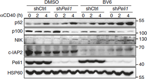
BV6 purchased from MedChemExpress. Usage Cited in: Nat Commun. 2018 Mar 19;9(1):1136. [Abstract]
Western Blot analysis of p52, p100, NIK, c-IAP2, Peli1, and HSP60 in total lysis of control and Peli1-knockdown M12 cells that pretreated with DMSO or smac mimetic BV6 for 4 h, and then stimulated with anti-CD40 (αCD40) at the indicated time points.
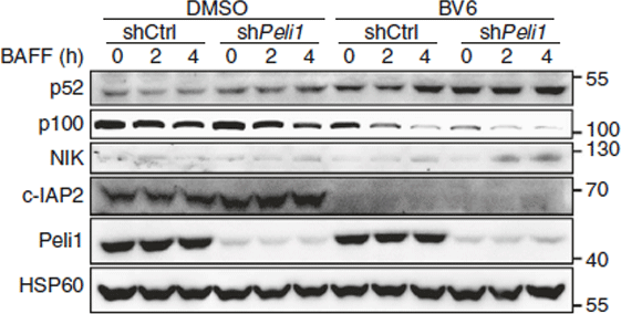
BV6 purchased from MedChemExpress. Usage Cited in: Nat Commun. 2018 Mar 19;9(1):1136. [Abstract]
Western Blot analysis of p52, p100, NIK, c-IAP2, Peli1, and HSP60 in total lysis of control and Peli1-knockdown M12 cells that pretreated with DMSO or smac mimetic BV6 for 4 h, and then stimulated with BAFF at the indicated time points.
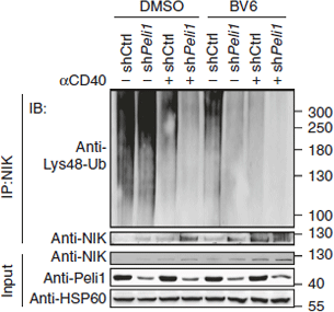
BV6 purchased from MedChemExpress. Usage Cited in: Nat Commun. 2018 Mar 19;9(1):1136. [Abstract]
Analysis of Lys48 ubiquitination of NIK in control and Peli1-knockdown M12 cells that pretreated with DMSO or smac mimetic BV6 for 4 h, then left unstimulated or stimulated with anti-CD40 (αCD40) for 4 h in the presence of MG132.
-
Cell Death Differ
Ubiquitin-binding domain in ABIN1 is critical for regulating cell death and inflammation during development. [Abstract]2022 Oct;29(10):2034-2045. PMID: 35430614 -
Cell Death Dis
Adipocyte-specific Mlkl knockout mitigates obesity-induced metabolic dysfunction by enhancing mitochondrial functions. [Abstract]2025 Oct 6;16(1):683. PMID: 41053093 -
Cell Death Dis
Polarity protein AF6 functions as a modulator of necroptosis by regulating ubiquitination of RIPK1 in liver diseases. [Abstract]2023 Oct 12;14(10):673. PMID: 37828052 -
J Orthop Translat
2024 Jun 7:47:15-28. PMID: 38957269 -
J Med Chem
Discovery of a 1 H-Pyrazol-3-Amine Derivative as a Novel, Selective, and Orally Available RIPK1 Inhibitor for the Treatment of Inflammatory Disease. [Abstract]2025 Oct 23;68(20):21766-21785. PMID: 41077763 -
-
Biomater Adv
Etoposide-loaded lipopolymer nanoparticles promote Smac minetic activity against inhibitor of apoptosis protein for glioblastoma treatment. [Abstract]2025 Jan 13:170:214185. PMID: 39879864 -
Neurobiol Dis
2019 Jul:127:570-581. PMID: 30981830 -
Chem Biol Interact
Formoterol, a clinically approved drug, inhibits ferroptosis by suppressing lipid peroxidation and attenuates APAP-induced acute liver injury. [Abstract]2025 Sep 4:111724. PMID: 40914538 -
Mol Neurobiol
Myeloid TBK1 Deficiency Induces Motor Deficits and Axon Degeneration Through Inflammatory Cell Infiltration. [Abstract]2021 May;58(5):2435-2446. PMID: 33439438 -
Biochim Biophys Acta Mol Basis Dis
Directed elimination of senescent cells attenuates development of osteoarthritis by inhibition of c-IAP and XIAP. [Abstract]2019 Jun 26;1865(10):2618-2632. PMID: 31251987 -
iScience
The necroptosis-inducing pseudokinase mixed lineage kinase domain-like regulates the adipogenic differentiation of pre-adipocytes. [Abstract]2022 Sep 19;25(10):105166. PMID: 36204273 -
Aging (Albany NY)
Ubiquitin-specific protease 4 promotes metastasis of hepatocellular carcinoma by increasing TGF-β signaling-induced epithelial-mesenchymal transition. [Abstract]2018 Oct 18;10(10):2783-2799. PMID: 30335615 -
J Cell Sci
FKBP12 mediates necroptosis by initiating RIPK1-RIPK3-MLKL signal transduction in response to TNF receptor 1 ligation. [Abstract]2019 May 20;132(10):jcs227777. PMID: 31028177 -
Exp Cell Res
Indole-3-carbinol ameliorates necroptosis and inflammation of intestinal epithelial cells in mice with ulcerative colitis by activating aryl hydrocarbon receptor. [Abstract]2021 Jul 15;404(2):112638. PMID: 34015312 -
-
-
-
-


Solvent & Solubility
DMSO : ≥ 100 mg/mL (82.95 mM; Hygroscopic DMSO has a significant impact on the solubility of product, please use newly opened DMSO)
* "≥" means soluble, but saturation unknown.
Please refer to the solubility information to select the appropriate solvent. Once prepared, please aliquot and store the solution to prevent product inactivation from repeated freeze-thaw cycles.
Storage method and period of stock solution: -80°C, 2 years; -20°C, 1 year. When stored at -80°C, please use it within 2 years. When stored at -20°C, please use it within 1 year.
Concentration (start) × Volume (start) = Concentration (final) × Volume (final)
Select the appropriate dissolution method based on your experimental animal and administration route.
- For the following dissolution methods, please ensure to first prepare a clear stock solution using an In Vitro approach and then sequentially add co-solvents:
- To ensure reliable experimental results, the clarified stock solution can be appropriately stored based on storage conditions. As for the working solution for In Vivo experiments, it is recommended to prepare freshly and use it on the same day.
- The percentages shown for the solvents indicate their volumetric ratio in the final prepared solution. If precipitation or phase separation occurs during preparation, heat and/or sonication can be used to aid dissolution.
Add each solvent one by one: 10% DMSO 40% PEG300 5% Tween-80 45% Saline
Solubility: ≥ 2.5 mg/mL (2.07 mM); Clear solution
This protocol yields a clear solution of ≥ 2.5 mg/mL (saturation unknown).
Taking 1 mL working solution as an example, add 100 μL DMSO stock solution (25.0 mg/mL) to 400 μL PEG300, and mix evenly; then add 50 μL Tween-80 and mix evenly; then add 450 μL Saline to adjust the volume to 1 mL.
Preparation of Saline: Dissolve 0.9 g sodium chloride in ddH₂O and dilute to 100 mL to obtain a clear Saline solution.
Add each solvent one by one: 10% DMSO 90% (20% SBE-β-CD in Saline)
Solubility: ≥ 2.5 mg/mL (2.07 mM); Clear solution
This protocol yields a clear solution of ≥ 2.5 mg/mL (saturation unknown).
Taking 1 mL working solution as an example, add 100 μL DMSO stock solution (25.0 mg/mL) to 900 μL 20% SBE-β-CD in Saline, and mix evenly.
Preparation of 20% SBE-β-CD in Saline (4°C, storage for one week): 2 g SBE-β-CD powder is dissolved in 10 mL Saline, completely dissolve until clear.
Please enter the basic information of animal experiments:
-
-
-
-
Recommended: Prepare an additional quantity of animals to account for potential losses during experiments.
Please enter your animal formula composition:
-
%DMSO +
Recommended: Keep the proportion of DMSO in working solution below 2% if your animal is weak.
-
%+
-
+%Tween-80 + +
-
%Saline +
The co-solvents required include: DMSO, . All of co-solvents are available by MedChemExpress (MCE). , Tween 80. All of co-solvents are available by MedChemExpress (MCE).
Working solution concentration: 0.22 mg/mL
Method for preparing stock solution: mg drug dissolved in μL DMSO. Stock solution concentration: mg/mL.
1. Take μL DMSO stock solution;
2. Add μL .
μL , mix evenly;
3. Then add μL Tween 80, mix evenly;
4. Then add μL
Please ensure that the stock solution in the first step is dissolved to a clear state, and add co-solvents in sequence. You can use ultrasonic heating (ultrasonic cleaner, recommended frequency 20-40 kHz), vortexing, etc. to assist dissolution.


Purity & Documentation
-
Data Sheet (284 KB)
-
SDS (393 KB)
- English - EN (393 KB)
- Français - FR (393 KB)
- Deutsch - DE (393 KB)
- Norwegian - NO (393 KB)
- Español - ES (393 KB)
- Swedish - SV (393 KB)
- Italian - IT (393 KB)
- Korean - KR (393 KB)
- Portuguese - PT (393 KB)
-
Handling Instructions (2659 KB)
References
[1]. Li W, et al. BV6, an IAP antagonist, activates apoptosis and enhances radiosensitization of non-small cell lung carcinoma in vitro. J Thorac Oncol. 2011 Nov;6(11):1801-9. [Content Brief]
[2]. Uegaki T, et al. Inhibitor of apoptosis proteins (IAPs) may be effective therapeutic targets for treating endometriosis. Hum Reprod. 2015 Jan;30(1):149-58. [Content Brief]


Complete Stock Solution Preparation Table
Please refer to the solubility information to select the appropriate solvent. Once prepared, please aliquot and store the solution to prevent product inactivation from repeated freeze-thaw cycles.
Storage method and period of stock solution: -80°C, 2 years; -20°C, 1 year. When stored at -80°C, please use it within 2 years. When stored at -20°C, please use it within 1 year.
| Optional Solvent | Concentration Solvent Mass | 1 mg | 5 mg | 10 mg | 25 mg |
|---|---|---|---|---|---|
| DMSO | 1 mM | 0.8295 mL | 4.1474 mL | 8.2948 mL | 20.7371 mL |
| 5 mM | 0.1659 mL | 0.8295 mL | 1.6590 mL | 4.1474 mL | |
| 10 mM | 0.0829 mL | 0.4147 mL | 0.8295 mL | 2.0737 mL | |
| 15 mM | 0.0553 mL | 0.2765 mL | 0.5530 mL | 1.3825 mL | |
| 20 mM | 0.0415 mL | 0.2074 mL | 0.4147 mL | 1.0369 mL | |
| 25 mM | 0.0332 mL | 0.1659 mL | 0.3318 mL | 0.8295 mL | |
| 30 mM | 0.0276 mL | 0.1382 mL | 0.2765 mL | 0.6912 mL | |
| 40 mM | 0.0207 mL | 0.1037 mL | 0.2074 mL | 0.5184 mL | |
| 50 mM | 0.0166 mL | 0.0829 mL | 0.1659 mL | 0.4147 mL | |
| 60 mM | 0.0138 mL | 0.0691 mL | 0.1382 mL | 0.3456 mL | |
| 80 mM | 0.0104 mL | 0.0518 mL | 0.1037 mL | 0.2592 mL |


 Powered by Bioz