


Ripasudil free base
Based on 6 publication(s) in Google Scholar
Ripasudil free base (K-115 free base) is a specific inhibitor of ROCK, with IC50s of 19 and 51 nM for ROCK2 and ROCK1, respectively.
For research use only. We do not sell to patients.
- CAS No.: 223645-67-8
- Formula: C15H18FN3O2S
- Molecular Weight:323.39
-
Storage:
Please store the product under the recommended conditions in the Certificate of Analysis.

To place orders, for customer services and technical support, please contact: MedChemExpress USA
Tel: 609-228-6898 E-mail: [email protected] [email protected]


-
 Biological Activity
-
Chemical Information
- Protocol
- Purity & Documentation
- References
-
 Help & FAQs


Publications Citing Use of MedChemExpress (MCE) Ripasudil free base
More Customer Validation & Images

-
In Vivo Imaging
-
Cell Imaging/Staining
-
WB


Biological Activity
|
ROCK2 19 nM (IC50) |
ROCK1 51 nM (IC50) |
CaMKIIa 370 nM (IC50) |
PKACa 2.1 μM (IC50) |
PKC 27 μM (IC50) |
|
Cell Line
|
Type | Value | Description | References |
|---|---|---|---|---|
| A7R5 | EC50 |
210 nM
Compound: ripasudil
|
Inhibition of ROCK-catalysed MLC Thr18/Ser19 phosphorylation in rat A7r5 cells after 90 mins by ELISA
Inhibition of ROCK-catalysed MLC Thr18/Ser19 phosphorylation in rat A7r5 cells after 90 mins by ELISA
|
[PMID: 25898023] |
Ripasudil (K-115) is a potent inhibitor of ROCK, with IC50s of 19 and 51 nM for ROCK2 and ROCK1, respectively. Ripasudil also shows less potent inhibitory activities against CaMKIIα, PKACα and PKC, with IC50s of 370 nM, 2.1 μM and 27 μM, respectively[1]. Ripasudil (K-115; 1, 10 μM) induces cytoskeletal changes, including retraction and cell rounding and reduced actin bundles of cultured trabecular meshwork (TM) cells. Ripasudil (5 μM) sifnificantly reduces transendothelial electrical resistance (TEER), and increases FITC-dextran permeability in Schlemm’s canal endothelial (SCE) cell monolayers[2].
MedChemExpress (MCE) has not independently confirmed the accuracy of these methods. They are for reference only.
MedChemExpress (MCE) has not independently confirmed the accuracy of these methods. They are for reference only.


Chemical Information
-
CAS No. 223645-67-8
-
Molecular Weight 323.39
-
Formula C15H18FN3O2S
-
SMILES
FC1=CN=CC2=C1C(S(N3CCCNC[C@@H]3C)(=O)=O)=CC=C2
-
Synonyms
K-115 free base
-
Shipping
Room temperature in continental US; may vary elsewhere.
-
Storage
Please store the product under the recommended conditions in the Certificate of Analysis.


Publications (6)
-
Journal Impact Factor
-
Most Recent
-
Science
2017 Dec 1;358(6367):eaan4368. PMID: 29191878 -
Adv Sci (Weinh)
Pharmacological Perturbation of Mechanical Contractility Enables Robust Transdifferentiation of Human Fibroblasts into Neurons. [Abstract]2022 May;9(13):e2104682. PMID: 35240008 -
Mol Ther
ROCK inhibition enhanced hepatocyte liver engraftment by retaining membrane CD59 and attenuating complement activation. [Abstract]2023 Jun 7;31(6):1846-1856. PMID: 36860134
Ripasudil free base purchased from MedChemExpress. Usage Cited in: Mol Ther. 2023 Jun 7;31(6):1846-1856. [Abstract]
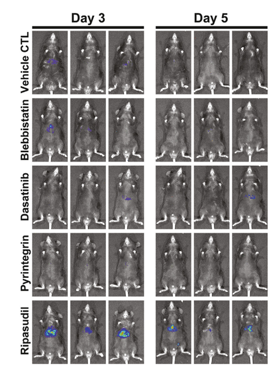
C57 mice were transplanted with luciferase-expressing hepatocytes. The effects of the ROCK inhibitor Ripasudil (17.3 mg/kg, i.p.), nonmuscle myosin II ATPase inhibitor Blebbistatin, β1-integrin agonist Pyrintegrin, and Src kinase inhibitor Dasatinib on hepatocyte liver engraftment were analyzed by bioluminescence imaging on days 3 and 5 after intrasplenic infusion.
Ripasudil free base purchased from MedChemExpress. Usage Cited in: Mol Ther. 2023 Jun 7;31(6):1846-1856. [Abstract]
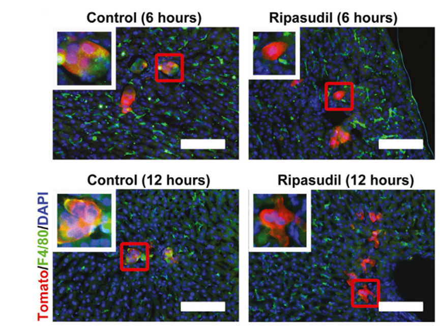
Ripasudil (17.3 mg/kg, i.p.) treatment significantly decreased the colocalization between transplanted hepatocytes and Kupffer cells to 11.38% at 6 h and 14.49% at 12 h after transplantation.
Ripasudil free base purchased from MedChemExpress. Usage Cited in: Mol Ther. 2023 Jun 7;31(6):1846-1856. [Abstract]
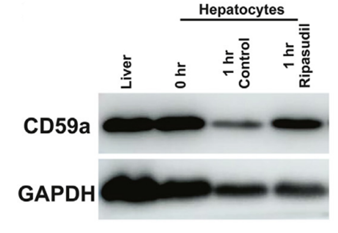
Western blot assays showed rapid loss of CD59a protein on isolated hepatocytes, which could be blocked by Ripasudil (17.3 mg/kg, i.p.) treatment.
-
Exp Eye Res
Laboratory exploration of the use of ripasudil in descemetorhexis with a human ex vivo model. [Abstract]2024 Jun 18:109977. PMID: 38901724 -
Curr Eye Res
Effects of Ripasudil, a Rho-Kinase Inhibitor, on Scar Formation in a Mouse Model of Filtration Surgery. [Abstract]2023 Sep;48(9):826-835. PMID: 37216470 -

Protocol
ROCK 1 (0.75 ng/mL) and ROCK 2 (0.5 ng/mL) are incubated with various concentrations of Ripasudil, Y-27632, or HA-1077 at 25°C for 90 min in 50 mM Tris-HCl buffer (pH 7.5) containing 100 mM KCl, 10 mM MgCl2, 0.1 mM EGTA, 30 mM Long S6 Kinase Substrate peptide, and 1 mM ATP in a total volume of 40 mL. PKACa, PKC, and CaMKIIa are also incubated with various concentrations of Ripasudil, Y-27632, or HA-1077. PKACa (0.0625 ng/mL) is incubated at 25°C for 30 min in 40 mM Tris-HCl buffer (pH 7.5) containing 20 mM MgCl2, 1 mg/ mL BSA, 5 mM Kemptide peptide substrate, and 1 mM ATP in a total volume of 40 mL. PKC (0.025 ng/mL) is incubated at 25°C for 80 min in 20 mM Tris-HCl buffer (pH 7.5) containing 20 mM MgCl2, 0.4 mM CaCl2, 0.1 mg/mL BSA, 0.25 mM EGTA, 25 ng/mL phosphatidylserine, 2.5 ng/mL diacylglycerol, 0.0075% Triton-X-100, 25 mM DTT, 10 mM Neurogranin (28-43) peptide substrate, and 1 mM ATP in a total volume of 40 mL. CaMKIIa (0.025 ng/mL) is incubated at 25°C for 90 min in 50 mM Tris-HCl buffer (pH 7.5) containing 10 mM MgCl2, 2 mM CaCl2, 0.04 mg/mL BSA, 16 mg/mL purified calmodulin from bovine testis, 500 mM DTT, 50 mM Autocamitide 2, and 1 mM ATP in a total volume of 40 mL. After incubation, 40 mL of KinaseGlo Luminescent Kinase Assay solution is added, and allowed to remain at 25°C for 10 min, and Relative Light Units (RLU) are measured using a luminometer. The RLU without test compound is set as 100% (Control value), and that without enzyme and compound is set as 0% (Normal value). The reaction rate (% of control) is then calculated from the RLU with addition of each concentration of test compounds, and the 50% inhibitory concentrations (IC50) are determined by logistic regression analysis using SAS[1].
MedChemExpress (MCE) has not independently confirmed the accuracy of these methods. They are for reference only.
Trabecular meshwork (TM) cells are plated on 6 well plates at a density of 1 × 10⁴ cells per well in DMEM containing 10% FBS. Following overnight culture, when cells have reached semiconfluence, 1 or 10 μM of Ripasudil, 10 μM of Y-27632, or 10 μM of fasudil are added to culture wells. PBS is used as a control vehicle. After 60 min, drug solutions are removed and replaced with DMEM containing 10% FBS. Cells are observed by phase-contrast microscopy and photographed 60 min after drug application and 2 h after drug removal. For immunohistochemistry, TM cells are plated on gelatin-coated 8 well chamber slides at a density of 1 × 10⁴ cells per well in DMEM containing 10% FBS. After overnight culture, when cells reach semiconfluence, cell are incubated in Ripasudil at 1 or 10 μM, Y-27632 at 10 μM, or fasudil at 10 μM for 60 min. PBS is used as a control vehicle. Drug solutions are removed and replaced with DMEM containing 10% FBS after 2 h. Cells are fixed with 4% paraformaldehyde in PBS for 15 min then washed with cytoskeletal buffer (10 mM MES, 150 mM NaCl, 5 mM EGTA, 5 mM MgCl2, 5 mM glucose, pH 6.1) and serum buffer (10% FBS in PBS). Cells are permeabilized with 0.5% Triton X-100 in PBS for 12 min at room temperature and blocked with serum buffer for at least 2 h at 4°C. Filamentous actin (F-actin) is labeled with 0.05 mg/mL Phalloidin-TRITC for 1 h at room temperature. After washing with PBS, cells are mounted with commercial mounting medium containing DAPI and observed using a fluorescence microscope. The exposure to take images for F-actin and DAPI are 0.1 and 0.05 sec, respectively[2].
MedChemExpress (MCE) has not independently confirmed the accuracy of these methods. They are for reference only.
Rabbits[1]
In the rabbit experiments, 50 mL of vehicle or Ripasudil at concentrations of 0.0625%, 0.125%, 0.25, or 0.5% is instilled into one eye. Intraocular pressure (IOP) is measured in both eyes before and 0.5, 1, 2, 3, 4, and 5 h after instillation. The contralateral eye is not treated. Animals are administered all concentrations of Ripasudil assigned using the Latin square method with intervals of at least 2 d.
Monkeys[1]
In the monkey experiments, 20 mL of Ripasudil at concentrations of 0.1%, 0.2%, or 0.4%, and latanoprost at a concentration of 0.005% are instilled into one eye. IOP is measured in both eyes before and 1, 2, 4, 6, and 8 h after instillation. The contralateral eye is not treated. Animals are arranged to receive all formulations with intervals of at least 1 week using the Latin square method. The IOPs are compared with the results for the instillation side at pre-dose and at each time point after instillation of Ripasudil, and are compared with both eyes at each time point.
MedChemExpress (MCE) has not independently confirmed the accuracy of these methods. They are for reference only.


Purity & Documentation
References
[1]. Isobe T, et al. Effects of K-115, a rho-kinase inhibitor, on aqueous humor dynamics in rabbits. Curr Eye Res. 2014 Aug;39(8):813-22. [Content Brief]
[2]. Kaneko Y, et al. Effects of K-115 (Ripasudil), a novel ROCK inhibitor, on trabecular meshwork and Schlemm's canal endothelial cells. Sci Rep. 2016 Jan 19;6:19640. [Content Brief]
[3]. Yamamoto K, et al. The novel Rho kinase (ROCK) inhibitor K-115: a new candidate drug for neuroprotective treatment in glaucoma. Invest Ophthalmol Vis Sci. 2014 Oct 2;55(11):7126-36. [Content Brief]

Calculators
Concentration (start) × Volume (start) = Concentration (final) × Volume (final)


 Powered by Bioz