


Sanguinarine
Based on 16 publication(s) in Google Scholar
Sanguinarine (Sanguinarin), a benzophenanthridine alkaloid derived from the root of Sanguinaria Canadensis, can stimulate apoptosis via activating the production of reactive oxygen species (ROS). Sanguinarine-induced apoptosis is associated with the activation of JNK and NF-κB.
For research use only. We do not sell to patients.
- CAS No.: 2447-54-3
- Formula: C20H14NO4+
- Molecular Weight:332.33
-
Storage:
Please store the product under the recommended conditions in the Certificate of Analysis.

To place orders, for customer services and technical support, please contact: MedChemExpress USA
Tel: 609-228-6898 E-mail: [email protected] [email protected]


-
 Biological Activity
-
Chemical Information
- Protocol
- Purity & Documentation
- References
-
 Help & FAQs


Publications Citing Use of MedChemExpress (MCE) Sanguinarine
More- J Adv Res. 2026 Feb 21:S2090-1232(26)00176-1. [Abstract]
- Adv Sci (Weinh). 2024 Jul 5:e2403451. [Abstract]
- Oncogene. 2025 May;44(16):1078-1092. [Abstract]
- Phytother Res. 2023 Oct;37(10):4771-4790. [Abstract]
- Am J Chin Med. 2025;53(5):1545-1571. [Abstract]
- PLoS Pathog. 2026 Mar 3;22(3):e1014032. [Abstract]
- PLoS Pathog. 2023 Dec 7;19(12):e1011796. [Abstract]
- Pestic Biochem Physiol. 2022 Nov:188:105259. [Abstract]
- Comp Biochem Physiol C Toxicol Pharmacol. 2022 Feb:252:109228. [Abstract]
- Antiviral Res. 2026 Jun:250:106417. [Abstract]
- Brain Behav Immun Health. 2022 Oct 29:26:100546. [Abstract]
- Toxicol Lett. 2021 Oct 10:350:71-80. [Abstract]
- Fishes. 2022, 7(5), 229.
- Planta Med. 2024 Jun;90(7-08):523-533. [Abstract]
- bioRxiv. 2024 Feb 5.
- Research Square Print. 2022 Jun.
 Customer Validation & Images

-
Cell Proliferation/Viability Assay
-
Cell Proliferation/Viability Assay
-
Bio/Physico-chemical Assay
-
Bio/Physico-chemical Assay
-
WB


Biological Activity
Apoptosis[1]
|
Cell Line
|
Type | Value | Description | References |
|---|---|---|---|---|
| A2780/Taxol | IC50 |
0.4 μM
Compound: 4
|
Antiproliferative activity against human A2780T cells assessed as reduction in cell viability incubated for 48 hrs by CCK-8 assay
Antiproliferative activity against human A2780T cells assessed as reduction in cell viability incubated for 48 hrs by CCK-8 assay
|
[PMID: 39213483] |
| A549 | IC50 |
610 nM
Compound: 1
|
Growth inhibition of human A549 cells measured every 2 hrs over 6 days by live cell imaging based method
Growth inhibition of human A549 cells measured every 2 hrs over 6 days by live cell imaging based method
|
[PMID: 28621943] |
| A549/TR | IC50 |
0.4 μM
Compound: 4
|
Antiproliferative activity against human A549/Taxol cells assessed as reduction in cell viability incubated for 48 hrs by CCK-8 assay
Antiproliferative activity against human A549/Taxol cells assessed as reduction in cell viability incubated for 48 hrs by CCK-8 assay
|
[PMID: 39213483] |
| K562 | IC50 |
2 μM
Compound: Sanguinarine
|
Inhibition of NF-kappaB transactivation in TNF-alpha-stimulated human K562 cells preincubated for 2 hrs followed by TNF-alpha challenge measured after 6 hrs by dual luciferase reporter gene assay
Inhibition of NF-kappaB transactivation in TNF-alpha-stimulated human K562 cells preincubated for 2 hrs followed by TNF-alpha challenge measured after 6 hrs by dual luciferase reporter gene assay
|
[PMID: 24775915] |
| LoVo | IC50 |
0.4 μM
Compound: 4
|
Antiproliferative activity against human LoVo cells assessed as reduction in cell viability incubated for 48 hrs by CCK-8 assay
Antiproliferative activity against human LoVo cells assessed as reduction in cell viability incubated for 48 hrs by CCK-8 assay
|
[PMID: 39213483] |
| MKN-45 | IC50 |
1.51 μM
Compound: Sanguinarine
|
Cytotoxicity against human MKN45 cells assessed as decrease in cell viability after 48 hrs by MTT assay
Cytotoxicity against human MKN45 cells assessed as decrease in cell viability after 48 hrs by MTT assay
|
[PMID: 27887841] |
| NB-4 | IC50 |
0.53 μM
Compound: Sanguinarine
|
Cytotoxicity against human NB4 cells assessed as decrease in cell viability after 48 hrs by MTT assay
Cytotoxicity against human NB4 cells assessed as decrease in cell viability after 48 hrs by MTT assay
|
[PMID: 27887841] |
| PC-3 | IC50 |
0.4 μM
Compound: 4
|
Antiproliferative activity against human PC-3 cells assessed as reduction in cell viability incubated for 24 hrs by CCK-8 assay
Antiproliferative activity against human PC-3 cells assessed as reduction in cell viability incubated for 24 hrs by CCK-8 assay
|
[PMID: 39213483] |
Sanguinarine (SANG)-induced apoptosis is associated with the activation of JNK and NF-κB signal pathways.To determine the effects of Sanguinarine on cell viability, 22B-cFluc cells are stimulated with different concentrations of Sanguinarine for 24 h, and then a CKK-8 assay is performed. The treatment with Sanguinarine decreases the proliferation of 22B cells in a dose-dependent manner. Meanwhile, the cytosolic extracts of 22B-cFluc cells treated with different dose of Sanguinarine are measured to detect cellular caspase-3 activity using Ac-DEVD-pNA, which is a validated caspase-3 substrate. The absorbance at 450 nm increases in a dose-dependent manner, indicating increased caspase-3 activity stimulated by Sanguinarine[1].
MedChemExpress (MCE) has not independently confirmed the accuracy of these methods. They are for reference only.
MedChemExpress (MCE) has not independently confirmed the accuracy of these methods. They are for reference only.


Chemical Information
-
CAS No. 2447-54-3
-
Molecular Weight 332.33
-
Formula C20H14NO4+
-
SMILES
C[N+]1=CC2=C3C(OCO3)=CC=C2C(C=CC4=C5)=C1C4=CC6=C5OCO6
-
Synonyms
Sanguinarin; Sanguinarium; Pseudochelerythrine
-
Structure Classification
-
Initial Source
-
Shipping
Room temperature in continental US; may vary elsewhere.
-
Storage
Please store the product under the recommended conditions in the Certificate of Analysis.


Publications (16)
-
Journal Impact Factor
-
Most Recent
-
J Adv Res
Identification of a novel EphB4 inhibitor, Sanguinarine, which attenuates β-catenin signaling to inhibit tumor proliferation and migration in lung cancer. [Abstract]2026 Feb 21:S2090-1232(26)00176-1. PMID: 41730417 -
Adv Sci (Weinh)
Short-Term Statin Therapy Induces Hepatic Insulin Resistance Through HNF4α/PAQR9/PPM1α Axis Regulated AKT Phosphorylation. [Abstract]2024 Jul 5:e2403451. PMID: 38970167 -
Oncogene
The protection of UCK2 protein stability by GART maintains pyrimidine salvage synthesis for HCC growth under glucose limitation. [Abstract]2025 May;44(16):1078-1092. PMID: 39865175 -
Phytother Res
NPLC0393 from Gynostemma pentaphyllum ameliorates Alzheimer's disease-like pathology in mice by targeting protein phosphatase magnesium-dependent 1A phosphatase. [Abstract]2023 Oct;37(10):4771-4790. PMID: 37434441 -
Am J Chin Med
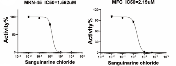
Sanguinarine Inhibits Gastric Cancer Progression by Targeting the NOS2/SOD1 Axis to Promote Ferroptosis. [Abstract]2025;53(5):1545-1571. PMID: 40663435
Sanguinarine purchased from MedChemExpress. Usage Cited in: Am J Chin Med. 2025;53(5):1545-1571. [Abstract]
MKN-45 and MFC cells were treated with different concentration of Sanguinarin chloride (0, 0.01, 0.1, 10, 100 μM) for 24h, and cell viability was measured by CCK-8 assays.
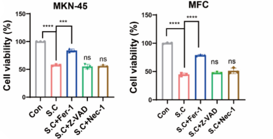
Sanguinarine purchased from MedChemExpress. Usage Cited in: Am J Chin Med. 2025;53(5):1545-1571. [Abstract]
Cell viability was measured in MKN-45 and MFC cells treated with Sanguinarin chloride (2 μM) with Fer-1 (10 μM), Z-VAD (10 μM), or Nec-1 (10 μM) for 24h.
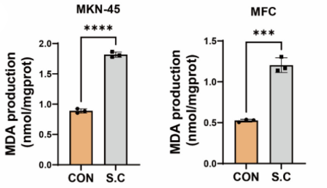
Sanguinarine purchased from MedChemExpress. Usage Cited in: Am J Chin Med. 2025;53(5):1545-1571. [Abstract]
MKN-45 and MFC cells were treated with Sanguinarin chloride (2 μM) for 24 h. MDA level was detected by MDA assay kit.
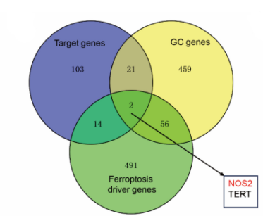
Sanguinarine purchased from MedChemExpress. Usage Cited in: Am J Chin Med. 2025;53(5):1545-1571. [Abstract]
Prediction of Sanguinarin chloride-targeted genes, GC-related proteins, and ferroptosis driver genes, respectively. Two overlapping proteins are displayed.
Sanguinarine purchased from MedChemExpress. Usage Cited in: Am J Chin Med. 2025;53(5):1545-1571. [Abstract]
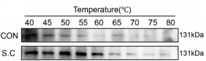
GETSA was performed after 24h of Sanguinarin chloride (2 μM) action, and the protein stability of NOS2 was examined by western at 40-80℃.
Sanguinarine purchased from MedChemExpress. Usage Cited in: Am J Chin Med. 2025;53(5):1545-1571. [Abstract]
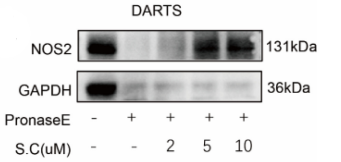
Cell lysates were incubated with Sanguinarin chloride (2 μM, 5 μM, 10 μM), digested with PronaseE, the reaction was terminated by the addition of allozyme inhibitor, and the protein level of NOS2 was detected by western.
Sanguinarine purchased from MedChemExpress. Usage Cited in: Am J Chin Med. 2025;53(5):1545-1571. [Abstract]
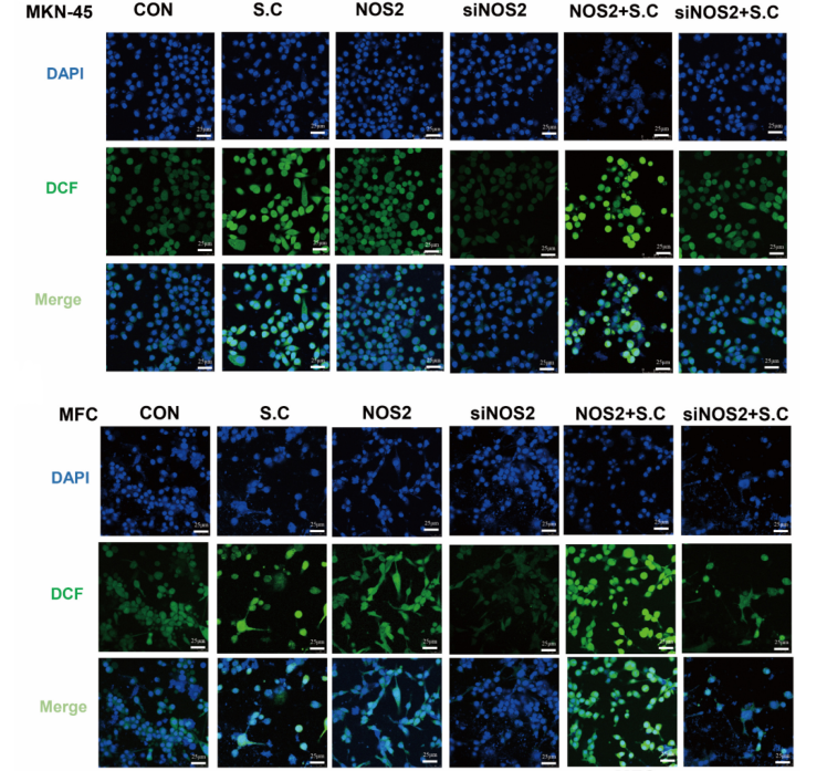
ROS level in MKN-45 and MFC cells transfected with NOS2 or siNOS2 incubated with Sanguinarin chloride was detected by DCFH-DA.
-
PLoS Pathog
The CD97-PPM1G axis dampens antiviral immunity by dephosphorylating IRF7 in type I interferon pathway. [Abstract]2026 Mar 3;22(3):e1014032. PMID: 41774756 -
PLoS Pathog
G-quadruplex in the TMV Genome Regulates Viral Proliferation and Acts as Antiviral Target of Photodynamic Therapy. [Abstract]2023 Dec 7;19(12):e1011796. PMID: 38060599 -
Pestic Biochem Physiol
Neurotoxicity of sanguinarine via inhibiting mitophagy and activating apoptosis in zebrafish and PC12 cells. [Abstract]2022 Nov:188:105259. PMID: 36464364 -
Comp Biochem Physiol C Toxicol Pharmacol
Cardiotoxicity of sanguinarine via regulating apoptosis and MAPK pathways in zebrafish and HL1 cardiomyocytes. [Abstract]2022 Feb:252:109228. PMID: 34744004 -
Antiviral Res
Repurposing screen using a robust human rhinovirus infectious clone identifies pyrvinium pamoate with antiviral activity. [Abstract]2026 Jun:250:106417. PMID: 42025967 -
Brain Behav Immun Health
Miltefosine as a PPM1A activator improves AD-like pathology in mice by alleviating tauopathy via microglia/neurons crosstalk. [Abstract]2022 Oct 29:26:100546. PMID: 36388134 -
Toxicol Lett
Developmental toxicity caused by sanguinarine in zebrafish embryos via regulating oxidative stress, apoptosis and wnt pathways. [Abstract]2021 Oct 10:350:71-80. PMID: 34252508 -
-
Planta Med
Novel Approaches for the Analysis and Isolation of Benzylisoquinoline Alkaloids in Chelidonium majus. [Abstract]2024 Jun;90(7-08):523-533. PMID: 38843792 -
-

Protocol
The caspase-3 activity is measured using a caspase-3 activity assay kit. Briefly, the cells treated by different concentrations of Sanguinarine (0.5 μM, 1 μM, 2 μM, 4 μM) or control DMSO are collected, washed and lysed in a lysis buffer for 30 min on ice. The supernatants are then collected by centrifuging at 1,2000 rpm for 10 min. The Ac-DEVD-pNA (2 mM) is added to each sample and incubated at 37°C for 1 h. The optical density (OD) of each sample is finally quantified at a wavelength of 405 nm using a spectrophotometer. The p-NA standard is used to calibrate the caspase-3 activity of each sample[1].
MedChemExpress (MCE) has not independently confirmed the accuracy of these methods. They are for reference only.
The cell viability of Sanguinarine is determined by CCK-8 assay using a cell counting kit-8. Briefly, 22B-cFluc cells are seeded in a 96-well plate (5×10³ cells/well) and treated with different concentrations of Sanguinarine (0.5 μM, 1 μM, 2 μM, 4 μM) for 24 h. Then 10 mL CKK-8 is added to each well for 4 h and the absorbance at 450 nm is measured with a microplate reader. The optical density (OD) values are determined to reflect the viable cell populations from each well[1].
MedChemExpress (MCE) has not independently confirmed the accuracy of these methods. They are for reference only.
Mice[1]
Xenografted tumor models are prepared by injection of 1×10⁷ 22B-cFluc cells suspended in PBS into nude mouse (n=6). After tumors reach a volume of approximately 100 mm3, Sanguinarine (10 mg/kg) is i.v. injected into mice. After injection for 24 h, 48 h and 72 h, mice are given a single i.p. dose of 150 mg/kg D-luciferin and bioluminescence imaging are performed using a Xenogen Lumina II system. The signal intensity in the region of interest is expressed using the Living Image software 4.1. For the anti-tumor therapy studies, one group of tumor-bearing mice (n=6) receive intravenously 10 mg/kg of Sanguinarine every other day throughout the experimental period, while the control group of mice (n=6) receive DMSO only. Tumor growth measurement is calculated[1].
MedChemExpress (MCE) has not independently confirmed the accuracy of these methods. They are for reference only.


Purity & Documentation
References

Calculators
Concentration (start) × Volume (start) = Concentration (final) × Volume (final)


 Powered by Bioz