


Atrosimab
Based on 1 Customer Validation
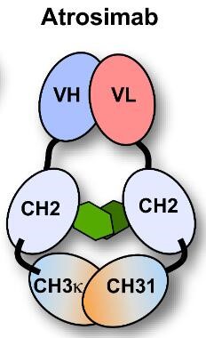
Atrosimab is an Fv-Fc1K fusion protein with an EC50 value of 0.37 nM against humans. Atrosimab inhibits TNF-induced TNFR1 activation, release of IL-6 and IL-8, and cell death, and alleviates neuroinflammation. Atrosimab is applicable to research related to inflammatory diseases, neurodegenerative diseases, acute and chronic inflammation, experimental arthritis, non-alcoholic steatohepatitis, and experimental autoimmune encephalomyelitis.
For research use only. We do not sell to patients.
- Purity: 99%
- Molecular Weight:72.338 kDa
-
Storage:
Please store the product under the recommended conditions in the Certificate of Analysis.


-
 Biological Activity
-
Chemical Information
- Purity & Documentation
- References
-
 Help & FAQs


Biological Activity
Fv-Fc
Human
Atrosimab (serial dilutions; 4-128 nM) binds purified human TNFR1-Fc with high affinity, and lacks binding to human complement protein C1q and human Fcγ receptors Ia, IIb, IIIa[1].
Atrosimab (100 nM; 1, 3, 7 days) exhibits good stability in human plasma for at least 7 days and has an aggregation temperature of 64°C[1].
Atrosimab (serial dilutions; 16-20 h, 24 h) potently inhibits TNF-mediated TNFR1 activation in HeLa, HT1080, and Kym-1 cells, with IC50 values of 54.5 nM, 24.2 nM, and 16.2 nM, respectively[1].
Atrosimab (serial dilutions; 16-20 h) does not exhibit TNFR1 agonistic activity in HT1080 cells even when cross-linked by three different goat anti-human IgG sera[1].
Atrosimab (24 h) does not affect spontaneous or glutamate-induced altered firing rates of huTNFR1-k/i mouse embryonic day 14 primary cortical neurons after 24 h incubation[2].
Atrosimab (100 μg; heated from 35°C to 80°C with 2 min equilibration per 1°C interval; 2-500 nM; up to 9 months) is highly thermally and long-term stable, binds selectively and with high affinity to human, rhesus, and cynomolgus TNFR1 (but not mouse/rat TNFR1 or any TNFR2), and recognizes an epitope on human TNFR1 that includes residues P23, L67, R68, H69, and Q24[3].
Atrosimab (up to 1000 nM; 16-20 h at 37°C, 5% CO2; 1-7 days at 37°C in 50% human serum) potently and dose-dependently inhibits TNF-induced IL-8 release in HT1080 cells with IC50 values of 21.9-34.9 nM, does not exhibit agonistic activity in these cells, and maintains functional stability after incubation in human serum at 37°C for up to 7 days[3].
Atrosimab (100 μg/mL; 24 h) inhibits TNF-induced VCAM-1 and ICAM-1 expression in hCMEC/D3 human brain microvascular endothelial cells[3].
MedChemExpress (MCE) has not independently confirmed the accuracy of these methods. They are for reference only.
-
Cell Line:HT1080 cells
-
Concentration:serial dilutions of Atrosimab; ~15.8 nM (constant anti-human IgG serum)
-
Incubation Time:16-20 h
-
Result:Showed no induction of IL-8 release across all tested concentrations, indicating no cross-linking-mediated TNFR1 agonistic activity.
| Species | Dose | Route | T1/2 (Distribution) | T1/2β | AUC0-t | C0 | Vss | CL |
|---|---|---|---|---|---|---|---|---|
| Mice[1] | 400 μg | i.v. | 2.2 h | 41.7 h | 5856.0 μg·h/mL | 324.7 μg/mL | 3.4 μg/mL | 0.29 μg/mL |
| Mice[3] | 30.0 mg/kg | i.v. | 2.26 h | 32.77 h | 6989.7 μg·h/mL | / | / | / |
| Mice[3] | 1.0 mg/kg | i.v. | 1.05 h | 31.37 h | 365.78 μg·h/mL | / | / | / |
| Mice[3] | 30.0 mg/kg | s.c. | / | 33.7 h | 6190.7 μg·h/mL | / | / | / |
| Mice[3] | 1.0 mg/kg | s.c. | / | 54.56 h | 321.83 μg·h/mL | / | / | / |
| Mice[3] | 30.0 mg/kg | i.v. | 0.66 h | 31.52 h | 7983.8 μg·h/mL | / | / | / |
| Mice[3] | 1.0 mg/kg | i.v. | 5.41 h | 9.52 h | 177.8 μg·h/mL | / | / | / |
| Mice[3] | 30.0 mg/kg | s.c. | / | 41 h | 6816.7 μg·h/mL | / | / | / |
| Mice[3] | 1.0 mg/kg | s.c. | / | 16.75 h | 214.07 μg·h/mL | / | / | / |
Atrosimab (30 mg/kg; i.v.; single dose) potently blocks acute TNF-induced inflammatory responses in huTNFR1-k/i mice[3].
Atrosimab (20-80 mg/kg; s.c.; twice weekly; 11 weeks) prevents arthritis development in a prophylactic setting and Atrosimab (5-45 mg/kg; s.c.; twice weekly; 6 weeks) alleviates established arthritis with dose-dependent efficacy in Tg197hTNFR1KI mice[3].
Atrosimab (45 mg/kg; i.p.; twice weekly; 8 weeks) alleviates liver fibrosis, steatosis, and apoptotic injury in a huTNFR1-k/i mouse model of NASH[3].
Atrosimab (30-60 mg/kg; i.p.; single doses on days 1, 4, 8, 12 of manifest disease) ameliorates motor symptoms, demyelination, and axonal injury in a huTNFR1-k/i mouse model of EAE, with 30 mg/kg being sufficient for maximal therapeutic activity[3].
MedChemExpress (MCE) has not independently confirmed the accuracy of these methods. They are for reference only.
-
Animal Model:huTNFR1-k/i (7-month-old male; C57Bl/6J background; NMDA-induced acute cholinergic neurodegeneration via stereotactic injection into NBM)[2]
-
Dosage:3 μg
-
Administration:stereotactic co-injection into NBM; infused at 0.1 μl/min at two dorsoventral depths: 0.3 μl at -0.44 mm and 0.3 μl at -0.46 mm relative to Bregma; total volume 0.6 μl
-
Result:Increased post-shock latency to 274.8 s in passive avoidance paradigm, indicating improved long-term associative memory.\nPrevented significant body weight loss, with mean body weight change of -2.43 g from surgery to perfusion.\nIncreased surface area of ChAT-positive cholinergic fibers in layer V of somatosensory cortex to 11.04%.\nIncreased ratio of cholinergic neurons in lesioned vs. non-lesioned NBM hemisphere to 0.14.\nIncreased ratio of NeuN-positive neuronal surface area in lesioned vs. non-lesioned somatosensory cortex layer V to 0.48.\nReduced volume of IBA-1-positive activated microglia/lesion size to 0.373 mm3.\nReduced ratio of CD68 mean intensity (lesioned vs. non-lesioned NBM hemisphere) to 1.22.\nReduced ratio of GFAP mean intensity (lesioned vs. non-lesioned NBM hemisphere) to 2.51. Showed no significant effect on short-term spatial memory, anxiety-like behavior, locomotor activity, or hippocampal synapsin-1 levels.
-
Animal Model:huTNFR1-k/i (C57BL/6 background)[3]
-
Dosage:30 mg/kg
-
Administration:i.v.; single dose
-
Result:Completely blocked TNF-induced body weight loss.\nSignificantly reduced serum IL-6 levels at 1 hour and 6 hours post-TNF injection to almost baseline levels.\nSignificantly reduced TNF-induced systemic CRP levels at 6 hours post-TNF injection compared to control-treated mice.
-
Animal Model:Tg197hTNFR1KI[3]
-
Dosage:20-80 mg/kg (prophylactic setting); 5-45 mg/kg (therapeutic setting)
-
Administration:s.c.; twice weekly; 11 weeks (prophylactic setting); 6 weeks (therapeutic setting)
-
Result:Prevented development of arthritic disease, prevented disease-associated inhibition of body weight gain, and resulted in no histopathological changes beyond the level of 4-week-old control mice (all doses, prophylactic setting).\nHalted disease progression (5 mg/kg, therapeutic setting).\nShowed improved therapeutic activity comparable to approved anti-TNF therapeutics (15 mg/kg, 45 mg/kg, therapeutic setting).\nImproved joint histopathology, with histopathological scores below the level of 9-week-old control mice (all doses, therapeutic setting).
-
Animal Model:huTNFR1-k/i (male)[3]
-
Dosage:45 mg/kg
-
Administration:i.p.; twice weekly; 8 weeks
-
Result:Significantly reduced liver fibrosis (assessed by Sirius Red staining).\nReduced liver steatosis (assessed by Oil Red O staining).\nDecreased activated caspase-3-positive cells (a marker of apoptotic liver injury) compared to saline-treated mice.
-
Animal Model:huTNFR1-k/i (female, 6-8 weeks old)[3]
-
Dosage:30-60 mg/kg
-
Administration:i.p.; single doses on days 1, 4, 8, 12 of manifest disease
-
Result:Ameliorated EAE motor symptoms.\nReduced cumulative EAE score.\nPrevented disease-associated weight loss.\nReduced cumulative weight loss.\nSignificantly reduced spinal cord demyelination (assessed by Luxol fast blue staining).\nSignificantly reduced axonal injury (assessed by amyloid precursor protein accumulation) compared to control-treated mice.\nShowed no efficacy difference between the 30 mg/kg and 60 mg/kg doses.
| NCT Number | Sponsor | Condition | Start Date |
Phase
|
|---|---|---|---|---|
| NCT01329991 | Plexxikon| | 2011-05 | PHASE1 |
Unconjugated
The product can be reconstituted/diluted with sterile PBS or saline.
-
Fv-Fc1κ
ELISA, FACS, Functional assay
-
 Loaded Atrosimab on ProA biosensor, can bind TNFR-1/CD120a Protein, Human (HEK293, His, HY-P700263) with an affinity constant of 2.076E-09 M as determined in BLI assay.


Chemical Information
-
Appearance Liquid
-
Molecular Weight 72.338 kDa
-
Color Colorless to light yellow
-
SMILES
[Atrosimab]
-
Synonyms
ATM-001
-
Shipping
Shipping with dry ice.
-
Formulation
Please refer to the lot-specific COA for specific buffer information.
-
Storage
Please store the product under the recommended conditions in the Certificate of Analysis.


Purity & Documentation
-
Data Sheet (268 KB)
-
SDS (251 KB)
- English - EN (251 KB)
- Français - FR (251 KB)
- Deutsch - DE (251 KB)
- Norwegian - NO (251 KB)
- Español - ES (251 KB)
- Swedish - SV (251 KB)
- Italian - IT (251 KB)
- Korean - KR (251 KB)
- Portuguese - PT (251 KB)
-
Inhibitory Antibodies User Guide (603 KB)
References
[1]. Richter F, et al. Improved monovalent TNF receptor 1-selective inhibitor with novel heterodimerizing Fc. MAbs. 2019;11(4):653-665. [Content Brief]
[2]. Ortí-Casañ N, et al. The TNFR1 antagonist Atrosimab reduces neuronal loss, glial activation and memory deficits in an acute mouse model of neurodegeneration. Sci Rep. 2023;13(1):10622. Published 2023 Jun 30. [Content Brief]
[3]. Richter F, et al. The TNFR1 Antagonist Atrosimab Is Therapeutic in Mouse Models of Acute and Chronic Inflammation. Front Immunol. 2021;12:705485. Published 2021 Jul 7. [Content Brief]

Calculators
Concentration (start) × Volume (start) = Concentration (final) × Volume (final)


 Powered by Bioz