



BWA-522

Based on 1 Customer Validation
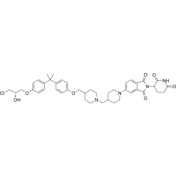
BWA-522 is an orally active PROTAC degrader targeting full-length androgen receptor (AR-FL) and androgen receptor splice variant 7 (AR-V7). BWA-522 antagonizes the N-terminal domain (AR-NTD) of the androgen receptor, suppresses AR downstream signaling proteins and induces cancer cells apoptosis. BWA-522 inhibits tumor growth in LNCaP xenograft mouse model. BWA-522 can be used for the research of prostate cancer.
(Pink: Androgen Receptor ligand (HY-109070); Blue: Cereblon ligand (HY-14658); Black: linker).
Nur für Forschungszwecke. Wir verkaufen nicht an Patienten.
- Reinheit : 98.04%
- CAS. Nr.: 3042820-12-9
- Formel: C43H51ClN4O7
- Molecular Weight:771.34
-
Speicherung:Powder -20°C, 3 years , 4°C, 2 years ; In solvent -80°C, 6 months , -20°C, 1 month

To place orders, for customer services and technical support, please contact: MedChemExpress USA
Tel: 609-228-6898 E-mail: [email protected] [email protected]


-
 Biologische Aktivität
-
Chemical Information
-
Lösungsmittel & Löslichkeit
- Protokoll
- Reinheit & Dokumentation
- Verweise
-
 Help & FAQs


Alle PROTACs Isoform-spezifische Produkte anzeigen
More

Biologische Aktivität
Beschreibung
IC50 & Target
[1]|
Cereblon |
Cellular Effect
|
Cell Line

|
Type | Value | Description | References |
|---|---|---|---|---|
| CWR22R | IC50 |
4.08 μM
Compound: 28; BWA-522
|
Antiproliferative activity against enzalutamide-resistant human 22Rv1 cells assessed as reduction in cell viability incubated for 6 days by CCK-8 assay
Antiproliferative activity against enzalutamide-resistant human 22Rv1 cells assessed as reduction in cell viability incubated for 6 days by CCK-8 assay
|
[PMID: 37556600] |
| LNCaP | IC50 |
1.07 μM
Compound: 28; BWA-522
|
Antiproliferative activity against human LNCaP cells assessed as reduction in cell viability incubated for 6 days by CCK-8 assay
Antiproliferative activity against human LNCaP cells assessed as reduction in cell viability incubated for 6 days by CCK-8 assay
|
[PMID: 37556600] |
| VCaP | IC50 |
5.59 μM
Compound: 28; BWA-522
|
Antiproliferative activity against human VCaP cells assessed as reduction in cell viability incubated for 6 days by CCK-8 assay
Antiproliferative activity against human VCaP cells assessed as reduction in cell viability incubated for 6 days by CCK-8 assay
|
[PMID: 37556600] |
In Vitro
BWA-522 (0.03-30 μM; 3-48 h) dose- and time-dependently degrades both AR-FL and AR-V7 in LNCaP, VCaP, and 22Rv1 prostate cancer cells via the ubiquitin proteasome system and CRBN/cullin 4A ubiquitin ligase, with DC50 values ranging from 0.67 to 3.45 μM[1].
BWA-522 (6 days) potently inhibits the growth of AR-dependent LNCaP, VCaP, and Enzalutamide (HY-70002)-resistant 22Rv1 prostate cancer cells with IC50 values ranging from 1.07 to 5.59 μM, while sparing AR-independent and normal prostate cells[1].
BWA-522 (1-10 μM; 2 weeks) dose-dependently inhibits long-term colony formation of LNCaP and 22Rv1 prostate cancer cells[1].
BWA-522 (1-10 μM; 48 h) dose-dependently suppresses AR downstream signaling proteins and induces apoptosis in LNCaP and VCaP prostate cancer cells[1].
BWA-522 has a low risk of cardiotoxicity, as it exhibits minimal inhibition of the hERG channel with an IC50 greater than 10 μM[1].
MedChemExpress (MCE) has not independently confirmed the accuracy of these methods. They are for reference only.
-
Cell Line:LNCaP, VCaP, and 22Rv1 prostate cancer cells
-
Concentration:10 μM
-
Incubation Time:3, 6, 9, 12, 24, 48 h
-
Result:Time-dependently degraded both AR-FL and AR-V7.
-
Cell Line:LNCaP, VCaP human prostate cancer cells
-
Concentration:1, 5, 10 μM
-
Incubation Time:48 h
-
Result:Reduced TMPRSS2, FKBP51, and PSA levels.
Induced significant cleavage of PARP-1 and caspase-3 .
Parmacokinetics
| Species | Dose | Route | T1/2 | Tmax | Cmax | AUC0-t | CL | F |
|---|---|---|---|---|---|---|---|---|
| Mice[1] | 2 mg/kg | i.v. | 8.2 ± 1.0 h | 0.1 ± 0.0 h | 782 ± 275 ng/mL | 3018 ± 326 ng·h/mL | 594 ± 49 mL/h/kg | / |
| Mice[1] | 10 mg/kg | p.o. | 7.9 ± 0.7 h | 4.7 ± 1.2 h | 376 ± 55 ng/mL | 5947 ± 729 ng·h/mL | / | 40.5 ± 4.6 % |
| Dog[1] | 1 mg/kg | i.v. | 9.9 ± 1.1 h | 0.1 ± 0.0 h | 150.0 ± 24.8 ng/mL | 919.0 ± 181.0 ng·h/mL | 932.0 ± 227.0 mL/h/kg | / |
| Dog[1] | 5 mg/kg | p.o. | 31.2 ± 15.9 h | 2.8 ± 2.2 h | 97.3 ± 56.4 ng/mL | 1411.0 ± 678.0 ng·h/mL | / | 69.3 ± 0.5 % |
In Vivo
MedChemExpress (MCE) has not independently confirmed the accuracy of these methods. They are for reference only.
-
Animal Model:LNCaP xenograft-bearing NOD SCID mice (6 weeks old)[1]
-
Dosage:20 mg/kg; 60 mg/kg
-
Administration:p.o.; daily; 28 days
-
Result:Achieved 26% tumor growth inhibition relative to vehicle control at 20 mg/kg.
Achieved 76% tumor growth inhibition relative to vehicle control at 60 mg/kg.
Caused no noticeable body weight loss throughout the study.
- Detection of Apoptosis
- TUNEL staining for apoptotic DNA fragmentation
- Annexin V plus membrane-impermeant dye apoptosis staining
- Research Protocol for Endocrine Diseases
- Patient-Derived Xenograft (PDX)
- Apoptosis Solutions
- Patient-Derived Orthotopic Xenograft (PDOX)
- Splenic/Portal-Vein Liver Metastasis Xenograft
- Subcutaneous Cell-Line-Derived Xenograft
- Orthotopic Cell-Line Xenograft
- Intraperitoneal/Peritoneal Dissemination Xenograft
- How to Choose the Right Model Animal


Chemical Information
-
CAS. Nr. 3042820-12-9
-
Appearance Solid
-
Molecular Weight 771.34
-
Formel C43H51ClN4O7
-
Color Light yellow to yellow
-
SMILES
CC(C1=CC=C(OC[C@H](O)CCl)C=C1)(C)C(C=C2)=CC=C2OCC3CCN(CC4CCN(C5=CC=C6C(C(N(C7CCC(NC7=O)=O)C6=O)=O)=C5)CC4)CC3
-
Versand
Room temperature in continental US; may vary elsewhere.
-
Speicherung
Powder -20°C 3 years 4°C 2 years In solvent -80°C 6 months -20°C 1 month


Lösungsmittel & Löslichkeit
In Vitro:
DMSO : 90 mg/mL (116.68 mM; Need ultrasonic; Hygroscopic DMSO has a significant impact on the solubility of product, please use newly opened DMSO)
Please refer to the solubility information to select the appropriate solvent. Once prepared, please aliquot and store the solution to prevent product inactivation from repeated freeze-thaw cycles.
Storage method and period of stock solution: -80°C, 6 months; -20°C, 1 month. When stored at -80°C, please use it within 6 months. When stored at -20°C, please use it within 1 month.
Please refer to the solubility information to select the appropriate solvent. Once prepared, please aliquot and store the solution to prevent product inactivation from repeated freeze-thaw cycles.
Storage method and period of stock solution: -80°C, 6 months; -20°C, 1 month. When stored at -80°C, please use it within 6 months. When stored at -20°C, please use it within 1 month.
Konzentration (Stammlösung) × Volumen (Stammlösung) = Konzentration (Ziellösung) × Volumen (Ziellösung)


Reinheit & Dokumentation
-
Data Sheet (270 KB)
-
SDS (254 KB)
- English - EN (254 KB)
- Français - FR (254 KB)
- Deutsch - DE (254 KB)
- Norwegian - NO (254 KB)
- Español - ES (254 KB)
- Swedish - SV (254 KB)
- Italian - IT (254 KB)
- Korean - KR (254 KB)
- Portuguese - PT (254 KB)
-
Handling Instructions (2659 KB)
Verweise


Complete Stock Solution Preparation Table
Please refer to the solubility information to select the appropriate solvent. Once prepared, please aliquot and store the solution to prevent product inactivation from repeated freeze-thaw cycles.
Storage method and period of stock solution: -80°C, 6 months; -20°C, 1 month. When stored at -80°C, please use it within 6 months. When stored at -20°C, please use it within 1 month.
| Optional Solvent | Concentration Solvent Mass | 1 mg | 5 mg | 10 mg | 25 mg |
|---|---|---|---|---|---|
| DMSO | 1 mM | 1.2964 mL | 6.4822 mL | 12.9645 mL | 32.4111 mL |
| 5 mM | 0.2593 mL | 1.2964 mL | 2.5929 mL | 6.4822 mL | |
| 10 mM | 0.1296 mL | 0.6482 mL | 1.2964 mL | 3.2411 mL | |
| 15 mM | 0.0864 mL | 0.4321 mL | 0.8643 mL | 2.1607 mL | |
| 20 mM | 0.0648 mL | 0.3241 mL | 0.6482 mL | 1.6206 mL | |
| 25 mM | 0.0519 mL | 0.2593 mL | 0.5186 mL | 1.2964 mL | |
| 30 mM | 0.0432 mL | 0.2161 mL | 0.4321 mL | 1.0804 mL | |
| 40 mM | 0.0324 mL | 0.1621 mL | 0.3241 mL | 0.8103 mL | |
| 50 mM | 0.0259 mL | 0.1296 mL | 0.2593 mL | 0.6482 mL | |
| 60 mM | 0.0216 mL | 0.1080 mL | 0.2161 mL | 0.5402 mL | |
| 80 mM | 0.0162 mL | 0.0810 mL | 0.1621 mL | 0.4051 mL | |
| 100 mM | 0.0130 mL | 0.0648 mL | 0.1296 mL | 0.3241 mL |


BWA-522 Related Classifications
HY-149433 Related Classifications
 Powered by Bioz