



C12 NBD Sphingomyelin

Based on 1 Customer Validation
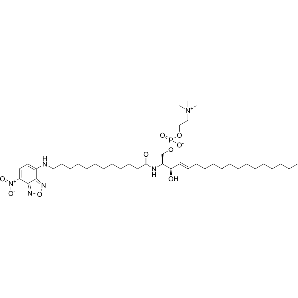
C12 NBD sphingomyelin is an active derivative of Sphingomyelin (HY-113498) that is tagged with fluorescent C12 nitrobenzoxadiazole (C12 NBD). C12 NBD sphingomyelin can be used as a sphingomyelinase substrate for studying the metabolism and transport of sphingomyelins (Ex=470 nm, Em=525 nm).
For research use only. We do not sell to patients.
- Purity : 99.0%
- CAS No.: 254117-01-6
- Formula: C41H73N6O9P
- Molecular Weight:825.03
-
Storage:
-20°C, protect from light, stored under nitrogen
* In solvent : -80°C, 6 months; -20°C, 1 month (protect from light, stored under nitrogen)

To place orders, for customer services and technical support, please contact: MedChemExpress USA
Tel: 609-228-6898 E-mail: [email protected] [email protected]


-
 Biological Activity
-
Chemical Information
- Purity & Documentation
- References
-
 Help & FAQs



All Phospholipase Isoforms
More

Biological Activity
Description
IC50 & Target
Sphingomyelinase[1]
In Vitro
Guide (The following is our recommended solution. This solution is merely a guideline and should be modified according to your specific needs)[2].
1. Preparation of C12 NBD-Sphingomyelin-BSA Complex (for Cell Labeling)
1.1 Dissolve lipids: Prepare a 1 mM C12 NBD-Sphingomyelin stock solution using anhydrous ethanol.
1.2 Dried Lipids: Add 50 μL of the 1 mM stock solution into a glass tube, then blow dry it with nitrogen, and subsequently perform vacuum drying for 1 hour.
1.3 Formation of complex with BSA: Reconstitute the dried lipid with 200 μL anhydrous ethanol; take 10 mL of HBSS/HEPES solution containing 0.34 mg/mL defatted BSA, and slowly add the above ethanol solution of NBD-Sphingomyelin while vigorously vortexing.
2. Cell Staining Procedure
2.1 Cell Washing: Use HBSS/HEPES buffer to wash the cells that are growing on the cover glass and have a confluence of 70-80%.
2.2 Incubation: Add a staining solution of NBD-Sphingomyelin/BSA complex at a concentration of 5 μM to the cells.
2.3 Temperature Control: Incubate at 4℃ for 30 minutes to allow the probe to penetrate the cell membrane while minimizing endocytosis within the cells.
2.4 Washing: Remove the culture medium and wash the cells multiple times with ice-cold culture medium to remove the unbound probes.
2.5 Imaging and Analysis: Observations are conducted using a fluorescence microscope (excitation wavelength Ex = 470 nm, emission wavelength Em = 525 nm); if uptake studies are to be carried out, the cells can be maintained at 37°C for further processing.
MedChemExpress (MCE) has not independently confirmed the accuracy of these methods. They are for reference only. .
Emission (Em)
525
Excitation (Ex)
485


Chemical Information
-
CAS No. 254117-01-6
-
Appearance Solid
-
Molecular Weight 825.03
-
Formula C41H73N6O9P
-
Color Yellow to orange
-
SMILES
C[N+](C)(C)CCOP(OC[C@@H]([C@H](O)/C=C/CCCCCCCCCCCCC)NC(CCCCCCCCCCCNC1=CC=C([N+]([O-])=O)C2=NON=C12)=O)([O-])=O
-
Shipping
Room temperature in continental US; may vary elsewhere.
-
Storage
-20°C, protect from light, stored under nitrogen
* In solvent : -80°C, 6 months; -20°C, 1 month (protect from light, stored under nitrogen)


Purity & Documentation
-
Data Sheet (267 KB)
-
SDS (252 KB)
- English - EN (252 KB)
- Français - FR (252 KB)
- Deutsch - DE (252 KB)
- Norwegian - NO (252 KB)
- Español - ES (252 KB)
- Swedish - SV (252 KB)
- Italian - IT (252 KB)
- Korean - KR (252 KB)
- Portuguese - PT (252 KB)
-
Handling Instructions (2659 KB)
References
[1]. Nakagawa, et al. Preparation of fluorescence-labeled GM1 and sphingomyelin by the reverse hydrolysis reaction of sphingolipid ceramide N-deacylase as substrates for assay of sphingolipid-degrading enzymes and for detection of sphingolipid-binding proteins. J. Biochem. 126(3), 601-611 (1999). [Content Brief]

Calculators
Concentration (start) × Volume (start) = Concentration (final) × Volume (final)


C12 NBD Sphingomyelin Related Classifications
HY-D1584 Related Classifications
 Powered by Bioz