



FW1256
Based on 1 publication(s) in Google Scholar
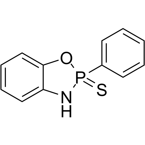
FW1256 is a phenyl analogue and a slow-releasing hydrogen sulfide (H2S) donor. FW1256 inhibits NF-κB activity and induces cell apoptosis. FW1256 exerts potent anti-inflammatory effects and has the potential for cancer and cardiovascular disease treatment.
For research use only. We do not sell to patients.
- Purity : 95.0%
- CAS No.: 117089-08-4
- Formula: C12H10NOPS
- Molecular Weight:247.25
-
Storage:
-20°C, sealed storage, away from moisture
* In solvent : -80°C, 2 years; -20°C, 1 year (sealed storage, away from moisture)

To place orders, for customer services and technical support, please contact: MedChemExpress USA
Tel: 609-228-6898 E-mail: [email protected] [email protected]


-
 Biological Activity
-
Chemical Information
-
Solvent & Solubility
- Protocols
- Purity & Documentation
- References
-
 Help & FAQs


Publications Citing Use of MedChemExpress (MCE) FW1256
More

Biological Activity
Description
Cellular Effect
|
Cell Line

|
Type | Value | Description | References |
|---|---|---|---|---|
| Hepatocyte | IC50 |
26.3 μM
Compound: 22, FW1256
|
Toxicity in transforming growth factor alpha transgenic mouse hepatocytes assessed as inhibition of cell proliferation after 24 hrs by CellTiter-Glo cell viability assay
Toxicity in transforming growth factor alpha transgenic mouse hepatocytes assessed as inhibition of cell proliferation after 24 hrs by CellTiter-Glo cell viability assay
|
[PMID: 26147240] |
| MCF7 | IC50 |
5.7 μM
Compound: 22, FW1256
|
Cytotoxicity against human MCF7 cells assessed as inhibition of cell proliferation after 4 days by crystal violet assay
Cytotoxicity against human MCF7 cells assessed as inhibition of cell proliferation after 4 days by crystal violet assay
|
[PMID: 26147240] |
| SK-OV-3 | IC50 |
6.12 μM
Compound: 22, FW1256
|
Cytotoxicity against human SKOV3 cells assessed as inhibition of cell proliferation after 4 days by crystal violet assay
Cytotoxicity against human SKOV3 cells assessed as inhibition of cell proliferation after 4 days by crystal violet assay
|
[PMID: 26147240] |
| SK-OV-3 | IC50 |
6.12 μM
Compound: 3; FW1256
|
Antiproliferative activity against human SKOV3 cells after 4 days by crystal violet staining based spectrophotometric method
Antiproliferative activity against human SKOV3 cells after 4 days by crystal violet staining based spectrophotometric method
|
[PMID: 28082040] |
| WI-38 | IC50 |
>50 μM
Compound: 22, FW1256
|
Toxicity in human WI38 cells assessed as inhibition of cell proliferation after 4 days by crystal violet assay
Toxicity in human WI38 cells assessed as inhibition of cell proliferation after 4 days by crystal violet assay
|
[PMID: 26147240] |
| WI-38 | IC50 |
>50 μM
Compound: 3; FW1256
|
Antiproliferative activity against human WI38 cells after 4 days by crystal violet staining based spectrophotometric method
Antiproliferative activity against human WI38 cells after 4 days by crystal violet staining based spectrophotometric method
|
[PMID: 28082040] |
In Vitro
FW1256 (200 μM; 24.5 hours; AW264.7 cells) treatment significantly reduces IL-1β, COX-2 and iNOS mRNA and protein in LPS-stimulated RAW264.7 macrophages[1].
FW1256 (200 μM; 24.5 hours; AW264.7 cells) treatment significantly reduces IL-1β, COX-2 and iNOS PROTE and protein in LPS-stimulated RAW264.7 macrophages[1].
FW1256 concentration dependently decreases TNF-α (IC50 of 61.2 μM), IL-6 (IC50 of 11.7 μM), PGE2 (IC50 of 25.5 μM) and NO (IC50 of 34.6 μM) generation in LPS-stimulated RAW264.7 macrophages and bone marrow-derived macrophages (BMDMs) (IC50s of 414.9 μM, 300.2 μM, 4 μM and 9.5 μM for TNF-α, IL-6, PGE2 and NO, respectively) [1].
FW1256 decreases NF-κB activation as evidenced by reduced cytosolic phospho-IκBα levels and reduces nuclear p65 levels in LPS-stimulated RAW264.7 macrophages treated with FW1256[1].
MedChemExpress (MCE) has not independently confirmed the accuracy of these methods. They are for reference only.
-
Cell Line:RAW264.7 cells
-
Concentration:200 µM
-
Incubation Time:24.5 hours
-
Result:Significantly reduced IL-1β, COX-2 and iNOS mRNA in LPS-stimulated RAW264.7 macrophages..
-
Cell Line:RAW264.7 cells
-
Concentration:200 µM
-
Incubation Time:24.5 hours
-
Result:Significantly reduced IL-1β, COX-2 and iNOS proteinin LPS-stimulated RAW264.7 macrophages..
In Vivo
MedChemExpress (MCE) has not independently confirmed the accuracy of these methods. They are for reference only.
-
Animal Model:Male C57BL/6 mice (20-25 g, 6-10 weeks) injected with E. coli lipopolysaccharide (LPS)[1]
-
Dosage:100 mg/kg
-
Administration:Intraperitoneal injection
-
Result:Reduced IL-1β, TNFα, nitrate/nitrite and PGE2 levels in LPS-treated mice.


Chemical Information
-
CAS No. 117089-08-4
-
Appearance Solid
-
Molecular Weight 247.25
-
Formula C12H10NOPS
-
Color White to off-white
-
SMILES
S=P1(OC2=CC=CC=C2N1)C3=CC=CC=C3
-
Shipping
Room temperature in continental US; may vary elsewhere.
-
Storage
-20°C, sealed storage, away from moisture
* In solvent : -80°C, 2 years; -20°C, 1 year (sealed storage, away from moisture)


Publications (1)
-
Journal Impact Factor
-
Most Recent
-
Am J Transl Res
Exosomes released from mesenchymal stem cells overexpressing microRNA-30e ameliorate heart failure in rats with myocardial infarction. [Abstract]2021 May 15;13(5):4007-4025. PMID: 34149995


Solvent & Solubility
In Vitro:
DMSO : ≥ 250 mg/mL (1011.12 mM; Hygroscopic DMSO has a significant impact on the solubility of product, please use newly opened DMSO)
* "≥" means soluble, but saturation unknown.
Please refer to the solubility information to select the appropriate solvent. Once prepared, please aliquot and store the solution to prevent product inactivation from repeated freeze-thaw cycles.
Storage method and period of stock solution: -80°C, 2 years; -20°C, 1 year (sealed storage, away from moisture). When stored at -80°C, please use it within 2 years. When stored at -20°C, please use it within 1 year.
Please refer to the solubility information to select the appropriate solvent. Once prepared, please aliquot and store the solution to prevent product inactivation from repeated freeze-thaw cycles.
Storage method and period of stock solution: -80°C, 2 years; -20°C, 1 year (sealed storage, away from moisture). When stored at -80°C, please use it within 2 years. When stored at -20°C, please use it within 1 year.
Concentration (start) × Volume (start) = Concentration (final) × Volume (final)


Purity & Documentation
-
Data Sheet (278 KB)
-
SDS (252 KB)
- English - EN (252 KB)
- Français - FR (252 KB)
- Deutsch - DE (252 KB)
- Norwegian - NO (252 KB)
- Español - ES (252 KB)
- Swedish - SV (252 KB)
- Italian - IT (252 KB)
- Korean - KR (252 KB)
- Portuguese - PT (252 KB)
-
Handling Instructions (2659 KB)
References
[1]. Huang CW, et al. A novel slow-releasing hydrogen sulfide donor, FW1256, exerts anti-inflammatory effects in mouse macrophages and in vivo. Pharmacol Res. 2016 Nov;113(Pt A):533-546. [Content Brief]
[2]. Feng W, et al. Discovery of New H2S Releasing Phosphordithioates and 2,3-Dihydro-2-phenyl-2-sulfanylenebenzo[d][1,3,2]oxazaphospholes with Improved Antiproliferative Activity. J Med Chem. 2015 Aug 27;58(16):6456-80. [Content Brief]


Complete Stock Solution Preparation Table
Please refer to the solubility information to select the appropriate solvent. Once prepared, please aliquot and store the solution to prevent product inactivation from repeated freeze-thaw cycles.
Storage method and period of stock solution: -80°C, 2 years; -20°C, 1 year (sealed storage, away from moisture). When stored at -80°C, please use it within 2 years. When stored at -20°C, please use it within 1 year.
| Optional Solvent | Concentration Solvent Mass | 1 mg | 5 mg | 10 mg | 25 mg |
|---|---|---|---|---|---|
| DMSO | 1 mM | 4.0445 mL | 20.2224 mL | 40.4449 mL | 101.1122 mL |
| 5 mM | 0.8089 mL | 4.0445 mL | 8.0890 mL | 20.2224 mL | |
| 10 mM | 0.4044 mL | 2.0222 mL | 4.0445 mL | 10.1112 mL | |
| 15 mM | 0.2696 mL | 1.3482 mL | 2.6963 mL | 6.7408 mL | |
| 20 mM | 0.2022 mL | 1.0111 mL | 2.0222 mL | 5.0556 mL | |
| 25 mM | 0.1618 mL | 0.8089 mL | 1.6178 mL | 4.0445 mL | |
| 30 mM | 0.1348 mL | 0.6741 mL | 1.3482 mL | 3.3704 mL | |
| 40 mM | 0.1011 mL | 0.5056 mL | 1.0111 mL | 2.5278 mL | |
| 50 mM | 0.0809 mL | 0.4044 mL | 0.8089 mL | 2.0222 mL | |
| 60 mM | 0.0674 mL | 0.3370 mL | 0.6741 mL | 1.6852 mL | |
| 80 mM | 0.0506 mL | 0.2528 mL | 0.5056 mL | 1.2639 mL | |
| 100 mM | 0.0404 mL | 0.2022 mL | 0.4044 mL | 1.0111 mL |


FW1256 Related Classifications
HY-121955 Related Classifications
 Powered by Bioz