



Other Analysis Dyes
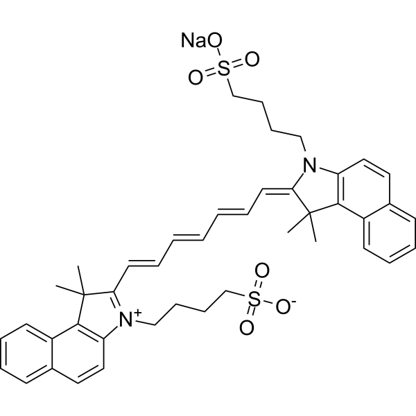
- Formula: C43H47N2NaO6S2
- Molecular Weight: 774.96
Indocyanine green (Foxgreen) is a low toxicic fluorescent agent that has been widely used in medical diagnostics, such as determining cardiac output, hepatic function, and liver blood flow, and for ophthalmic angiography (Ex/Em = 785/813 nm).
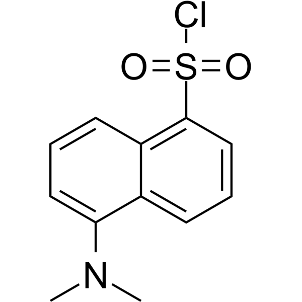
- Formula: C12H12ClNO2S
- Molecular Weight: 269.75
Dansyl chloride is a reagent that produces stable blue or blue-green fluorescent sulfonamide adducts in the reaction of aliphatic and aromatic amines with primary amino groups, and is widely used for modified amino acids, protein sequencing and amino acid analysis.
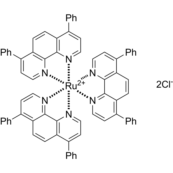
- Formula: C72H48Cl2N6Ru
- Molecular Weight: 1169.17
Tris(4,7-diphenyl-1,10-phenanthroline)ruthenium(II) dichloride ([Ru(dpp)3]2+) is an electrochemiluminescent (ECL) probe and oxygen-sensitive sensor. Tris(4,7-diphenyl-1,10-phenanthroline)ruthenium(II) dichloride can be used to modify electrode surfaces for the detection of sulfates (S2O82-) and oxalates, based on electrochemical reactions that generate excited-state species, releasing photons through irreversible redox reactions. Tris(4,7-diphenyl-1,10-phenanthroline)ruthenium(II) dichloride utilizes the oxygen quenching of fluorescence mechanism, where fluorescence intensity reflects the metabolic rate of living microorganisms or oxygen levels within cells/tumors, allowing for real-time detection. Tris(4,7-diphenyl-1,10-phenanthroline)ruthenium(II) dichloride's main applications include microbial detection, antibacterial activity studies, and tumor microenvironment research.
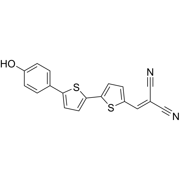
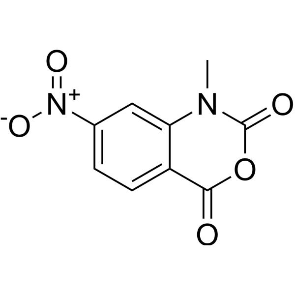
- Formula: C9H6N2O5
- Molecular Weight: 222.15
1-Methyl-7-nitroisatoic anhydride (1M7) is a reagent that detects local nucleotide flexibility, for probing 2'-hydroxyl reactivity, can be used for RNA structure analysis.

- Formula: C41H44ClIN2O2
- Molecular Weight: 759.16
HS-CyBz is a Fluorescent probe for H₂S detection, enabling ratiometric optical/photoacoustic dual-modality in/ex vivo imaging. Its detection mechanism relies on nucleophilic substitution of its benzoic ester group by HS⁻, which releases an enolic meso-hydroxyltricarboheptamethine cyanine that then undergoes keto-enol tautomerization to form Cy-ketone; this tautomerization causes distinct shifts in absorption and emission spectra, producing a ratiometric response that reduces interferences from tissue scattering, autofluorescence, and probe concentration. In its initial state, HS-CyBz has an excitation wavelength of 595 nm, with emission bands centered at 805 nm (main) and 630 nm (minor); upon reaction with H₂S, the 805 nm emission band decreases while the 630 nm band drastically increases, and its absorption spectrum shows a sharp band at 775 nm and a shoulder band at 708 nm, which decrease upon H₂S reaction with a minor increase at 850 nm and an isosbestic point at 825 nm. For in vivo optical imaging, excitation at 560 nm is used with emission channels at 620 nm and 790 nm, while in vivo photoacoustic imaging uses excitation at 775 nm and 825 nm. The detection limit of HS-CyBz for H₂S is 0.5 μM, and it shows high selectivity, with only H₂S inducing a distinct enhancement of the emission ratio F₆₃₀/F₈₀₅ and PA ratio PA₈₂₅/PA₇₇₅, while other biochemical species including cations, anions, reactive oxygen species, biothiols, and carboxylesterase trigger only minor changes and do not interfere with H₂S sensing. Tail intravenous injection of HS-CyBz leads to accumulation in the liver of mice, and it can be used to verify endogenous H₂S upregulation triggered by S-adenosyl-L-methionine via ratiometric optical/photoacoustic imaging.

- Formula: C28H21ClN8O3
- Molecular Weight: 552.97
CMTDP is a Fluorescent probe for detecting local polarity changes around the Cys34 domain of bovine serum albumin during pH-induced N→B conformational transition. It is a thiol-specific and polarity-sensitive probe; it covalently labels the free cysteine residue at position 34 (Cys34) of bovine serum albumin, and its fluorescence maximum emission wavelength shifts in response to solvent polarity, but not to pH and temperature. When labeled to BSA, as the local polarity around the Cys34 domain increases with rising pH, the probe shows a bathchromic shift in fluorescence emission wavelength and a decrease in fluorescence intensity, which allows quantification of the local polarity change via the relationship between emission wavelength shift and dielectric constant. CMTDP itself has absorption peaks at ~392 and 508 nm, and CMTDP-labeled BSA has excitation maxima at 376 and 470 nm at pH 6.0, with the shorter-wavelength excitation peak red-shifting as pH increases while the longer one remains stable; when excited at 470 nm, the fluorescence emission maximum shifts from 570 nm at pH 6.0 to 577 nm at pH 9.1, while free CMTDP at pH 7.4 has an emission maximum at 621 nm. The excitation/emission wavelengths for CMTDP-labeled BSA are Ex/Em = 376/570 nm at pH 6.0, Ex/Em = ~376+/573 nm at pH 7.4, Ex/Em = ~376+/575 nm at pH 8.0, Ex/Em = ~376+/577 nm at pH 9.1, and for free CMTDP at pH 7.4, Ex/Em (when excited at 470 nm) = 470/621 nm[1].

- Formula: C27H32BF3N4O3
- Molecular Weight: 528.37
BDL-E5 is a fluorescent probe for detecting live induced pluripotent stem cells at the early reprogramming stage. Its detection mechanism involves specific binding to authentic reprogramming cells, which exhibit increased expression of pluripotency and epithelial genes and decreased expression of mesenchymal genes compared to non-reprogramming cells; it appears to co-localize more significantly with the Golgi complex than other organelle markers in reprogramming cells, and it does not require washing after staining to reduce background signals; fluorescence is generated upon binding to these early reprogramming cells, with positive cells appearing 7 days before iPS colonies are visible and stain positive for the conventional pluripotent marker TRA-1-60, and sorting BDL-E5-positive cells enriches populations that generate higher numbers and quality of iPS colonies. Its detection wavelength is Ex/Em = 578/599 nm.
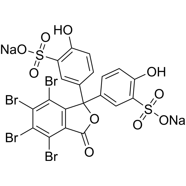
- Formula: C20H8Br4Na2O10S2
- Molecular Weight: 838.00
Sulfobromophthalein (Bromosulfophthalein) disodium salt is an organic anion dye used in the study of a variety of membrane carriers expressed in animal tissues and involved in transport of agents and metabolites.
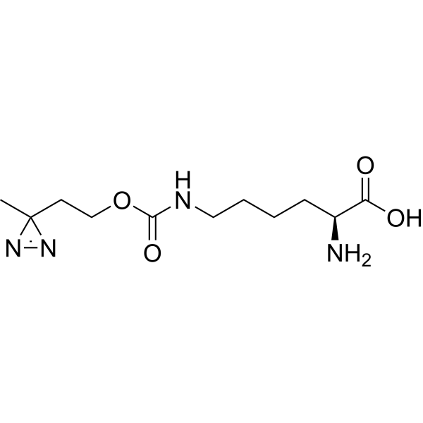
- Formula: C11H20N4O4
- Molecular Weight: 272.30
DiAzKs (H-L-Photo-lysine) is a diazirine-containing lysine amino acid and is a photo-cross-linker. DiAzKs can site-selective incorporated into proteins and is used to crosslink protein-protein interactions in vitro and in living cells. DiAzKs acts as a UV light-activated photo-crosslinking probe.
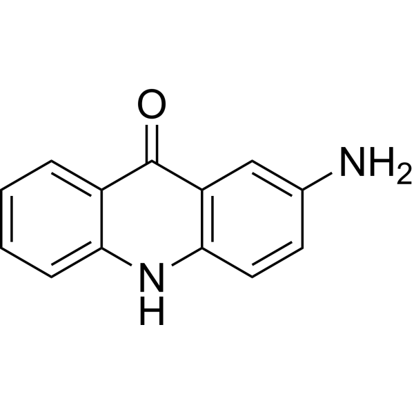
- Formula: C13H10N2O
- Molecular Weight: 210.24
2-Aminoacridone is a widely used fluorophore (λexc=428 nm, λem=525 nm).
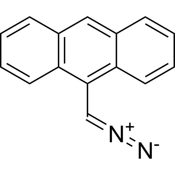
- Formula: C15H10N2
- Molecular Weight: 218.25
9-Anthryldiazomethane is a fluorescent labeling reagent, which can be used for detecting fatty acids and derivatives.


- Formula: C31H24IN3S2
- Molecular Weight: 629.58
MBCB is a two-photon Fluorescent probe for dual-detection of mitochondrial SO₂ derivatives and viscosity. For SO₂ derivatives detection, the probe utilizes a Michael addition mechanism: nucleophilic addition of SO₂ derivatives to the C=C bond between the carbazole skeleton and 3-methylbenzothiazolium moiety destroys the strong intramolecular charge transfer (ICT) system between these groups, while enhancing the weak ICT system between the benzothiazole group and carbazole framework; this causes the red emission at 600 nm to decrease and the blue emission at 434 nm to increase, creating a ratiometric response based on the I₄₃₄ₙₘ/I₆₀₀ₙₘ intensity ratio. For viscosity detection, in low-viscosity environments, steric hindrance creates a twisted ICT (TICT) system with weak fluorescence, while in high-viscosity environments, intramolecular rotation is blocked, the TICT state is disrupted, and the strong ICT system is recovered, leading to a strong red emission at 567 nm with negligible change to the short-wavelength emission at 415 nm, creating a ratiometric response based on the I₅₆₇ₙₘ/I₄₁₅ₙₘ intensity ratio that has a logarithmic linear relationship with viscosity. The probe has excitation/emission wavelengths of Ex/Em = 351/434, 600 nm for SO₂ derivatives detection and Ex/Em = 351/567 nm for viscosity detection, with two-photon excitation at 740 nm for bioimaging; it also exhibits good mitochondrial targeting ability with a Pearson's colocalization coefficient of 0.93 when paired with Mito-Tracker Green. The probe shows high sensitivity and selectivity for SO₂ derivatives, has low cell cytotoxicity, and can be applied to detect exogenous/endogenous HSO₃⁻ in living cells and in vivo, as well as visualize mitochondrial viscosity changes induced by nystatin[1].

- Formula: C32H26INO
- Molecular Weight: 567.46
HOTPy is a mitochondria-targeted fluorescent viscosity probe with aggregation-induced emission properties, which responds to microenvironmental viscosity. HOTPy can recognize the abnormal increase in mitochondrial viscosity and capture dynamic viscosity changes in acute alcoholic liver injury models at both cellular and in vivo levels. The maximum absorption peak of HOTPy in PBS solution is at 379 nm; its maximum emission peak reaches 611 nm in a high-viscosity glycerol system. HOTPy can be used for the research of acute alcoholic liver injury.

- Formula: C30H36BrN7OS2
- Molecular Weight: 654.69
Biotin-V is a fluorescent probe targeting the biotin receptor (BiR), which can be used to detect changes in intracellular viscosity in cancer cells and tumor tissues. Biotin-V consists of a viscosity-responsive TICT (twisted internal charge transfer) fluorophore and a biotin-targeting moiety. In a high-viscosity environment, intramolecular rotation is restricted, which reduces non-radiative transitions and enhances fluorescence intensity and fluorescence lifetime. Biotin-V enters BiR-positive cancer cells via biotin receptor-mediated uptake, and mainly localizes to mitochondria, enabling dual-modal viscosity imaging (fluorescence intensity imaging and fluorescence lifetime imaging) (Ex/Em = 540/608 nm; cell imaging Ex/Em = 561/570-620 nm).

- Formula: C37H30N6O2Se
- Molecular Weight: 669.63
MC-PSE is a fluorescent probe that can be used for glutathione (GSH) detection, NIR-II fluorescence imaging and ratiometric photoacoustic imaging. MC-PSE spontaneously forms J-aggregates in aqueous solution through π-π stacking, and its phenylselenyl group acts as a fluorescence quencher and GSH recognition receptor, thereby eliminating intrinsic fluorescence and generating a maximum absorption peak at 1000 nm. The excitation/emission wavelengths of MC-PSE are Ex = 808 nm and Em = 940 nm. MC-PSE can be applied in tumor-related research.

- Formula: C38H49N7O3
- Molecular Weight: 651.84
Tz‑IFDL is a chemiluminescent probe for in vivo imaging of N‑methyl‑D‑aspartate receptors (NMDAR), which specifically binds to the NR2B subunit of NMDAR. Tz‑IFDL undergoes a bioorthogonal reaction with probe CL‑NC (HY-D3370), triggering molecular fragment release and chemiluminescence. The CL‑NC/Tz‑IFDL system composed of Tz‑IFDL exhibits a chemiluminescence emission wavelength of 710 nm. When combined with CL‑NC, Tz‑IFDL can be used for deep tissue imaging to distinguish NMDAR expression levels between Alzheimer's disease model mice and normal mice.

- Formula: C20H21N5O9PtS2
- Molecular Weight: 734.62
Pt-Luc is a bioluminescent probe used for real-time, non-invasive monitoring of Pt (IV) prodrug reduction in living cells and animal models. The detection mechanism of Pt-Luc relies on the intracellular reduction mediated by active cysteine residues in proteins. Pt-Luc can penetrate the membrane of living cells; after entering cells, active cysteine thiolates in high-molecular-weight protein components reduce the Pt (IV) complex, thereby cleaving the caged aminoluciferin substrate. The released free aminoluciferin is then oxidized by luciferase in the presence of ATP to generate a bioluminescent signal, whose intensity is directly correlated with the reduction degree of Pt (IV).

- Formula: C35H28B2N2O9
- Molecular Weight: 642.23
Rhobo6 is a cell-impermeable glycan-binding, fluorescence turn-on imaging agent with a Kd of 53 µM for glycans. Rhobo6 reversibly binds target glycans and enables wash-free live ECM visualization. Rhobo6 can be used for fluorescent labeling of ECM in living samples or decellularized tissues (Ex/Em = 488/561 nm).



