



MCB-22-174

Based on 1 Customer Validation
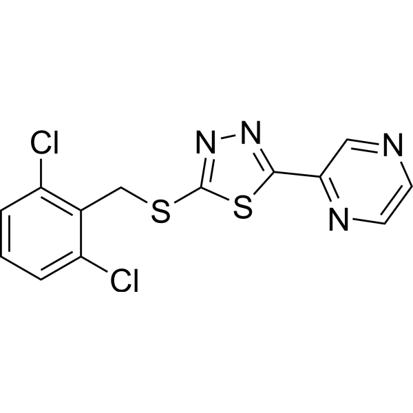
MCB-22-174 is a deuterated Piezo1 agonist, with an EC50 value of 6.28 μM. MCB-22-174 remarkably activates the CaMKII/ERK signaling pathway and initiates Ca2+ influx in rMSCs. MCB-22-174 significantly decreases the expression of chondrogenesis markers (Comp, Acan) and adipogenesis markers (Lpl, Fabp4) in MSCs. MCB-22-174 can effectively improve bone quality in hind-limb unloading (HU) model rats. MCB-22-174 can be used for the study of disuse osteoporosis (OP).
For research use only. We do not sell to patients.
- Purity : 99.78%
- CAS No.: 3058199-58-6
- Formula: C16H14DCl2N5OS2
- Molecular Weight:429.37
-
Storage:Powder -20°C, 3 years ; In solvent -80°C, 6 months , -20°C, 1 month


To place orders, for customer services and technical support, please contact: MedChemExpress USA
Tel: 609-228-6898 E-mail: [email protected] [email protected]


-
 Biological Activity
-
Chemical Information
- Protocols
- Purity & Documentation
- References
-
 Help & FAQs




Biological Activity
Description
In Vitro
MCB-22-174 (0-40 μM) exhibits potent agonistic activity on Piezo1 in C3H10T1/2 cells with an EC50 value of 6.28 μM[1].
MCB-22-174 (3-5 μM, 72 h) significantly upregulates the mRNA level of Runx2 in rMSCs and promotes osteogenesis more effectively than Yoda1 (HY-18723)[1].
MCB-22-174 (5 μM, 5-15 min) remarkably upregulates p-ERK and p-CaMKII in rMSCs, and activates the CaMKII/ERK signaling pathway more efficiently than Yoda1[1].
MCB-22-174 (5 μM) significantly decreases the expression of chondrogenesis markers (Comp, Acan) and adipogenesis markers (Lpl, Fabp4) in MSCs, showing a more significant suppression of chondrogenesis and adipogenesis than Yoda1[1].
MedChemExpress (MCE) has not independently confirmed the accuracy of these methods. They are for reference only. Further protocols information, click here.
In Vivo
MedChemExpress (MCE) has not independently confirmed the accuracy of these methods. They are for reference only.
-
Animal Model:SD rats (2-month-old) were subjected to hind-limb unloading by tail suspension, with their tails fixed on the upper lids of cages to detach the hind limbs from the ground (forelimbs unaffected), simulating weightlessness to induce disuse osteoporosis[1]
-
Dosage:2 mg/kg (5 μmol/kg)
-
Administration:Intraperitoneally injected on days 1, 4, 7, 10, and 13
-
Result:Improved bone quality in HU model rats.
Upregulated bone volume ratio (BV/TV) and trabecular thickness (Tb.Th.).
Downregulated trabecular separation (Tb.Sp.).
Increased the fluorescent area of alkaline phosphatase (ALP).
Promoted osteogenesis, with better efficacy than Yoda1.
Application
1. This compound can be used as a tracer
2. This compound can be used as an internal standard for quantitative analysis by NMR, GC-MS, or LC-MS.


Chemical Information
-
CAS No. 3058199-58-6
-
Unlabeled CAS 3087134-90-2
-
Appearance Solid
-
Molecular Weight 429.37
-
Formula C16H14DCl2N5OS2
-
Color Off-white to light yellow
-
SMILES
ClC(C=CC=C1Cl)=C1C([2H])SC2=NN=C(C3=NC=C(NC[C@H](O)C)N=C3)S2
-
Shipping
Room temperature in continental US; may vary elsewhere.
-
Storage
Powder -20°C 3 years In solvent -80°C 6 months -20°C 1 month

Protocols
-
RNA extraction experimental
By lysing cells, releasing RNA, and removing impurities such as proteins and DNA, high-purity RNA products are finally obtained. The commonly used traditional method is the guanidine isothiocyanate/phenol/chloroform method (Trizol), which is suitable for a variety of animal materials including animal tissues, microorganisms, cultured cells, etc., and most plant materials.
-
3T3-L1 preadipocyte-to-adipocyte differentiation
3T3-L1 preadipocytes are induced to differentiate after growth arrest using adipogenic media containing insulin, dexamethasone, and IBMX; differentiation is assessed by lipid-droplet accumulation, triglyceride increase, Oil Red O staining, and adipocyte-marker induction such as PPARγ and C/EBPα.
-
Mesenchymal stromal/stem cell chondrogenic differentiation
MSC chondrogenic differentiation is commonly induced by culturing bone marrow-derived mesenchymal stromal/stem cells as high-density three-dimensional pellets or micromass aggregates in defined chondrogenic medium containing TGF-β family stimulation; the readout is formation of cartilage-like extracellular matrix, especially sulfated proteoglycans, aggrecan, and type II collagen. The assay detects chondrogenesis by pellet enlargement, metachromatic or Alcian blue/Safranin O staining of proteoglycan-rich matrix, immunodetection of type II collagen and aggrecan, and gene-expression changes in cartilage matrix markers; hypertrophic or fibrocartilaginous drift can be assessed by collagen X and collagen I readouts when included.
-
Ca2+ Staining Technique
Ca2+ staining is an experimental technique that utilizes specific fluorescent probes (such as Fluo-4 AM, Fura-2, etc.) to qualitatively or quantitatively detect dynamic changes in intracellular Ca2+ concentrations; this is achieved by monitoring the changes in fluorescent signals generated when these probes bind to free intracellular calcium ions. The underlying principle relies primarily on the presence of chelating groups within the probe's molecular structure that possess high affinity for calcium ions.
-
Lipid Droplets: Oil Red O/Sudan Dye Lipid Staining
Lipid droplets are intracellular organelles with a neutral-lipid core that stores triacylglycerols and sterol esters, and Oil Red O or Sudan dyes detect these hydrophobic lipid deposits by partitioning into retained lipids in fresh or frozen specimens. Oil Red O stains neutral triglycerides and lipids in frozen tissue sections or air-dried cytologic preparations, while Sudan Black B has also been used as a histochemical fat stain for lipid-rich tissue structures.


Purity & Documentation
-
Data Sheet (273 KB)
-
SDS (251 KB)
- English - EN (251 KB)
- Français - FR (251 KB)
- Deutsch - DE (251 KB)
- Norwegian - NO (251 KB)
- Español - ES (251 KB)
- Swedish - SV (251 KB)
- Italian - IT (251 KB)
- Korean - KR (251 KB)
- Portuguese - PT (251 KB)
-
Handling Instructions (2659 KB)
References

Calculators
Concentration (start) × Volume (start) = Concentration (final) × Volume (final)


MCB-22-174 Related Classifications
HY-165607S Related Classifications
 Powered by Bioz