



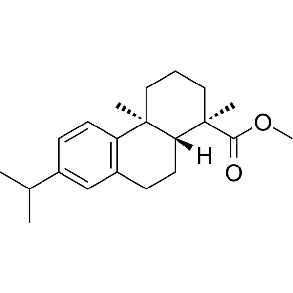
Methyl dehydroabietate

Based on 1 Customer Validation
Methyl dehydroabietate is an orally active resin acid. Methyl dehydroabietate disrupts microbial cell walls and exhibits significant antibacterial activity. Methyl dehydroabietate induces the expression of PPARα in the liver and PPARγ in adipose tissue, and promotes adipocyte differentiation. Methyl dehydroabietate improves insulin resistance, reduces TNFα levels, and alleviates adipocyte hypertrophy and hepatic steatosis in obese mice. Methyl dehydroabietate can be used in research related to obesity, insulin resistance and hepatic steatosis.
Nos produits utilisent uniquement pour la recherche. Nous ne vendons pas aux patients.
- Pureté : 99.89%
- CAS No.: 1235-74-1
- Formule: C21H30O2
- Masse moléculaire:314.46
-
Stockage:Powder -20°C, 3 years , 4°C, 2 years ; In solvent -80°C, 6 months , -20°C, 1 month

To place orders, for customer services and technical support, please contact: MedChemExpress USA
Tel: 609-228-6898 E-mail: [email protected] [email protected]


-
 Activité biologique
-
Chemical Information
-
Solvant et solubilité
- Protocole
- Pureté et documentation
- Références
-
 Help & FAQs




Activité biologique
Description
Cellular Effect
|
Cell Line

|
Type | Value | Description | References |
|---|---|---|---|---|
| A549 | GI50 |
15 μM
Compound: 8
|
Antiproliferative activity against human A549 cells
Antiproliferative activity against human A549 cells
|
[PMID: 30840453] |
| A549 | IC50 |
41.25 μM
Compound: 6
|
Cytotoxic activity against human A549 cells assessed as reduction in cell viability after 72 hrs by SRB assay
Cytotoxic activity against human A549 cells assessed as reduction in cell viability after 72 hrs by SRB assay
|
[PMID: 28011223] |
| HBL-100 | GI50 |
19 μM
Compound: 8
|
Antiproliferative activity against human HBL100 cells
Antiproliferative activity against human HBL100 cells
|
[PMID: 30840453] |
| HeLa | GI50 |
15 μM
Compound: 8
|
Antiproliferative activity against human HeLa cells
Antiproliferative activity against human HeLa cells
|
[PMID: 30840453] |
| HeLa | IC50 |
28 μg/mL
Compound: 3, methyl dehydroabietate
|
Cytotoxicity against human HeLa cells after 48 hrs by MTT assay
Cytotoxicity against human HeLa cells after 48 hrs by MTT assay
|
[PMID: 19892441] |
| Jurkat | IC50 |
21 μg/mL
Compound: 3, methyl dehydroabietate
|
Cytotoxicity against human Jurkat cells after 48 hrs by MTT assay
Cytotoxicity against human Jurkat cells after 48 hrs by MTT assay
|
[PMID: 19892441] |
| PC-3 | IC50 |
37.7 μM
Compound: 6
|
Cytotoxic activity against human PC3 cells assessed as reduction in cell viability after 72 hrs by SRB assay
Cytotoxic activity against human PC3 cells assessed as reduction in cell viability after 72 hrs by SRB assay
|
[PMID: 28011223] |
| SK-OV-3 | IC50 |
51.31 μM
Compound: 6
|
Cytotoxic activity against human SKOV3 cells assessed as reduction in cell viability after 72 hrs by SRB assay
Cytotoxic activity against human SKOV3 cells assessed as reduction in cell viability after 72 hrs by SRB assay
|
[PMID: 28011223] |
| SW1573 | GI50 |
22 μM
Compound: 8
|
Antiproliferative activity against human SW1573 cells
Antiproliferative activity against human SW1573 cells
|
[PMID: 30840453] |
| T47D | GI50 |
16 μM
Compound: 8
|
Antiproliferative activity against human T47D cells
Antiproliferative activity against human T47D cells
|
[PMID: 30840453] |
| Vero | IC50 |
20 μg/mL
Compound: 18
|
Concentration required for 50% inhibition of herpes simplex virus 2(HSV-2) using standard plaque reduction assay in vero cells
Concentration required for 50% inhibition of herpes simplex virus 2(HSV-2) using standard plaque reduction assay in vero cells
|
10.1016/S0960-894X(01)80236-5 |
| Vero | IC50 |
36 μg/mL
Compound: 3, methyl dehydroabietate
|
Cytotoxicity against african green monkey Vero cells after 48 hrs by MTT assay
Cytotoxicity against african green monkey Vero cells after 48 hrs by MTT assay
|
[PMID: 19892441] |
| WiDr | GI50 |
10 μM
Compound: 8
|
Antiproliferative activity against human WiDr cells
Antiproliferative activity against human WiDr cells
|
[PMID: 30840453] |
In Vitro
Methyl dehydroabietate exhibits potent antibacterial activity against a wide range of Gram-positive and Gram-negative bacteria, as well as against certain antibiotic-resistant microorganisms (e.g., methicillin-resistant Staphylococcus aureus, enterococci, etc.); it also demonstrates antifungal activity against common dermatophytes (i.e., the primary pathogens responsible for fungal skin diseases)[1].
Methyl dehydroabietate (10-50 μM; 13-14 days) promotes adipocyte differentiation in 3T3-L1 preadipocytes in vitro in a dose-dependent manner, with significant induction observed at 50 μM[2].
MedChemExpress (MCE) has not independently confirmed the accuracy of these methods. They are for reference only. Further protocols information, click here.
-
Cell Line:mouse 3T3-L1 preadipocytes
-
Concentration:10 μM; 50 μM
-
Incubation Time:13 days (10 μM); 14 days (50 μM)
-
Result:Increased neutral lipid accumulation significantly relative to vehicle control at 50 μM.
Exhibited significantly higher differentiation potency than dehydroabietic acid at 50 μM.
Did not produce a statistically significant change in adipocyte differentiation at 10 μM.
In Vivo
Methyl dehydroabietate (50 mg/kg; p.o.; daily; for 8 weeks) exerts no significant effects on body weight, organ weight, or plasma metabolic/inflammatory biomarkers in healthy male C57BL/6J mice, but upregulates the protein expression of PPARα in the liver and PPARγ in adipose tissue, respectively[2].
MedChemExpress (MCE) has not independently confirmed the accuracy of these methods. They are for reference only.
-
Animal Model:C57BL/6J (male, 5 weeks old, 18-21 g, HFD-induced obesity)[2]
-
Dosage:50 mg/kg
-
Administration:p.o.; once daily; 8 weeks
-
Result:Reduced final mean body weight to 27.6 g, significantly lower than the HFD-only group's 31.0 g.
Reduced mean epididymal adipose tissue weight to 0.99 g, significantly lower than the HFD-only group's 1.44 g.
Significantly reduced HFD-induced elevated plasma glucose, insulin, leptin, and TNFα levels.
Reduced HFD-induced adipocyte hypertrophy in epididymal adipose tissue and decreased the number of large hepatic lipid droplets (attenuated hepatic steatosis).
Increased PPARγ and aP2 protein expression in epididymal adipose tissue, increased PPARα protein expression in the liver, and reduced HFD-induced perilipin-2 protein expression in the liver.
-
Animal Model:C57BL/6J (male, 5 weeks old, 18-21 g, healthy)[2]
-
Dosage:50 mg/kg
-
Administration:p.o.; once daily; 8 weeks
-
Result:Resulted in a final mean body weight of 25.4 g, which was not significantly different from the normal diet control group's 25.3 g.
Showed mean liver weight of 0.87 g (slightly increased vs.
control's 0.82 g), mean kidney weight of 0.26 g, and mean epididymal adipose tissue weight of 0.24 g; none of these differed significantly from control values.
Plasma glucose, insulin, leptin, and TNFα levels were not significantly different from normal diet control values.
Adipocyte size and liver morphology were comparable to normal diet control mice.
Increased PPARγ and aP2 protein expression in epididymal adipose tissue and increased PPARα protein expression in the liver relative to normal diet control mice.


Chemical Information
-
CAS No. 1235-74-1
-
Appearance Solid
-
Masse moléculaire 314.46
-
Formule C21H30O2
-
Color White to off-white
-
SMILES
O=C([C@]1(C)CCC[C@]2(C)C3=C(CC[C@@]12[H])C=C(C(C)C)C=C3)OC
-
Structure Classification
-
Initial Source
-
Livraison
Room temperature in continental US; may vary elsewhere.
-
Stockage
Powder -20°C 3 years 4°C 2 years In solvent -80°C 6 months -20°C 1 month


Solvant et solubilité
In Vitro:
DMSO : 50 mg/mL (159.00 mM; Need ultrasonic; Hygroscopic DMSO has a significant impact on the solubility of product, please use newly opened DMSO)
Please refer to the solubility information to select the appropriate solvent. Once prepared, please aliquot and store the solution to prevent product inactivation from repeated freeze-thaw cycles.
Storage method and period of stock solution: -80°C, 6 months; -20°C, 1 month. When stored at -80°C, please use it within 6 months. When stored at -20°C, please use it within 1 month.
Please refer to the solubility information to select the appropriate solvent. Once prepared, please aliquot and store the solution to prevent product inactivation from repeated freeze-thaw cycles.
Storage method and period of stock solution: -80°C, 6 months; -20°C, 1 month. When stored at -80°C, please use it within 6 months. When stored at -20°C, please use it within 1 month.
Concentration (start) × Volume (start) = Concentration (final) × Volume (final)
In Vivo:
Select the appropriate dissolution method based on your experimental animal and administration route.
- For the following dissolution methods, please ensure to first prepare a clear stock solution using an In Vitro approach and then sequentially add co-solvents:
- To ensure reliable experimental results, the clarified stock solution can be appropriately stored based on storage conditions. As for the working solution for In Vivo experiments, it is recommended to prepare freshly and use it on the same day.
- The percentages shown for the solvents indicate their volumetric ratio in the final prepared solution. If precipitation or phase separation occurs during preparation, heat and/or sonication can be used to aid dissolution.
In Vivo Dissolution Calculator
Please enter the basic information of animal experiments:
-
-
-
-
Recommended: Prepare an additional quantity of animals to account for potential losses during experiments.
Please enter your animal formula composition:
-
%DMSO +
Recommended: Keep the proportion of DMSO in working solution below 2% if your animal is weak.
-
%+
-
+%Tween-80 + +
-
%Saline +
The co-solvents required include: DMSO, . All of co-solvents are available by MedChemExpress (MCE). , Tween 80. All of co-solvents are available by MedChemExpress (MCE).
Working solution concentration: 0.22 mg/mL
Method for preparing stock solution: mg drug dissolved in μL DMSO. Stock solution concentration: mg/mL.
1. Take μL DMSO stock solution;
2. Add μL .
μL , mix evenly;
3. Then add μL Tween 80, mix evenly;
4. Then add μL
Please ensure that the stock solution in the first step is dissolved to a clear state, and add co-solvents in sequence. You can use ultrasonic heating (ultrasonic cleaner, recommended frequency 20-40 kHz), vortexing, etc. to assist dissolution.

Protocole
-
Research Protocol for Infectious Diseases
Infectious-disease experiments test how pathogens interact with host barriers, innate immune receptors, inflammatory signaling, pathogen replication, and tissue injury; pattern-recognition receptors such as TLRs, RIG-I-like receptors, NOD-like receptors, and inflammasomes detect microbial molecules and activate NF-κB, interferon, and cytokine responses. The central hypothesis is that infection severity reflects the balance between pathogen burden and host response: protective inflammation restricts pathogen growth, whereas excessive or mislocalized inflammation contributes to tissue damage and disease phenotype. Unresolved questions include which host pathways are protective versus pathogenic, why some infection models fail to translate to human disease, and which combined readouts best predict clinically relevant infection outcomes.
-
RNA extraction experimental
By lysing cells, releasing RNA, and removing impurities such as proteins and DNA, high-purity RNA products are finally obtained. The commonly used traditional method is the guanidine isothiocyanate/phenol/chloroform method (Trizol), which is suitable for a variety of animal materials including animal tissues, microorganisms, cultured cells, etc., and most plant materials.
-
3T3-L1 preadipocyte-to-adipocyte differentiation
3T3-L1 preadipocytes are induced to differentiate after growth arrest using adipogenic media containing insulin, dexamethasone, and IBMX; differentiation is assessed by lipid-droplet accumulation, triglyceride increase, Oil Red O staining, and adipocyte-marker induction such as PPARγ and C/EBPα.
-
Large-size fat particle sorting
Large-size fat particle sorting is widely used to isolate cells up to 200 μm in diameter. Single-cell flow sorting will allow greater insight into adipocyte heterogeneity by identifying gene expression, protein composition, and metabolic signatures at the single-cell level.
-
Research Protocol for Endocrine Diseases
Endocrine diseases often arise from disrupted hormone production, hormone signaling, or target-tissue responsiveness; for diabetes-focused endocrine disease models, insulin signaling regulates glucose uptake, hepatic glucose output, lipid metabolism, and β-cell compensation. Type 2 diabetes develops through interacting defects in insulin resistance, β-cell dysfunction, adipose inflammation, hepatic glucose overproduction, altered incretin signaling, and ectopic lipid metabolism. A major unresolved question is whether endocrine dysfunction is driven primarily by target-tissue insulin resistance, intrinsic β-cell failure, immune/inflammatory stress, or combined multi-organ failure that differs by disease stage.
-
Bacterial live/dead nucleic-acid viability staining
The LIVE/DEAD bacterial viability staining method is based on differential permeability of nucleic-acid-binding fluorescent dyes, most commonly SYTO 9 and propidium iodide (PI), which enables discrimination of bacterial populations with intact versus compromised cytoplasmic membranes. SYTO 9 penetrates both intact and damaged bacterial membranes and binds nucleic acids to produce green fluorescence, whereas propidium iodide penetrates only cells with compromised membranes and fluoresces red while also reducing SYTO 9 signal through competitive binding and fluorescence interactions. The resulting fluorescence pattern is interpreted as a proxy for membrane integrity, which is widely used as an indicator of bacterial viability in microscopy, flow cytometry, and spectroscopic platforms. However, mechanistic studies show that SYTO 9 and PI interactions involve displacement and fluorescence resonance energy transfer effects, which can influence signal interpretation depending on dye ratios a
-
Research Protocol for Metabolic Diseases
AMP-activated protein kinase, AMPK, is a conserved cellular energy sensor that responds to reduced cellular energy status and coordinates metabolism by increasing ATP-generating catabolic pathways while suppressing ATP-consuming anabolic processes. In metabolic disease research, the AMPK pathway is experimentally relevant because it regulates hepatic lipid synthesis, fatty acid oxidation, glucose production, skeletal-muscle glucose disposal, mTORC1-linked biosynthesis, autophagy, mitochondrial homeostasis, and whole-body energy balance. The central pathway logic is that energy stress, metformin, exercise-like stimulation, or direct AMPK activators increase AMPKα Thr172 phosphorylation and downstream substrate phosphorylation, including ACC and RAPTOR. Phosphorylation of ACC suppresses lipogenesis and supports fatty acid oxidation, whereas phosphorylation of RAPTOR suppresses mTORC1 signaling and links cellular energy status to growth and protein synthesis control. The pathway is linked
-
Liver Histomorphometry
Liver histomorphometry is a quantitative histological approach used to measure structural alterations in hepatic tissue, including parenchymal loss, steatosis, fibrosis, and vascular remodeling, by combining stained tissue section analysis with stereological or computerized image-based measurements. Classical morphometric frameworks quantify volume fractions of liver compartments and fibrotic regions using systematic sampling and image analysis, enabling objective comparison of pathological changes across experimental groups. These approaches are widely applied in liver cirrhosis and fibrosis studies to reduce subjectivity in histological scoring and improve reproducibility of tissue evaluation. Recent methodological advances integrate automated image analysis and radiomics-based extraction of histological features from standard liver stains (e. g. , H&E and fibrotic stains), enabling quantitative correlation between morphometric features and fibrosis stages in non-alcoholic fatty live


Pureté et documentation
-
Fiche technique (302 KB)
-
SDS (254 KB)
- English - EN (254 KB)
- Français - FR (254 KB)
- Deutsch - DE (254 KB)
- Norwegian - NO (254 KB)
- Español - ES (254 KB)
- Swedish - SV (254 KB)
- Italian - IT (254 KB)
- Korean - KR (254 KB)
- Portuguese - PT (254 KB)
-
Instruction de manipulation (2659 KB)
Références
[1]. Burčová Z, et al. Antibacterial and antifungal activity of phytosterols and methyl dehydroabietate of Norway spruce bark extracts. J Biotechnol. 2018;282:18-24. [Content Brief]
[2]. Yoshioka H, et al. Methyl dehydroabietate counters high fat diet-induced insulin resistance and hepatic steatosis by modulating peroxisome proliferator-activated receptor signaling in mice. Biomed Pharmacother. 2018;99:214-219. [Content Brief]


Complete Stock Solution Preparation Table
Please refer to the solubility information to select the appropriate solvent. Once prepared, please aliquot and store the solution to prevent product inactivation from repeated freeze-thaw cycles.
Storage method and period of stock solution: -80°C, 6 months; -20°C, 1 month. When stored at -80°C, please use it within 6 months. When stored at -20°C, please use it within 1 month.
| Optional Solvent | Concentration Solvent Mass | 1 mg | 5 mg | 10 mg | 25 mg |
|---|---|---|---|---|---|
| DMSO | 1 mM | 3.1801 mL | 15.9003 mL | 31.8005 mL | 79.5014 mL |
| 5 mM | 0.6360 mL | 3.1801 mL | 6.3601 mL | 15.9003 mL | |
| 10 mM | 0.3180 mL | 1.5900 mL | 3.1801 mL | 7.9501 mL | |
| 15 mM | 0.2120 mL | 1.0600 mL | 2.1200 mL | 5.3001 mL | |
| 20 mM | 0.1590 mL | 0.7950 mL | 1.5900 mL | 3.9751 mL | |
| 25 mM | 0.1272 mL | 0.6360 mL | 1.2720 mL | 3.1801 mL | |
| 30 mM | 0.1060 mL | 0.5300 mL | 1.0600 mL | 2.6500 mL | |
| 40 mM | 0.0795 mL | 0.3975 mL | 0.7950 mL | 1.9875 mL | |
| 50 mM | 0.0636 mL | 0.3180 mL | 0.6360 mL | 1.5900 mL | |
| 60 mM | 0.0530 mL | 0.2650 mL | 0.5300 mL | 1.3250 mL | |
| 80 mM | 0.0398 mL | 0.1988 mL | 0.3975 mL | 0.9938 mL | |
| 100 mM | 0.0318 mL | 0.1590 mL | 0.3180 mL | 0.7950 mL |


Methyl dehydroabietate Related Classifications
HY-W109613 Related Classifications
 Powered by Bioz