



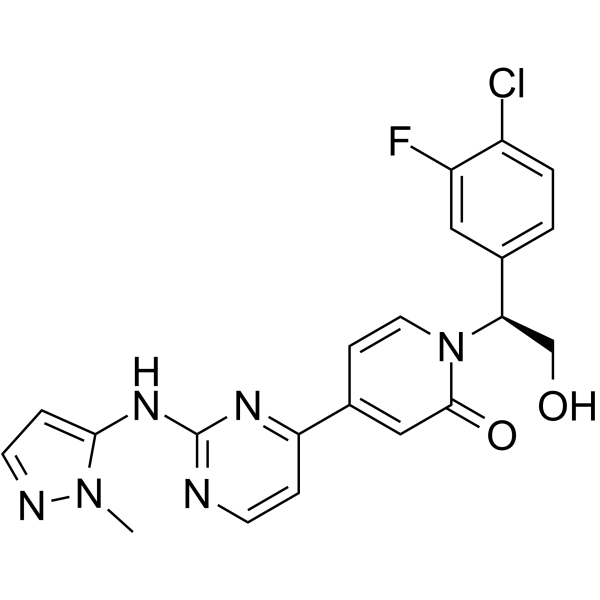
Ravoxertinib GMP is Ravoxertinib (HY-15947) produced by using GMP guidelines. GMP small molecules works appropriately as an auxiliary reagent for cell therapy manufacture. Ravoxertinib (GDC-0994) is an orally active ERK1/2 inhibitor. Ravoxertinib inhibits the ERK1/2 MAPK signaling pathway and reduces the expression levels of c-Myc, HK2 and LDHA. Ravoxertinib decreases mammosphere formation, and exerts additive and/or superadditive cytotoxicity when combined with Ipatasertib (HY-15186) in 3D tumor sphere models. Ravoxertinib can be used in research related to various cancers including breast cancer, melanoma, head and neck cancer, non-small cell lung cancer, ovarian cancer and Merkel cell carcinoma.
Nos produits utilisent uniquement pour la recherche. Nous ne vendons pas aux patients.
- CAS No.: 1453848-26-4
- Formule: C21H18ClFN6O2
- Masse moléculaire:440.86
-
Stockage:
Please store the product under the recommended conditions in the Certificate of Analysis.


To place orders, for customer services and technical support, please contact: MedChemExpress USA
Tel: 609-228-6898 E-mail: [email protected] [email protected]


-
 Activité biologique
-
Chemical Information
- Protocole
- Pureté et documentation
- Références
-
 Help & FAQs


-
HY-15947
RavoxertinibERK1/2 Inhibitor -
HY-50846
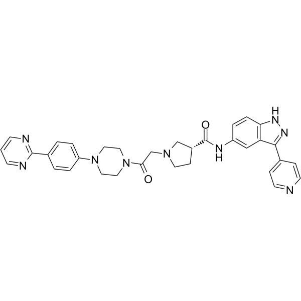
SCH772984ERK1/2 Inhibitor -
HY-15816
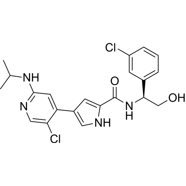
UlixertinibERK1/2 Inhibitor -
HY-101494
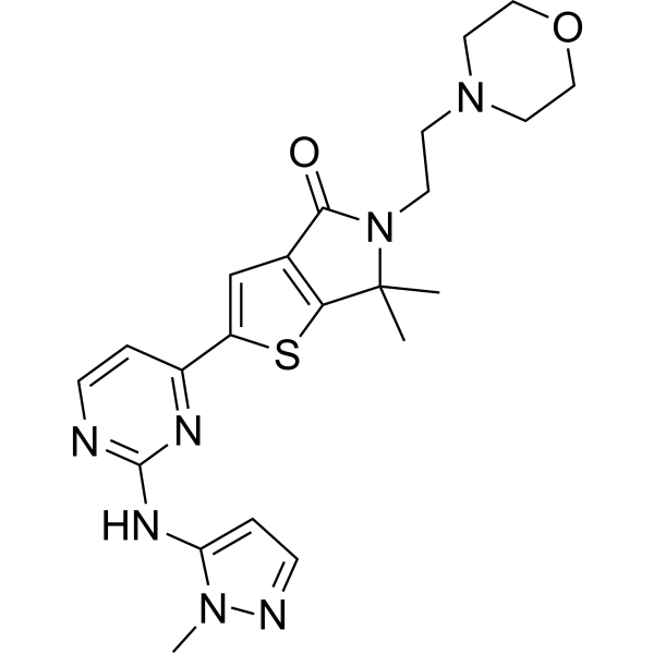
TemuterkibERK1/ERK2 Inhibitor -
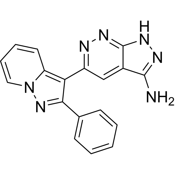
HY-12275
FR 180204ERK Inhibitor -
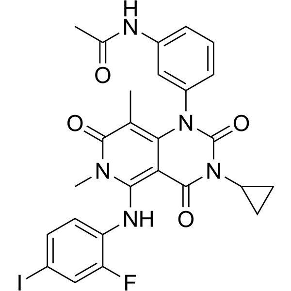
HY-10999
TrametinibMEK Inhibitor -
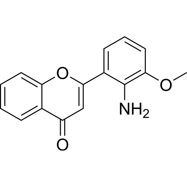
HY-12028
PD98059MEK Inhibitor -
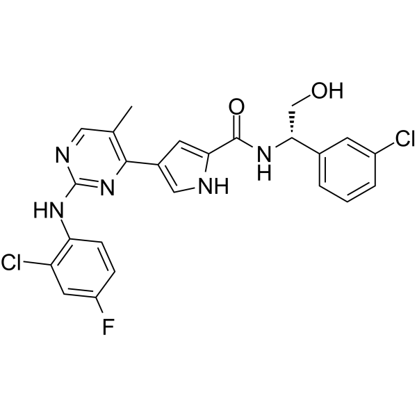
HY-14178
VX-11eERK Inhibitor -
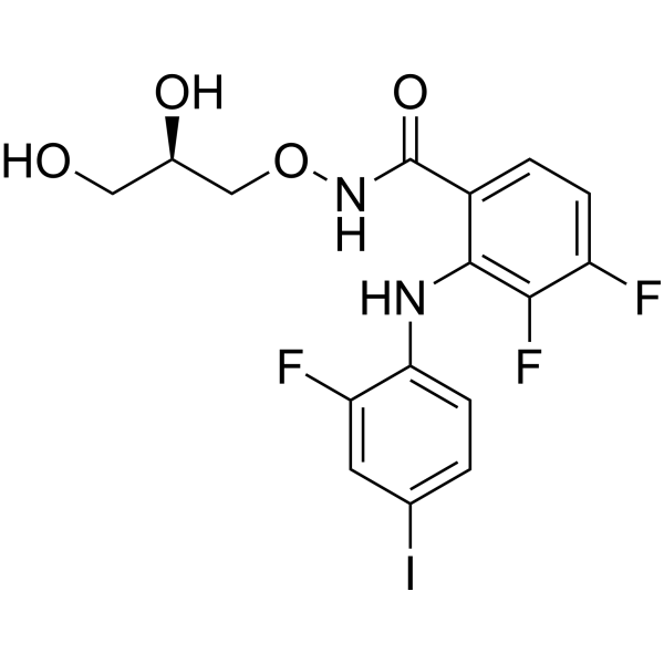
HY-10254
MirdametinibMEK Inhibitor -

HY-15816R
Ulixertinib (Standard)ERK1/2 Inhibitor -
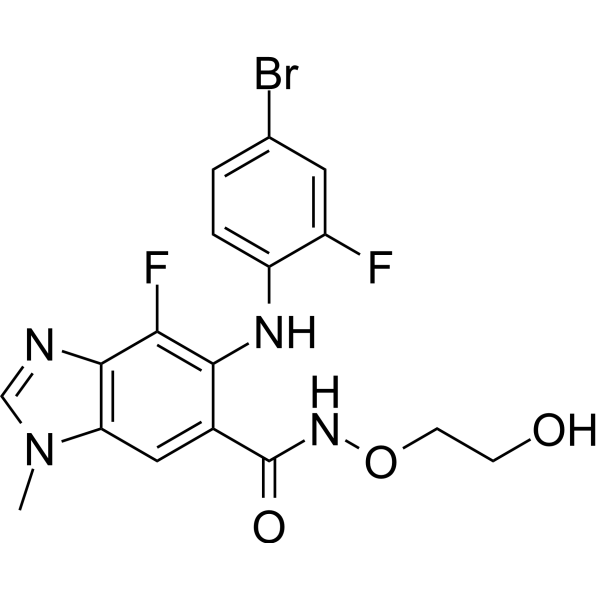
HY-15202
BinimetinibMEK1/2 Inhibitor -
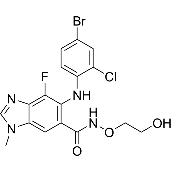
HY-50706
SelumetinibMEK1/2 Inhibitor


Activité biologique
Description
IC50 & Target
|
ERK1 |
ERK2 |
Cellular Effect
|
Cell Line

|
Type | Value | Description | References |
|---|---|---|---|---|
| HepG2 | IC50 |
12 nM
Compound: 22; GDC-0994
|
Inhibition of ERK in PMA-stimulated human HepG2 cells assessed as inhibition of p90RSK serine-380 phosphorylation
Inhibition of ERK in PMA-stimulated human HepG2 cells assessed as inhibition of p90RSK serine-380 phosphorylation
|
[PMID: 27227380] |
In Vitro
In relevant in vitro experiments on stem cell-like human glioma stem cells, Ravoxertinib GMP completely abrogates Humanin (HY-P1928)-induced Temozolomide (HY-17364) resistance, restores the growth inhibitory effect of Temozolomide on tumor cells, and inhibits the Humanin-induced upregulation of Hus1 protein expression[1].
Combination of Ravoxertinib GMP (0.1-10 μM; 7 d) and ipatasertib induces additive to superadditive cytotoxicity in multiple human tumor organoid spheres, with over 2-log cell killing achieved in the 417821-307-R-J1 lung squamous cell carcinoma organoid spheres[2].
Ravoxertinib GMP (1 μM; 72 h) does not significantly inhibit the LMCD1-mediated enhancement of cell proliferation in human dermal fibroblasts[3].
MedChemExpress (MCE) has not independently confirmed the accuracy of these methods. They are for reference only. Further protocols information, click here.
-
Cell Line:417821-307-R-J1 lung squamous-cell carcinoma, LG0567-F671 NSCLC, 138582-337-R-J1 Merkel cell tumor, 845751-090-R-J2 HNSCC, 171881-019-R-J1 breast carcinoma, 327498-153-R-J2 uterine sarcoma, 349418-098-R NSCLC, 989133-093-R-J1 NSCLC, MDA-MB-231 breast carcinoma, SK-OV-3 ovarian carcinoma (in multi-cell type 3D tumor spheroids with HUVECs and hMSCs)
-
Concentration:0.1-10 μM (in combination with ipatasertib)
-
Incubation Time:7 days
-
Result:Produced more than 2-logs of cytotoxicity in 417821-307-R-J1 lung squamous-cell carcinoma mct-spheroids.
Resulted in additive cytotoxicity with 1- to 2-logs of cytotoxicity in 9 tested cell lines.
Showed sub-additive to additive effects in LG0567-F671 NSCLC mct-spheroids via surface plot analysis.
Exhibited regions of greater-than-additive cytotoxicity across ipatasertib concentrations in 138582-337-R-J1 Merkel cell tumor, 845751-090-R-J2 HNSCC, and 171881-019-R-J1 breast carcinoma mct-spheroids.
In Vivo
Ravoxertinib GMP (20-30 mg/kg; p.o.) is well tolerated at 20 mg/kg and 30 mg/kg in NSG mice bearing 845751-090-R-J2 head and neck squamous cell carcinoma PDX, but its combination with ipatasertib does not control tumor growth, leading to progressive disease[2].
MedChemExpress (MCE) has not independently confirmed the accuracy of these methods. They are for reference only.
-
Animal Model:NOD-scid gamma mice (NSG) (female, 6 weeks old, ~26 g, breast carcinoma PDX model)[2]
-
Dosage:20 mg/kg; 30 mg/kg
-
Administration:p.o.
-
Result:Was well tolerated at 20 mg/kg and 30 mg/kg.
Resulted in progressive disease when combined with ipatasertib (60 mg/kg) at 30 mg/kg.
-
Animal Model:NOD-scid gamma mice (NSG) (female, 6 weeks old, ~26 g, head and neck squamous cell carcinoma PDX model)[2]
-
Dosage:20 mg/kg; 30 mg/kg
-
Administration:p.o.
-
Result:Was well tolerated at 20 mg/kg and 30 mg/kg.
Resulted in progressive disease when combined with ipatasertib (60 mg/kg) at 30 mg/kg.


Chemical Information
-
CAS No. 1453848-26-4
-
Masse moléculaire 440.86
-
Formule C21H18ClFN6O2
-
SMILES
O=C1C=C(C2=NC(NC3=CC=NN3C)=NC=C2)C=CN1[C@@H](C4=CC=C(Cl)C(F)=C4)CO
-
Synonyms
GDC-0994 (GMP)
-
Livraison
Room temperature in continental US; may vary elsewhere.
-
Stockage
Please store the product under the recommended conditions in the Certificate of Analysis.

Protocole
-
RNA extraction experimental
By lysing cells, releasing RNA, and removing impurities such as proteins and DNA, high-purity RNA products are finally obtained. The commonly used traditional method is the guanidine isothiocyanate/phenol/chloroform method (Trizol), which is suitable for a variety of animal materials including animal tissues, microorganisms, cultured cells, etc., and most plant materials.
-
Cell Cytotoxicity Assay
Cytotoxicity assays are usually based on the assessment of cell membrane damage, which can also be indirectly detected by measuring cell viability. Detection methods include MTT assay, CKK-8 assay, LDH assay and ATP assay, etc.
-
Mammalian live/dead viability and cytotoxicity staining
Live/dead viability and cytotoxicity staining assays are based on the simultaneous detection of intracellular esterase activity in metabolically active (viable) cells and membrane integrity loss in non-viable cells. In commonly used dual-staining approaches, membrane-permeant fluorogenic substrates are converted by intracellular esterases into fluorescent products in live cells, while impermeant DNA-binding dyes selectively enter cells with compromised plasma membranes and label nucleic acids in dead or dying cells, enabling discrimination between viable and non-viable populations by fluorescence microscopy or flow cytometry.
-
Breast Cancer Modeling
Breast cancer is a heterogeneous cancer, and it has been distinguished into four subtypes: luminal A, luminal B, HER2-positive and basal-like. Molecular mutations, epigenetic alterations, hormone exposure and immune microenvironment are related to the progression of breast cancer.
-
Cell Viability Determination by MTT Colorimetric Assay
The following protocol uses the MTT colorimetric assay as a classic literature-established method for assessing cell viability/metabolic activity in cultured mammalian cells. MTT[3-(4,5-dimethylthiazol-2-yl)-2,5-diphenyltetrazolium bromide] is reduced by metabolically active cells to a colored formazan product; the amount of formazan is quantified spectrophotometrically and provides an indirect measure of metabolically active viable cells. Importantly, MTT reduction reflects cellular oxidoreductase/metabolic activity rather than an absolute direct count of living cells, so changes in cellular metabolism can alter the signal independently of cell number.


Pureté et documentation
Références
[2]. Teicher BA, et al. Targeted therapy combinations with ipatasertib in multi-cell type 3D tumor spheroid models. Acad Oncol. 2025;2(2):10.20935/acadonco7726. [Content Brief]
[3]. Ye Z, et al. LMCD1 facilitates the induction of pluripotency via cell proliferation, metabolism, and epithelial-mesenchymal transition. Cell Biol Int. 2022;46(9):1409-1422. [Content Brief]
[4]. Hunt BG, et al. RON-augmented cholesterol biosynthesis in breast cancer metastatic progression and recurrence. Oncogene. 2023;42(21):1716-1727. [Content Brief]

Calculators
Concentration (start) × Volume (start) = Concentration (final) × Volume (final)


Ravoxertinib (GMP) Related Classifications
HY-15947G Related Classifications
 Powered by Bioz