



RDN2150
Based on 1 publication(s) in Google Scholar
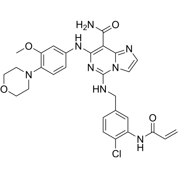
RDN2150 is a ZAP-70 inhibitor with an IC50 of 14.6 nM, and it exhibits selectivity for Syk over other kinases. RDN2150 inhibits signal transduction and activation of T cells/CAR-T cells, reduces the phosphorylation level of Erk1/2, suppresses the induction of CD69 and IL-2, and downregulates phosphotyrosine signaling pathways including hnRNP sites in T cells. RDN2150 can be used for psoriasis-related research.
For research use only. We do not sell to patients.
- CAS No.: 2839429-51-3
- Formula: C28H29ClN8O4
- Molecular Weight:577.03
-
Storage:
Please store the product under the recommended conditions in the Certificate of Analysis.

To place orders, for customer services and technical support, please contact: MedChemExpress USA
Tel: 609-228-6898 E-mail: [email protected] [email protected]


-
 Biological Activity
-
Chemical Information
- Protocols
- Purity & Documentation
- References
-
 Help & FAQs


Publications Citing Use of MedChemExpress (MCE) RDN2150
More

Biological Activity
Description
IC50 & Target
14.6 nM (ZAP-70)[1]
Cellular Effect
|
Cell Line

|
Type | Value | Description | References |
|---|---|---|---|---|
| Splenocyte | IC50 |
64 nM
Compound: 25; RDN2150
|
Immunosuppressive activity in BALB/c mouse Splenocyte assessed as inhibition of concanavalin A induced T-cell proliferation incubated for 48 hrs by [3H]-thymidine incorporation based beta scintillation counter analysis
Immunosuppressive activity in BALB/c mouse Splenocyte assessed as inhibition of concanavalin A induced T-cell proliferation incubated for 48 hrs by [3H]-thymidine incorporation based beta scintillation counter analysis
|
[PMID: 37594408] |
In Vitro
RDN2150 (0.1-10 μM; 3 h) blocks LAT Y220 phosphorylation, disrupts the LAT/SLP76 signalosome, and inhibits downstream Erk1/2 and PLCγ1 activation in Jurkat T cells[2].
RDN2150 (0.1-10 μM; 25 h) inhibits T cell activation markers in Jurkat T cells. In soluble antibody, APC-pMHC/TCR and CD19-CAR/Raji stimulation models, concentrations of 0.1-10 μM reduce CD69 expression and IL-2 secretion, while a concentration of 10 μM completely abrogates soluble antibody-induced CD69 expression[2].
MedChemExpress (MCE) has not independently confirmed the accuracy of these methods. They are for reference only.
-
Cell Line:Jurkat T cells (JOT1 and CD19-CAR Jurkat sublines)
-
Concentration:0.1 μM; 1 μM; 10 μM; 100 nM
-
Incubation Time:3 h
-
Result:Reduced basal Erk1/2 phosphorylation at 100 nM.
Impaired phosphorylation of LATY220 across all three stimulation methods at 10 μM.
Left phosphorylation of LckY394 unaffected at any concentration.
Significantly reduced phosphorylation of Erk1T202Y204/Erk2T185Y187 at 1 μM and 10 μM after 2.5 min of stimulation across all activation models.
Significantly enhanced PLCγ1Y783 induction after soluble antibody stimulation at low concentrations (0.1 μM, 1 μM).
In Vivo
MedChemExpress (MCE) has not independently confirmed the accuracy of these methods. They are for reference only.
-
Animal Model:BALB/c (female, 6-8 weeks old, 18-20 g, Imiquimod-induced psoriasis-like dermatitis)[1]
-
Dosage:2% (62.5 mg per application)
-
Administration:topical; once daily; 7 days
-
Result:Significantly reduced cumulative psoriasis severity total score (0-12 scale) compared to vehicle/ointment base controls, with statistically significant differences observed from day 2 onward.
Significantly decreased individual scores for scales, skin thickness, and erythema (0-4 scales each) with statistically significant differences from day 2 onward.
Significantly reduced splenomegaly, resulting in a lower spleen index (spleen weight/body weight) compared to the vehicle group.


Chemical Information
-
CAS No. 2839429-51-3
-
Molecular Weight 577.03
-
Formula C28H29ClN8O4
-
SMILES
O=C(C1=C(N=C(N2C1=NC=C2)NCC3=CC(NC(C=C)=O)=C(C=C3)Cl)NC4=CC(OC)=C(C=C4)N5CCOCC5)N
-
Shipping
Room temperature in continental US; may vary elsewhere.
-
Storage
Please store the product under the recommended conditions in the Certificate of Analysis.


Publications (1)
-
Journal Impact Factor
-
Most Recent


Purity & Documentation
References
[1]. Rao D, et al. Discovery and Structural Optimization of Covalent ZAP-70 Kinase Inhibitors against Psoriasis. J Med Chem. 2023;66(17):12018-12032. [Content Brief]
[2]. Callahan A, et al. Phosphotyrosine proteomics reveals novel Zap70 and Itk pathway targets downstream of TCR and CAR in Jurkat T cells. Sci Rep. 2026;16(1):16482. Published 2026 Apr 6. [Content Brief]

Calculators
Concentration (start) × Volume (start) = Concentration (final) × Volume (final)


RDN2150 Related Classifications
HY-155978 Related Classifications
 Powered by Bioz