



Carnosic acid
Based on 10 publication(s) in Google Scholar
Carnosic acid is an orally active lipid absorption inhibitor. Carnosic acid has demonstrated inhibition of oxidative stress and inflammation, suppression of cell proliferation, and antibacterial activity.
Nos produits utilisent uniquement pour la recherche. Nous ne vendons pas aux patients.
- Pureté : 98.08%
- CAS No.: 3650-09-7
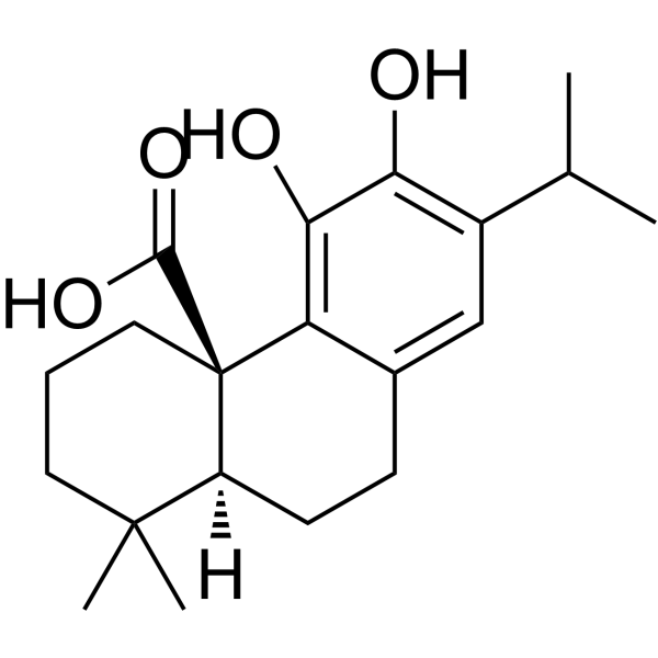
- Formule: C20H28O4
- Masse moléculaire:332.43
-
Stockage:
-80°C, stored under nitrogen

To place orders, for customer services and technical support, please contact: MedChemExpress USA
Tel: 609-228-6898 E-mail: [email protected] [email protected]


-
 Activité biologique
-
Chemical Information
-
Solvant et solubilité
- Protocole
- Pureté et documentation
- Références
-
 Help & FAQs


-
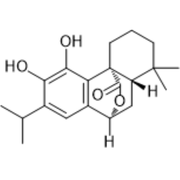
HY-N0643
CarnosolEndogenous Metabolite -
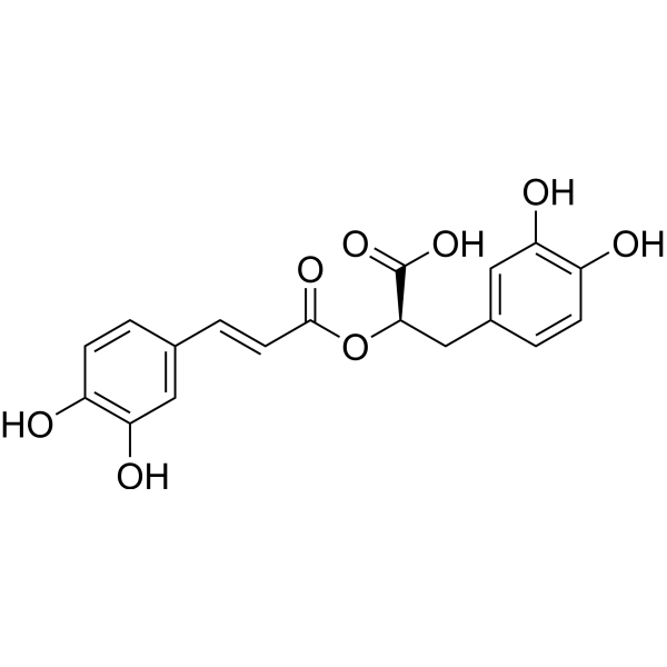
HY-N0529
Rosmarinic acidAnti-Inflammatory Agent -
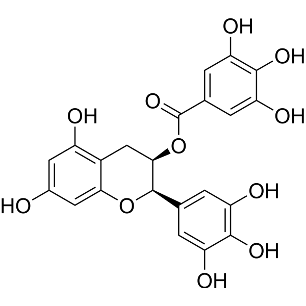
HY-13653
(-)-Epigallocatechin GallateAnticancer Agent -
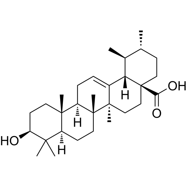
HY-N0140
Ursolic acidAutophagy Inducer -
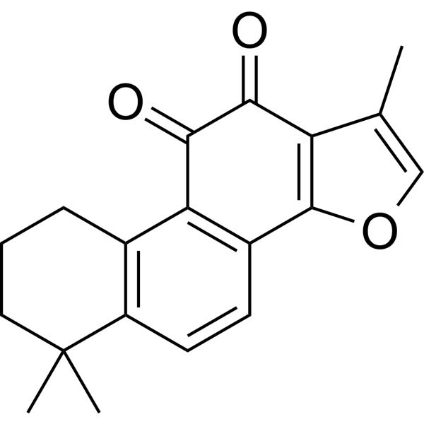
HY-N0135
Tanshinone IIAVEGFR Inhibitor -
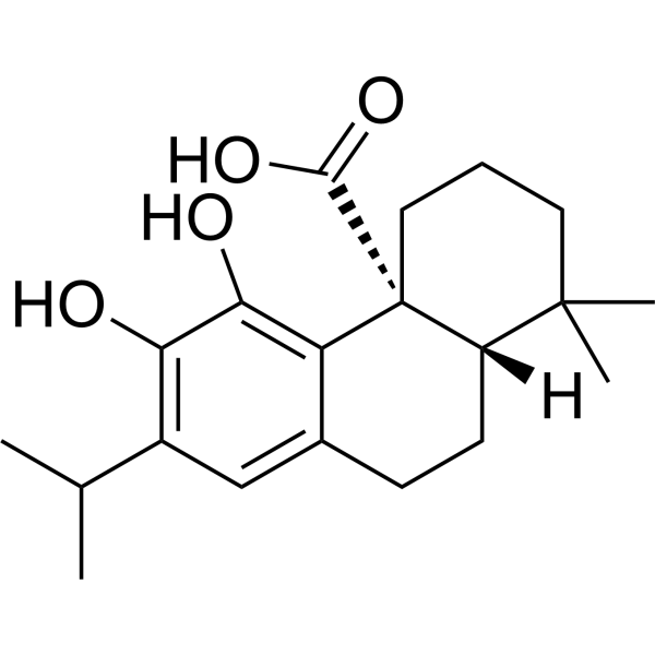
HY-N0644R
Carnosic acid (Standard)Bacterial Inhibitor -
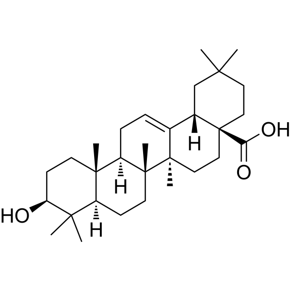
HY-N0156
Oleanolic acidAutophagy Inducer -
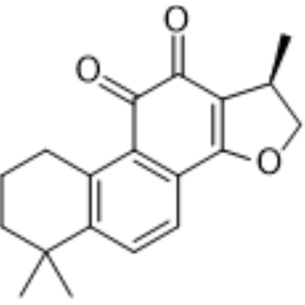
HY-N0174
CryptotanshinoneAutophagy Inducer -
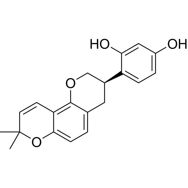
HY-N0393
GlabridinPPARγ Activator -
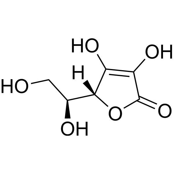
HY-B0166
L-Ascorbic acidAntioxidant -
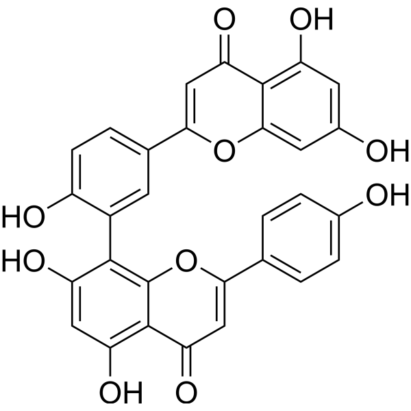
HY-N0662
AmentoflavoneGABA(A) Negative Modulator -
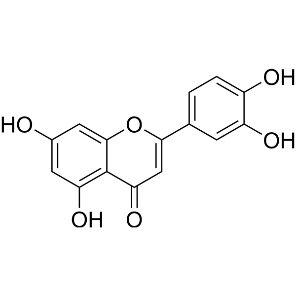
HY-N0162
LuteolinNrf2 Inhibitor
Publications Citing Use of MedChemExpress (MCE) Carnosic acid
More- Adv Sci (Weinh). 2026 Jun 11:e76080. [Abstract]
- PLoS Biol. 2024 Jun 27;22(6):e3002672. [Abstract]
- Nutrients. 2024 Apr 24;16(9):1257. [Abstract]
- Eur J Pharmacol. 2026 Mar 28:1019:178690. [Abstract]
- Int J Mol Sci. 2024 Feb 6;25(4):1950. [Abstract]
- Hum Mol Genet. 2022 Oct 10;31(20):3521-3538. [Abstract]
- Hereditas. 2025 Jul 23;162(1):139. [Abstract]
- SSRN. 2026 May 19.
- SSRN. 2025 Aug 26.
- bioRxiv. 2024 Apr 3:2023.06.02.542933. [Abstract]
 Customer Validation & Images

-
Cell Proliferation/Viability Assay
-
Flow Cytometry
-
WB
-
Cell Imaging/Staining
-
Cell Proliferation/Viability Assay


Activité biologique
Description
Cellular Effect
|
Cell Line

|
Type | Value | Description | References |
|---|---|---|---|---|
| A549 | IC50 |
5 μM
Compound: 8
|
Inhibition of microsomal PGES1 in ILbeta-stimulated human A549 cells using PGH2 as substrate preincubated for 15 mins followed by substrate addition measured after 1 min by RP-HPLC analysis
Inhibition of microsomal PGES1 in ILbeta-stimulated human A549 cells using PGH2 as substrate preincubated for 15 mins followed by substrate addition measured after 1 min by RP-HPLC analysis
|
10.1039/C5MD00278H |
| AGS | IC50 |
89.8 μM
Compound: 1
|
Cytotoxicity against human AGS cells after 24 hrs by neutral red uptake assay
Cytotoxicity against human AGS cells after 24 hrs by neutral red uptake assay
|
[PMID: 20359186] |
| Hepatocyte | EC50 |
94.8 μM
Compound: CA, Carnosic acid
|
Cytotoxicity against human hepatocytes assessed as reduction in cell viability after 24 hrs by ATP detection based assay
Cytotoxicity against human hepatocytes assessed as reduction in cell viability after 24 hrs by ATP detection based assay
|
[PMID: 22531045] |
| Hepatocyte | EC50 |
95.7 μM
Compound: CA, Carnosic acid
|
Cytotoxicity against human hepatocytes assessed as reduction in cell viability after 4 hrs by ATP detection based assay
Cytotoxicity against human hepatocytes assessed as reduction in cell viability after 4 hrs by ATP detection based assay
|
[PMID: 22531045] |
| HepG2 | IC50 |
41.3 μM
Compound: 1
|
Cytotoxicity against human HepG2 cells after 24 hrs by neutral red uptake assay
Cytotoxicity against human HepG2 cells after 24 hrs by neutral red uptake assay
|
[PMID: 20359186] |
| MIA PaCa-2 | EC50 |
5.5 μM
Compound: CA
|
Cytotoxicity against human MIAPaCa2 cells assessed as decrease in cell growth measured after 72 hrs by MTT assay
Cytotoxicity against human MIAPaCa2 cells assessed as decrease in cell growth measured after 72 hrs by MTT assay
|
[PMID: 31725297] |
| MRC5 | IC50 |
21.8 μM
Compound: 1
|
Cytotoxicity against human MRC5 cells after 24 hrs by neutral red uptake assay
Cytotoxicity against human MRC5 cells after 24 hrs by neutral red uptake assay
|
[PMID: 20359186] |
| PANC-1 | EC50 |
150 μM
Compound: CA
|
Cytotoxicity against human PANC1 cells assessed as decrease in cell growth measured after 72 hrs by MTT assay
Cytotoxicity against human PANC1 cells assessed as decrease in cell growth measured after 72 hrs by MTT assay
|
[PMID: 31725297] |
In Vitro
Carnosic acid, a phenolic diterpene, is enriched in the leaves of Lamiaceae plants, and is particularly high in dried leaves of Rosmarinus officinalis L. and Salvia officinalis. Carnosic acid has demonstrated inhibition of oxidative stress and inflammation, suppression of cell proliferation, and antibacterial activity. It is widely reported that Carnosic acid has a therapeutic potential in different types of cancer, mostly based on in vitro experiments. Carnosic acid has also shown neuroprotective effects in experimental models of neurodegenerative diseases, mainly through activation of the antioxidant NRF2/ARE pathway[1].
MedChemExpress (MCE) has not independently confirmed the accuracy of these methods. They are for reference only.
- Research Protocol for Infectious Diseases
- ROS/oxidative-stress fluorescent staining
- BrdU Incorporation Assay
- Somatic Cell Culture
- CFSE Dye Dilution Proliferation Assay
- Dye-dilution cell tracking and proliferation staining
- CCK-8/WST-8 Cell Proliferation Assay
- Protocol for Cell Cycle
- Research Protocol for Inflammation-related Diseases
- Cell Counting-Based Growth Curve Assay
- Colony Formation (Clonogenic) Assay
- Bacterial live/dead nucleic-acid viability staining
- Cell Viability Determination by MTT Colorimetric Assay
- EdU Incorporation Assay (Click Chemistry-Based DNA Synthesis Measurement)
- Ki-67 Immunostaining Proliferation Assay
- PCNA Immunodetection Proliferation Assay
- Protocol for Cell Counting and Cell Density Analysis
- MTT Cell Proliferation Assay


Chemical Information
-
CAS No. 3650-09-7
-
Appearance Solid
-
Masse moléculaire 332.43
-
Formule C20H28O4
-
Color White to light yellow
-
SMILES
OC([C@@]([C@@]1([H])CCC2=CC(C(C)C)=C3O)(CCCC1(C)C)C2=C3O)=O
-
Structure Classification
-
Livraison
Shipping with dry ice.
-
Stockage
-80°C, stored under nitrogen


Publications (10)
-
Journal Impact Factor
-
Most Recent
-
Adv Sci (Weinh)
Multi-Targeting Carnosic Acid Kills Drug-Resistant Helicobacter pylori With Narrow-Spectrum Activity. [Abstract]2026 Jun 11:e76080. PMID: 42272345 -
PLoS Biol
2024 Jun 27;22(6):e3002672. PMID: 38935621 -
Nutrients
Inhibition of Prostate Cancer Cell Survival and Proliferation by Carnosic Acid Is Associated with Inhibition of Akt and Activation of AMPK Signaling. [Abstract]2024 Apr 24;16(9):1257. PMID: 38732504
Carnosic acid purchased from MedChemExpress. Usage Cited in: Nutrients. 2024 Apr 24;16(9):1257. [Abstract]
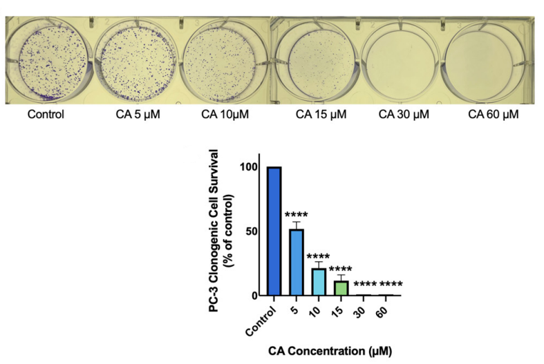
Carnosic acid (CA) dose-dependently Inhibits prostate cancer cell survival. PC-3 prostate cancer cells were treated without (Control) or with the indicated concentrations of Carnosic acid (CA) for 7 days followed by staining with methylene blue and colony counting.
Carnosic acid purchased from MedChemExpress. Usage Cited in: Nutrients. 2024 Apr 24;16(9):1257. [Abstract]
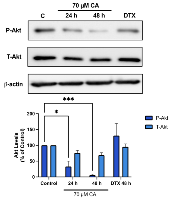
Carnosic acid (CA, 70 μM) inhibited Akt. PC-3 cells were treated without (Control) or with the indicated concentrations of CA or DTX for 24 or 48 h followed by whole cell lysate preparation and total protein yield assessment. Lysates (20 µg of protein) were resolved by SDS-PAGE and immunoblotted with specific antibodies against total or phosphorylated Akt (Ser473) or β-actin.
-
Eur J Pharmacol
Carnosic acid protects against mitochondrial dysfunction and ferroptosis in myocardial ischemia/reperfusion injury through mediating Mfn2. [Abstract]2026 Mar 28:1019:178690. PMID: 41765272
Carnosic acid purchased from MedChemExpress. Usage Cited in: Eur J Pharmacol. 2026 Mar 28:1019:178690. [Abstract]
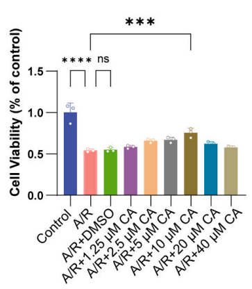
CCK-8 was used to assess the viability of A/R-induced H9c2 cells treated with different concentrations of Carnosic acid (CA) (1.25–40 μM; 48 h). The cell viability of H9c2 cells treated by anoxic/reoxygenation (A/R) was reversed after CA pretreatment at different concentrations.
Carnosic acid purchased from MedChemExpress. Usage Cited in: Eur J Pharmacol. 2026 Mar 28:1019:178690. [Abstract]
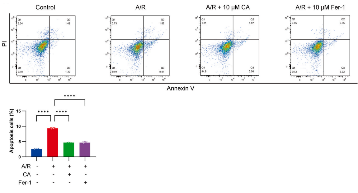
The apoptotic rate was measured using Annexin V-FITC/PI detected by flow cytometry. H9c2 cells in the CA + A/R group were treated with 10 μM CA for 48 h H9c2 cells in the er-1 + A/R group were pretreated with 10 μM Fer-1 for 2 h. CA, Carnosic acid; A/R, anoxic/reoxygenation; Fer-1, ferrostatin-1.
Carnosic acid purchased from MedChemExpress. Usage Cited in: Eur J Pharmacol. 2026 Mar 28:1019:178690. [Abstract]
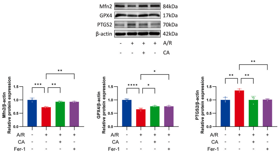
Western blots were used to analyze the expression of ferroptosis-related proteins and Mfn2 in A/R-induced cells after Carnosic acid (CA) or Fer-1 pretreatment. H9c2 cells in the CA + A/R group were treated with 10 μM CA for 48 h; H9c2 cells in the Fer-1 + A/R group were pretreated with 10 μM Fer-1 for 2 h. The protein level of PTGS2 was higher, and that of GPX4 was lower in the A/R group than in the control group. However, pretreatment with CA and Fer-1 prevented these changes.
Carnosic acid purchased from MedChemExpress. Usage Cited in: Eur J Pharmacol. 2026 Mar 28:1019:178690. [Abstract]
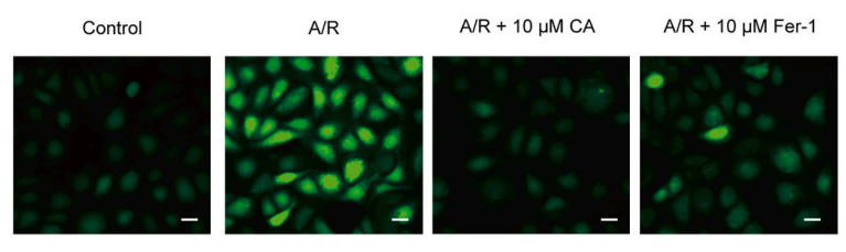
DCFH-DA-stained images for the detection of ROS (magnification, x400; scale bar, 20 μm). H9c2 cells in the CA + A/R group were treated with 10 μM CA for 48 h H9c2 cells in the Fer-1 + A/R group were pretreated with 10 μM Fer-1 for 2 h. CA, Carnosic acid; A/R, anoxic/reoxygenation; Fer-1, ferrpstatin-1.
-
Int J Mol Sci
Carnosic Acid against Lung Cancer: Induction of Autophagy and Activation of Sestrin-2/LKB1/AMPK Signalling. [Abstract]2024 Feb 6;25(4):1950. PMID: 38396629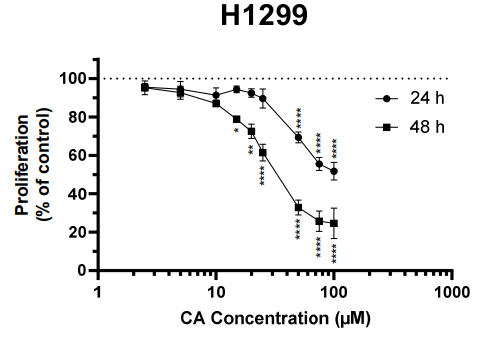
Carnosic acid purchased from MedChemExpress. Usage Cited in: Int J Mol Sci. 2024 Feb 6;25(4):1950. [Abstract]
Carnosic acid (CA) caused concentration- and time-dependent inhibition of H1299 cell proliferation with an IC50 of 47.3 µM and 27.1 µM for 24 h and 48 h, respectively.
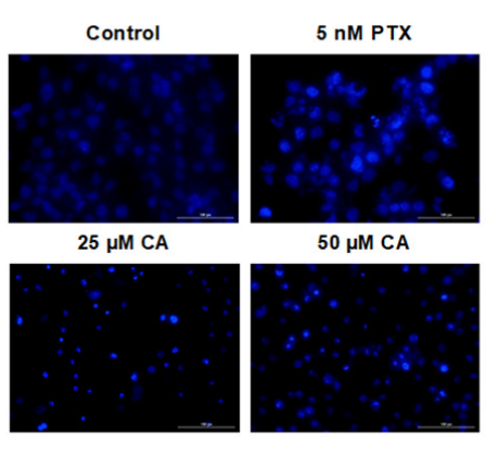
Carnosic acid purchased from MedChemExpress. Usage Cited in: Int J Mol Sci. 2024 Feb 6;25(4):1950. [Abstract]
Carnosic acid (CA) induced apoptosis of H1299 NSCLC cells. H1299 cells were treated for 24 h with 5 nM paclitaxel (PTX), 25 µM CA, or 50 µM CA, and nuclear morphology was assessed qualitatively using Hoechst 33342 and a BioTek Cytation 5 plate reader. CA-treated cells had noticeably smaller nuclei, which was consistent with the nuclear condensation that occurred during apoptosis.
-
Hum Mol Genet
Carnosol, a diterpene present in rosemary, increases ELP1 levels in familial Dysautonomia (FD) patient-derived cells and healthy adults: a possible therapy for FD. [Abstract]2022 Oct 10;31(20):3521-3538. PMID: 35708500 -
Hereditas
Carnosic acid enhances cisplatin sensitivity and suppresses gastric cancer progression via the TP53/SLC7A11/ALOX12 axis. [Abstract]2025 Jul 23;162(1):139. PMID: 40696490 -
-
-
bioRxiv
An efficient behavioral screening platform classifies natural products and other chemical cues according to their chemosensory valence in C. elegans. [Abstract]2024 Apr 3:2023.06.02.542933. PMID: 37333363


Solvant et solubilité
In Vitro:
DMSO : 130 mg/mL (391.06 mM; Need ultrasonic; Hygroscopic DMSO has a significant impact on the solubility of product, please use newly opened DMSO)
Please refer to the solubility information to select the appropriate solvent. Once prepared, please aliquot and store the solution to prevent product inactivation from repeated freeze-thaw cycles.
Storage method and period of stock solution: -80°C, 6 months (stored under nitrogen). When stored at -80°C, please use it within 6 months.
Please refer to the solubility information to select the appropriate solvent. Once prepared, please aliquot and store the solution to prevent product inactivation from repeated freeze-thaw cycles.
Storage method and period of stock solution: -80°C, 6 months (stored under nitrogen). When stored at -80°C, please use it within 6 months.
Concentration (start) × Volume (start) = Concentration (final) × Volume (final)
In Vivo:
Select the appropriate dissolution method based on your experimental animal and administration route.
- For the following dissolution methods, please ensure to first prepare a clear stock solution using an In Vitro approach and then sequentially add co-solvents:
- To ensure reliable experimental results, the clarified stock solution can be appropriately stored based on storage conditions. As for the working solution for In Vivo experiments, it is recommended to prepare freshly and use it on the same day.
- The percentages shown for the solvents indicate their volumetric ratio in the final prepared solution. If precipitation or phase separation occurs during preparation, heat and/or sonication can be used to aid dissolution.
Add each solvent one by one: 10% DMSO 40% PEG300 5% Tween-80 45% Saline
Solubility: ≥ 2.5 mg/mL (7.52 mM); Clear solution
This protocol yields a clear solution of ≥ 2.5 mg/mL (saturation unknown).
Taking 1 mL working solution as an example, add 100 μL DMSO stock solution (25.0 mg/mL) to 400 μL PEG300, and mix evenly; then add 50 μL Tween-80 and mix evenly; then add 450 μL Saline to adjust the volume to 1 mL.
Preparation of Saline: Dissolve 0.9 g sodium chloride in ddH₂O and dilute to 100 mL to obtain a clear Saline solution.
Add each solvent one by one: 10% DMSO 90% (20% SBE-β-CD in Saline)
Solubility: 2.08 mg/mL (6.26 mM); Suspended solution; Need ultrasonic
This protocol yields a suspended solution of 2.08 mg/mL. Suspended solution can be used for oral and intraperitoneal injection.
Taking 1 mL working solution as an example, add 100 μL DMSO stock solution (20.8 mg/mL) to 900 μL 20% SBE-β-CD in Saline, and mix evenly.
Preparation of 20% SBE-β-CD in Saline (4°C, storage for one week): 2 g SBE-β-CD powder is dissolved in 10 mL Saline, completely dissolve until clear.
In Vivo Dissolution Calculator
Please enter the basic information of animal experiments:
-
-
-
-
Recommended: Prepare an additional quantity of animals to account for potential losses during experiments.
Please enter your animal formula composition:
-
%DMSO +
Recommended: Keep the proportion of DMSO in working solution below 2% if your animal is weak.
-
%+
-
+%Tween-80 + +
-
%Saline +
The co-solvents required include: DMSO, . All of co-solvents are available by MedChemExpress (MCE). , Tween 80. All of co-solvents are available by MedChemExpress (MCE).
Working solution concentration: 0.22 mg/mL
Method for preparing stock solution: mg drug dissolved in μL DMSO. Stock solution concentration: mg/mL.
1. Take μL DMSO stock solution;
2. Add μL .
μL , mix evenly;
3. Then add μL Tween 80, mix evenly;
4. Then add μL
Please ensure that the stock solution in the first step is dissolved to a clear state, and add co-solvents in sequence. You can use ultrasonic heating (ultrasonic cleaner, recommended frequency 20-40 kHz), vortexing, etc. to assist dissolution.


Pureté et documentation
-
Fiche technique (293 KB)
-
SDS (393 KB)
- English - EN (393 KB)
- Français - FR (393 KB)
- Deutsch - DE (393 KB)
- Norwegian - NO (393 KB)
- Español - ES (393 KB)
- Swedish - SV (393 KB)
- Italian - IT (393 KB)
- Korean - KR (393 KB)
- Portuguese - PT (393 KB)
-
Instruction de manipulation (2659 KB)
Références
[1]. Yesil-Celiktas O, et al. Inhibitory effects of rosemary extracts, carnosic acid and rosmarinic acid on the growth of various human cancer cell lines. Plant Foods Hum Nutr. 2010 Jun;65(2):158-63. [Content Brief]
[2]. Kiyofumi Ninomiya, et al. Carnosic acid, a new class of lipid absorption inhibitor from sage. Bioorg Med Chem Lett. 2004 Apr 19;14(8):1943-6. [Content Brief]


Complete Stock Solution Preparation Table
Please refer to the solubility information to select the appropriate solvent. Once prepared, please aliquot and store the solution to prevent product inactivation from repeated freeze-thaw cycles.
Storage method and period of stock solution: -80°C, 6 months (stored under nitrogen). When stored at -80°C, please use it within 6 months.
| Optional Solvent | Concentration Solvent Mass | 1 mg | 5 mg | 10 mg | 25 mg |
|---|---|---|---|---|---|
| DMSO | 1 mM | 3.0082 mL | 15.0408 mL | 30.0815 mL | 75.2038 mL |
| 5 mM | 0.6016 mL | 3.0082 mL | 6.0163 mL | 15.0408 mL | |
| 10 mM | 0.3008 mL | 1.5041 mL | 3.0082 mL | 7.5204 mL | |
| 15 mM | 0.2005 mL | 1.0027 mL | 2.0054 mL | 5.0136 mL | |
| 20 mM | 0.1504 mL | 0.7520 mL | 1.5041 mL | 3.7602 mL | |
| 25 mM | 0.1203 mL | 0.6016 mL | 1.2033 mL | 3.0082 mL | |
| 30 mM | 0.1003 mL | 0.5014 mL | 1.0027 mL | 2.5068 mL | |
| 40 mM | 0.0752 mL | 0.3760 mL | 0.7520 mL | 1.8801 mL | |
| 50 mM | 0.0602 mL | 0.3008 mL | 0.6016 mL | 1.5041 mL | |
| 60 mM | 0.0501 mL | 0.2507 mL | 0.5014 mL | 1.2534 mL | |
| 80 mM | 0.0376 mL | 0.1880 mL | 0.3760 mL | 0.9400 mL | |
| 100 mM | 0.0301 mL | 0.1504 mL | 0.3008 mL | 0.7520 mL |


Carnosic acid Related Classifications
HY-N0644 Related Classifications
 Powered by Bioz