



Peretinoin
Based on 7 publication(s) in Google Scholar
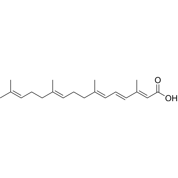
Peretinoin is an oral acyclic retinoid with a vitamin A-like structure that targets retinoid nuclear receptors such as retinoid X receptor (RXR) and retinoic acid receptor (RAR). Peretinoin reduces the mRNA level of sphingosine kinase 1 (SPHK1) in vitro by downregulating a transcription factor, Sp1. Peretinoin prevents the progression of non-alcoholic steatohepatitis (NASH) and the development of hepatocellular carcinoma (HCC) through activating the autophagy pathway by increased Atg16L1 expression. Peretinoin inhibits HCV RNA amplification and virus release by altering lipid metabolism with a EC50 of 9 μM.
For research use only. We do not sell to patients.
- Purity : 98.95%
- CAS No.: 81485-25-8
- Formula: C20H30O2
- Molecular Weight:302.45
-
Storage:
-20°C, protect from light, stored under nitrogen
* In solvent : -80°C, 6 months; -20°C, 1 month (protect from light, stored under nitrogen)

To place orders, for customer services and technical support, please contact: MedChemExpress USA
Tel: 609-228-6898 E-mail: [email protected] [email protected]


-
 Biological Activity
-
Chemical Information
-
Solvent & Solubility
- Protocols
- Purity & Documentation
- References
-
 Help & FAQs


-
HY-A0122
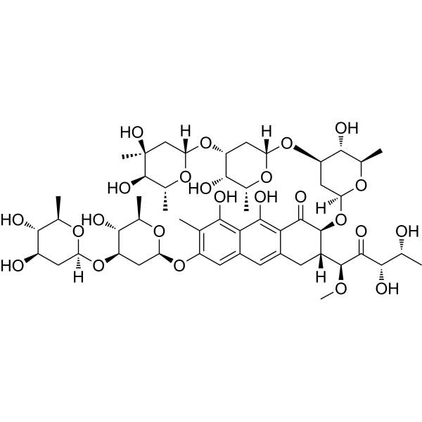
PlicamycinSp1 Inhibitor -
HY-15425
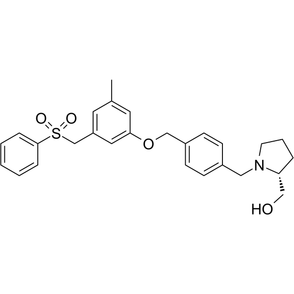
PF-543SPHK1 Inhibitor -
HY-15127
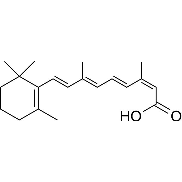
IsotretinoinAnti-acne Agent -
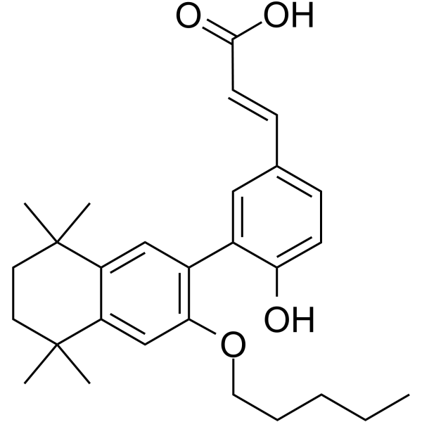
HY-107500
UVI 3003RXR Inhibitor -
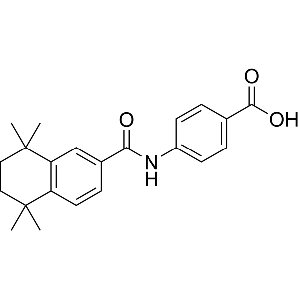
HY-10475
AM580RARα Agonist -
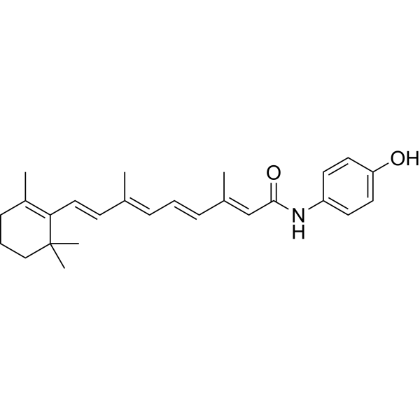
HY-15373
FenretinideAutophagy Inducer -
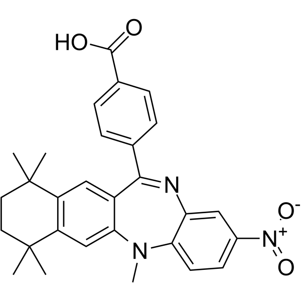
HY-108521
HX531RXR Antagonist -
HY-10291
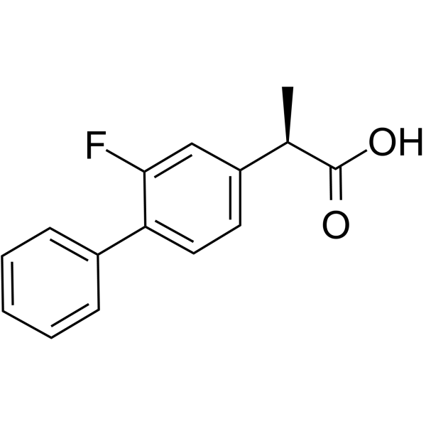
TarenflurbilEnantiomer of Flurbiprofen -
HY-15682
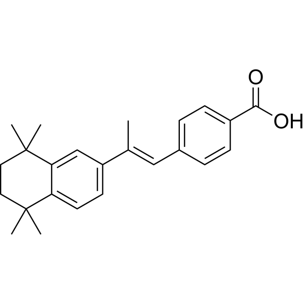
TTNPBApoptosis Inducer -
HY-14652
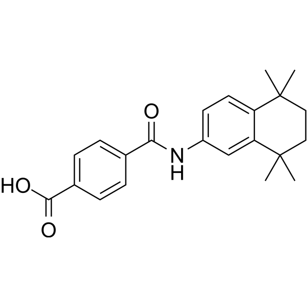
TamibaroteneApoptosis Inducer -

HY-100008R
Peretinoin (Standard)Acyclic Retinoid -
HY-14531
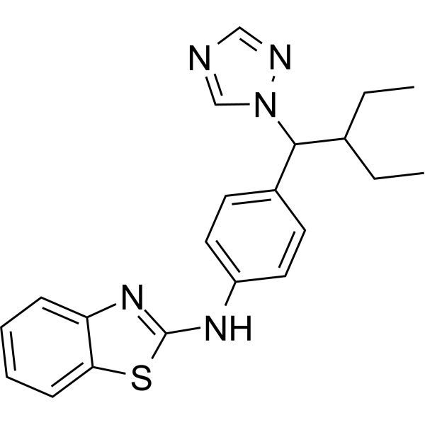
TalarozoleCYP26 Inhibitor/RAMBA
Publications Citing Use of MedChemExpress (MCE) Peretinoin
More- Adv Mater. 2026 Apr;38(19):e20475. [Abstract]
- Clin Mol Hepatol. 2024 Jul;30(3):515-538. [Abstract]
- Cell Death Dis. 2024 Jan 18;15(1):66. [Abstract]
- Arterioscler Thromb Vasc Biol. 2020 Mar;40(3):656-669. [Abstract]
- Antiviral Res. 2019 Oct:170:104570. [Abstract]
- J Biol Chem. 2024 Oct;300(10):107765. [Abstract]
- Patent. US20250295620A1.
 Customer Validation & Images

-
WB

-
IHC

-
Histological Imaging/Staining

-
RT-PCR

-
Bio/Physico-chemical Assay


Biological Activity
Description
In Vitro
Peretinoin (10-40 μM; 12-72 hours) exhibits suppressed SPHK1 expression after 24 h treatment, even at 10 μM and more prominent after 72 h peretinoin treatment [1].
Peretinoin (5 μM; 24 hours) up-regulates the expression of LC3B-II and increases autophagy flux in mouse primary hepatocytes[2].
MedChemExpress (MCE) has not independently confirmed the accuracy of these methods. They are for reference only.
-
Cell Line:Mouse primary hepatocytes (MPH) and the human HCC HepG2 cell line
-
Concentration:5 μM
-
Incubation Time:24 hours
-
Result:Up-regulated the expression of LC3B-II and increased autophagy flux.
-
Cell Line:Human hepatoma (Huh-7) cells
-
Concentration:10, 20 and 40 μM
-
Incubation Time:12, 24, 48 and 72 hours
-
Result:Exhibited suppressed SPHK1 expression after 24 h treatment, even at 10 μM.
Clinical Trial
| NCT Number | Sponsor | Condition | Start Date |
Phase

|
|---|---|---|---|---|
| NCT01329991 | Plexxikon| | 2011-05 | PHASE1 |


Chemical Information
-
CAS No. 81485-25-8
-
Appearance Solid
-
Molecular Weight 302.45
-
Formula C20H30O2
-
Color Light yellow to yellow
-
SMILES
C/C(C)=C/CC/C(C)=C/CC/C(C)=C/C=C/C(C)=C/C(O)=O
-
Synonyms
NIK333
-
Shipping
Room temperature in continental US; may vary elsewhere.
-
Storage
-20°C, protect from light, stored under nitrogen
* In solvent : -80°C, 6 months; -20°C, 1 month (protect from light, stored under nitrogen)


Publications (7)
-
Journal Impact Factor
-
Most Recent
-
Adv Mater
Piezoelectric Ceramic Nanofiber Aerogels Direct Neutrophil Fate for Diabetic Tissue Regeneration. [Abstract]2026 Apr;38(19):e20475. PMID: 41789514 -
Clin Mol Hepatol
2024 Jul;30(3):515-538. PMID: 38726504
Peretinoin purchased from MedChemExpress. Usage Cited in: Clin Mol Hepatol. 2024 Jul;30(3):515-538. [Abstract]
Hepatic ATG16L1 protein expression levels treated with Peretinoin (0.03%).
Peretinoin purchased from MedChemExpress. Usage Cited in: Clin Mol Hepatol. 2024 Jul;30(3):515-538. [Abstract]
Hepatic F4/80+ cell immunohistochemistry treated with Peretinoin (0.03 %).
Peretinoin purchased from MedChemExpress. Usage Cited in: Clin Mol Hepatol. 2024 Jul;30(3):515-538. [Abstract]
Representative eWAT H&E staining and F4/80 immunohistochemistry image of HFHCD-fed WT mice treated with or without Peretinoin (0.03%).
Peretinoin purchased from MedChemExpress. Usage Cited in: Clin Mol Hepatol. 2024 Jul;30(3):515-538. [Abstract]
Hepatic Tnfa, Il6, and Il1b gene expression levels treated with Peretinoin (0.03%).
Peretinoin purchased from MedChemExpress. Usage Cited in: Clin Mol Hepatol. 2024 Jul;30(3):515-538. [Abstract]
The levels of serum ALT were significantly lower in WT MASH mice treated with Peretinoin (0.03%) than in untreated mice fed the same diet.
-
Cell Death Dis
2024 Jan 18;15(1):66. PMID: 38238307 -
Arterioscler Thromb Vasc Biol
Retinoids Repress Human Cardiovascular Cell Calcification With Evidence for Distinct Selective Retinoid Modulator Effects. [Abstract]2020 Mar;40(3):656-669. PMID: 31852220 -
Antiviral Res
Antiviral candidates against the hepatitis E virus (HEV) and their combinations inhibit HEV growth in in vitro. [Abstract]2019 Oct:170:104570. PMID: 31362004 -
J Biol Chem
PPARγ and C/EBPα enable adipocyte differentiation upon inhibition of histone methyltransferase PRC2 in malignant tumors. [Abstract]2024 Oct;300(10):107765. PMID: 39276936 -


Solvent & Solubility
In Vitro:
DMSO : 100 mg/mL (330.63 mM; Need ultrasonic; Hygroscopic DMSO has a significant impact on the solubility of product, please use newly opened DMSO)
Please refer to the solubility information to select the appropriate solvent. Once prepared, please aliquot and store the solution to prevent product inactivation from repeated freeze-thaw cycles.
Storage method and period of stock solution: -80°C, 6 months; -20°C, 1 month (protect from light, stored under nitrogen). When stored at -80°C, please use it within 6 months. When stored at -20°C, please use it within 1 month.
Please refer to the solubility information to select the appropriate solvent. Once prepared, please aliquot and store the solution to prevent product inactivation from repeated freeze-thaw cycles.
Storage method and period of stock solution: -80°C, 6 months; -20°C, 1 month (protect from light, stored under nitrogen). When stored at -80°C, please use it within 6 months. When stored at -20°C, please use it within 1 month.
Concentration (start) × Volume (start) = Concentration (final) × Volume (final)
In Vivo:
Select the appropriate dissolution method based on your experimental animal and administration route.
- For the following dissolution methods, please ensure to first prepare a clear stock solution using an In Vitro approach and then sequentially add co-solvents:
- To ensure reliable experimental results, the clarified stock solution can be appropriately stored based on storage conditions. As for the working solution for In Vivo experiments, it is recommended to prepare freshly and use it on the same day.
- The percentages shown for the solvents indicate their volumetric ratio in the final prepared solution. If precipitation or phase separation occurs during preparation, heat and/or sonication can be used to aid dissolution.
Add each solvent one by one: 10% DMSO 40% PEG300 5% Tween-80 45% Saline
Solubility: ≥ 2.5 mg/mL (8.27 mM); Clear solution
This protocol yields a clear solution of ≥ 2.5 mg/mL (saturation unknown).
Taking 1 mL working solution as an example, add 100 μL DMSO stock solution (25.0 mg/mL) to 400 μL PEG300, and mix evenly; then add 50 μL Tween-80 and mix evenly; then add 450 μL Saline to adjust the volume to 1 mL.
Preparation of Saline: Dissolve 0.9 g sodium chloride in ddH₂O and dilute to 100 mL to obtain a clear Saline solution.
In Vivo Dissolution Calculator
Please enter the basic information of animal experiments:
-
-
-
-
Recommended: Prepare an additional quantity of animals to account for potential losses during experiments.
Please enter your animal formula composition:
-
%DMSO +
Recommended: Keep the proportion of DMSO in working solution below 2% if your animal is weak.
-
%+
-
+%Tween-80 + +
-
%Saline +
The co-solvents required include: DMSO, . All of co-solvents are available by MedChemExpress (MCE). , Tween 80. All of co-solvents are available by MedChemExpress (MCE).
Working solution concentration: 0.22 mg/mL
Method for preparing stock solution: mg drug dissolved in μL DMSO. Stock solution concentration: mg/mL. * In solvent : -80°C, 6 months; -20°C, 1 month (protect from light, stored under nitrogen)
1. Take μL DMSO stock solution;
2. Add μL .
μL , mix evenly;
3. Then add μL Tween 80, mix evenly;
4. Then add μL
Please ensure that the stock solution in the first step is dissolved to a clear state, and add co-solvents in sequence. You can use ultrasonic heating (ultrasonic cleaner, recommended frequency 20-40 kHz), vortexing, etc. to assist dissolution.


Purity & Documentation
-
Data Sheet (270 KB)
-
SDS (393 KB)
- English - EN (393 KB)
- Français - FR (393 KB)
- Deutsch - DE (393 KB)
- Norwegian - NO (393 KB)
- Español - ES (393 KB)
- Swedish - SV (393 KB)
- Italian - IT (393 KB)
- Korean - KR (393 KB)
- Portuguese - PT (393 KB)
-
Handling Instructions (2659 KB)
References
[1]. Honda M, et al. Peretinoin, an acyclic retinoid, improves the hepatic gene signature of chronic hepatitis C following curative therapy of hepatocellular carcinoma. BMC Cancer. 2013 Apr 15;13:191. [Content Brief]
[2]. Okada H, et al. Peretinoin, an acyclic retinoid, suppresses steatohepatitis and tumorigenesis by activating autophagy in mice fed an atherogenic high-fat diet. Oncotarget. 2017 Jun 20;8(25):39978-39993. [Content Brief]
[3]. Funaki M, et al. Peretinoin, an acyclic retinoid, inhibits hepatocarcinogenesis by suppressing sphingosine kinase 1 expression in vitro and in vivo. Sci Rep. 2017 Dec 5;7(1):16978. [Content Brief]


Complete Stock Solution Preparation Table
Please refer to the solubility information to select the appropriate solvent. Once prepared, please aliquot and store the solution to prevent product inactivation from repeated freeze-thaw cycles.
Storage method and period of stock solution: -80°C, 6 months; -20°C, 1 month (protect from light, stored under nitrogen). When stored at -80°C, please use it within 6 months. When stored at -20°C, please use it within 1 month.
| Optional Solvent | Concentration Solvent Mass | 1 mg | 5 mg | 10 mg | 25 mg |
|---|---|---|---|---|---|
| DMSO | 1 mM | 3.3063 mL | 16.5317 mL | 33.0633 mL | 82.6583 mL |
| 5 mM | 0.6613 mL | 3.3063 mL | 6.6127 mL | 16.5317 mL | |
| 10 mM | 0.3306 mL | 1.6532 mL | 3.3063 mL | 8.2658 mL | |
| 15 mM | 0.2204 mL | 1.1021 mL | 2.2042 mL | 5.5106 mL | |
| 20 mM | 0.1653 mL | 0.8266 mL | 1.6532 mL | 4.1329 mL | |
| 25 mM | 0.1323 mL | 0.6613 mL | 1.3225 mL | 3.3063 mL | |
| 30 mM | 0.1102 mL | 0.5511 mL | 1.1021 mL | 2.7553 mL | |
| 40 mM | 0.0827 mL | 0.4133 mL | 0.8266 mL | 2.0665 mL | |
| 50 mM | 0.0661 mL | 0.3306 mL | 0.6613 mL | 1.6532 mL | |
| 60 mM | 0.0551 mL | 0.2755 mL | 0.5511 mL | 1.3776 mL | |
| 80 mM | 0.0413 mL | 0.2066 mL | 0.4133 mL | 1.0332 mL | |
| 100 mM | 0.0331 mL | 0.1653 mL | 0.3306 mL | 0.8266 mL |


Peretinoin Related Classifications
HY-100008 Related Classifications
 Powered by Bioz