



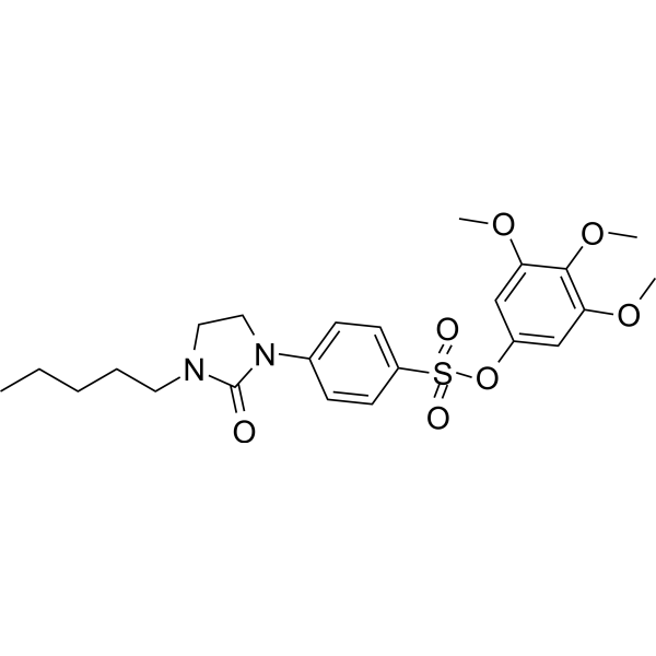
CYP1A1-IN-2
CYP1A1-IN-2 (Compound 14) is a competitive inhibitor of CYP1A1 (Ki: 1.4 μM). CYP1A1-IN-2 exhibits potent antimitotic activity and arrests cell in the G2/M phase. CYP1A1-IN-2 disrupts the microtubule and the cytoskeleton in CYP1A1-expressing breast cancer cells.
For research use only. We do not sell to patients.
- CAS No.: 1422527-87-4
- Formula: C23H30N2O7S
- Molecular Weight:478.56
-
Storage:
Please store the product under the recommended conditions in the Certificate of Analysis.

To place orders, for customer services and technical support, please contact: MedChemExpress USA
Tel: 609-228-6898 E-mail: [email protected] [email protected]


-
 Biological Activity
-
Chemical Information
- Protocols
- Purity & Documentation
- References
-
 Help & FAQs




Biological Activity
Description
IC50 & Target
[1]|
CYP1A1 1.4 μM (Ki) |
Cellular Effect
|
Cell Line

|
Type | Value | Description | References |
|---|---|---|---|---|
| HT-1080 | IC50 |
30 nM
Compound: 14
|
Antiproliferative activity against human HT-1080 cells expressing CYP1A1 assessed as cell growth inhibition incubated for 48 hrs by sulforhodamine B assay
Antiproliferative activity against human HT-1080 cells expressing CYP1A1 assessed as cell growth inhibition incubated for 48 hrs by sulforhodamine B assay
|
[PMID: 36780426] |
| HT-1080 | IC50 |
4440 nM
Compound: 14
|
Antiproliferative activity against human HT-1080 cells assessed as cell growth inhibition incubated for 48 hrs by sulforhodamine B assay
Antiproliferative activity against human HT-1080 cells assessed as cell growth inhibition incubated for 48 hrs by sulforhodamine B assay
|
[PMID: 36780426] |
| HT-1080 | IC50 |
5900 nM
Compound: 14
|
Antiproliferative activity against human HT-1080 cells transfected with empty vector assessed as cell growth inhibition incubated for 48 hrs by sulforhodamine B assay
Antiproliferative activity against human HT-1080 cells transfected with empty vector assessed as cell growth inhibition incubated for 48 hrs by sulforhodamine B assay
|
[PMID: 36780426] |
| HT-29 | IC50 |
>12500 nM
Compound: 14
|
Antiproliferative activity against human HT-29 cells assessed as cell growth inhibition incubated for 48 hrs by sulforhodamine B assay
Antiproliferative activity against human HT-29 cells assessed as cell growth inhibition incubated for 48 hrs by sulforhodamine B assay
|
[PMID: 36780426] |
| M21 | IC50 |
>12500 nM
Compound: 14
|
Antiproliferative activity against human M21 cells assessed as cell growth inhibition incubated for 48 hrs by sulforhodamine B assay
Antiproliferative activity against human M21 cells assessed as cell growth inhibition incubated for 48 hrs by sulforhodamine B assay
|
[PMID: 36780426] |
| MCF7 | IC50 |
200 nM
Compound: 14
|
Antiproliferative activity against human MCF7 cells expressing CYP1A1 assessed as cell growth inhibition incubated for 48 hrs by sulforhodamine B assay
Antiproliferative activity against human MCF7 cells expressing CYP1A1 assessed as cell growth inhibition incubated for 48 hrs by sulforhodamine B assay
|
[PMID: 36780426] |
| MCF7 | IC50 |
38 nM
Compound: 14
|
Antiproliferative activity against ER/PR/HER2-positive human MCF7 cells assessed as cell growth inhibition incubated for 72 hrs by sulforhodamine B assay
Antiproliferative activity against ER/PR/HER2-positive human MCF7 cells assessed as cell growth inhibition incubated for 72 hrs by sulforhodamine B assay
|
[PMID: 36780426] |
| MDA-MB-231 | IC50 |
8600 nM
Compound: 14
|
Antiproliferative activity against ER/PR/HER2-negative human MDA-MB-231 cells assessed as cell growth inhibition incubated for 72 hrs by sulforhodamine B assay
Antiproliferative activity against ER/PR/HER2-negative human MDA-MB-231 cells assessed as cell growth inhibition incubated for 72 hrs by sulforhodamine B assay
|
[PMID: 36780426] |
| MDA-MB-468 | IC50 |
21 nM
Compound: 14
|
Antiproliferative activity against ER/PR/HER2-negative human MDA-MB-468 cells assessed as cell growth inhibition incubated for 72 hrs by sulforhodamine B assay
Antiproliferative activity against ER/PR/HER2-negative human MDA-MB-468 cells assessed as cell growth inhibition incubated for 72 hrs by sulforhodamine B assay
|
[PMID: 36780426] |
| SK-BR-3 | IC50 |
3.2 nM
Compound: 14
|
Antiproliferative activity against ER/PR-negative HER2-positive human SK-BR-3 cells assessed as cell growth inhibition incubated for 72 hrs by sulforhodamine B assay
Antiproliferative activity against ER/PR-negative HER2-positive human SK-BR-3 cells assessed as cell growth inhibition incubated for 72 hrs by sulforhodamine B assay
|
[PMID: 36780426] |
| T47D | IC50 |
1 μM
Compound: 14
|
Antiproliferative activity against ER/PR-positive HER2-negative human T47D cells assessed as cell growth inhibition incubated for 72 hrs by sulforhodamine B assay
Antiproliferative activity against ER/PR-positive HER2-negative human T47D cells assessed as cell growth inhibition incubated for 72 hrs by sulforhodamine B assay
|
[PMID: 36780426] |
In Vitro
CYP1A1-IN-2 (Compound 14, 48 h) inhibits MCF-7 cell growth, with an IC50 of 200 nM, and is inactive on MDA-MB-231 (IC50 >8600 nM)[1].
CYP1A1-IN-2 inhibits MDA-MB-468, SK-BR cell growth with IC50s of 21 nM, 3.2 nM[1].
CYP1A1-IN-2 (50 nM, 48 h) causes accumulation of the cells in the G2/M phase by 42%[1].
CYP1A1-IN-2 shows high affinity for CYP1A1 (Ki: 1.4 μM)[1].
CYP1A1-IN-2 selectively inhibits HT-1080 cell proliferation, with IC50s of 30 nM for HT-1080 transfected withCYP1A1[1].
MedChemExpress (MCE) has not independently confirmed the accuracy of these methods. They are for reference only.
-
Cell Line:MCF-7 cell
-
Concentration:50 nM
-
Incubation Time:48 h
-
Result:Arrested cell in G2/M phase.
In Vivo
MedChemExpress (MCE) has not independently confirmed the accuracy of these methods. They are for reference only.
-
Animal Model:Chick embryos grafted with HT-1080CYP1A1 cells[1]
-
Dosage:1 μg/egg
-
Administration:Applied to chick chorioallantoic membrane.
-
Result:Reduced tumor weight by 49%.


Chemical Information
-
CAS No. 1422527-87-4
-
Molecular Weight 478.56
-
Formula C23H30N2O7S
-
SMILES
CCCCCN1CCN(C2=CC=C(S(=O)(OC3=CC(OC)=C(C(OC)=C3)OC)=O)C=C2)C1=O
-
Shipping
Room temperature in continental US; may vary elsewhere.
-
Storage
Please store the product under the recommended conditions in the Certificate of Analysis.


Purity & Documentation
References
[1]. Chavez Alvarez AC, et al. Homologation of the Alkyl Side Chain of Antimitotic Phenyl 4-(2-Oxo-3-alkylimidazolidin-1-yl)benzenesulfonate Prodrugs Selectively Targeting CYP1A1-Expressing Breast Cancers Improves Their Stability in Rodent Liver Microsomes. J Med Chem. 2023 Feb 23;66(4):2477-2497. [Content Brief]

Calculators
Concentration (start) × Volume (start) = Concentration (final) × Volume (final)


CYP1A1-IN-2 Related Classifications
HY-148595 Related Classifications
 Powered by Bioz