



Alcudacigib
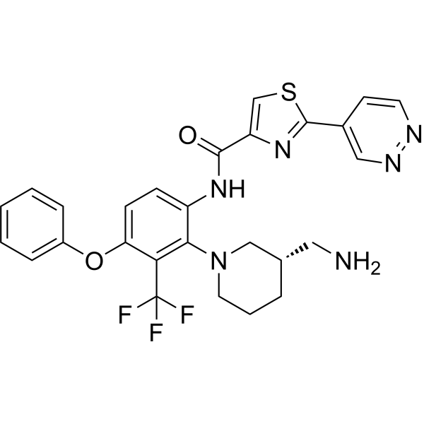
Alcudacigib (ASP1570; DGKζ-IN-1) is an orally active diacylglycerol kinase ζ (DGKζ) inhibitor, with an IC50 of 4.7 nM against human DGKζ and an IC50 of 3.0 nM against mouse DGKζ. Alcudacigib selectively inhibits the kinase activity of DGKζ and induces proteasome-dependent degradation of DGKζ protein. Alcudacigib enhances the anti-tumor functions of T cells and NK cells. Alcudacigib can be used for the research of advanced/metastatic solid tumors.
For research use only. We do not sell to patients.
- CAS No.: 2660218-70-0
- Formula: C27H25F3N6O2S
- Molecular Weight:554.59
-
Storage:
Please store the product under the recommended conditions in the Certificate of Analysis.

To place orders, for customer services and technical support, please contact: MedChemExpress USA
Tel: 609-228-6898 E-mail: [email protected] [email protected]


-
 Biological Activity
-
Chemical Information
- Protocols
- Purity & Documentation
- References
-
 Help & FAQs




Biological Activity
Description
IC50 & Target
[2]|
ERK1 |
ERK2 |
IL-2 |
In Vitro
Alcudacigib (0.1 μM; 5-30 min) enhances DAG-mediated ERK phosphorylation in DX5+ NK cells isolated from the spleens of wild-type mice stimulated with anti-NK1.1[1].
Alcudacigib (0.001-1 μM; 5 h) dose-dependently enhances IFN-γ production and degranulation in CD4/8-DX5+NKp46+ NK cells from wild-type mouse spleens stimulated with anti-NK1.1[1].
Alcudacigib (0.1-1000 nM; 5.5-24.5 h) degrades the DGKζ protein in Jurkat and B16F1 cells through a proteasome-dependent mechanism[2].
Alcudacigib (0.0003-1 μM; 30 min) enhances the downstream signaling pathway of DAG in TCR-stimulated Jurkat cells, which is evidenced by statistically significant increases in the phosphorylation levels of PKD and ERK1/2[2].
Alcudacigib (0.0003-1 μM; 24 h) enhances IL-2 production in TCR-stimulated Jurkat cells[2].
Alcudacigib (0.0001-1 μM; 3 days) enhances the proliferation, IL-2 production and IFN-γ production of human primary CD8+ T cells activated via TCR[2].
Alcudacigib (0.0001-1 μM; 3 days) restores and enhances the activation of primary human CD8+ T cells (including cell viability, IL-2 production, and IFN-γ production) that is inhibited by TGF-β1, PGE2 or AMP[2].
Alcudacigib (0.0001-1 μM; 3 days) enhances proliferation, IL-2 production and IFN-γ production of primary mouse CD8+ T cells stimulated by TCR[2].
Alcudacigib (0.0003-3 μM; 24 h) completely reverses PD-1-mediated T cell suppression and partially reverses CTLA-4- and TIGIT-mediated T cell suppression in cellular assays[2].
MedChemExpress (MCE) has not independently confirmed the accuracy of these methods. They are for reference only. Further protocols information, click here.
-
Cell Line:Jurkat (human T-cell line), B16F1 (mouse melanoma cell line)
-
Concentration:0.1, 1, 10, 100 and 1000 nM (Jurkat cells); 1 μM (B16F1 cells)
-
Incubation Time:24 h (Jurkat cells); 5 h (B16F1 cells, with 30 min pre-incubation with MG-132 or DMSO)
-
Result:Induced dose-dependent degradation of DGKζ protein in Jurkat cells.
Induced degradation of DGKζ protein in B16F1 cells, which was suppressed by pre-treatment with MG-132, a proteasome inhibitor.
In Vivo
Alcudacigib (0.1-3 mg/kg; p.o.; once daily or twice daily for 9-10 consecutive days) inhibits the growth of CPI-resistant B16F1 melanoma in female C57BL/6J mice[2].
Alcudacigib (3 mg/kg; p.o.; once daily for 10 consecutive days) exerted its antitumor effect against B16F10 melanoma in female C57BL/6J mice in a CD8+T cell-dependent manner[2].
Alcudacigib (3 mg/kg; p.o.; single administration) enhances the expression of T cell activation markers (CD25 and CD69) in splenic CD4+ and CD8+ T cells of healthy female C57BL/6J mice[2].
MedChemExpress (MCE) has not independently confirmed the accuracy of these methods. They are for reference only.
-
Animal Model:C57BL/6J (5-week-old female, subcutaneously inoculated with 2×10⁵ MC38 colon adenocarcinoma cells)[2]
-
Dosage:0.1 mg/kg; 0.3 mg/kg; 1 mg/kg; 3 mg/kg; 10 mg/kg; 5 mg/kg (twice daily)
-
Administration:p.o.; once daily; 10 days; twice daily; 10 days
-
Result:Significantly inhibited tumor growth at doses of 0.3 mg/kg and higher with no effect on mouse body weight.
Showed comparable or superior antitumor efficacy to anti-PD-1 antibody treatment at 5 mg/kg twice daily.
Increased the percentage of intratumoral CD45+, CD3+, CD4+, CD8+, and Treg cells, along with the absolute number of these cell populations, and significantly increased the CD8+/Treg ratio at 3 mg/kg once daily.
Significantly increased the frequency of Ki-67+, CD107a+, CD137+, and PD-1+ cells in both intratumoral CD4+ and CD8+ T cells.
-
Animal Model:C57BL/6J (5- to 6-week-old female, subcutaneously inoculated with 1×10⁵ B16F1 melanoma cells)[2]
-
Dosage:5 mg/kg (twice daily); 0.1 mg/kg; 0.3 mg/kg; 1 mg/kg; 3 mg/kg
-
Administration:p.o.; twice daily; 10 days; once daily; 9 days
-
Result:Significantly inhibited B16F1 tumor growth at 5 mg/kg twice daily while anti-PD-1 antibody showed no efficacy.
Significantly inhibited tumor growth at doses of 0.3 mg/kg and higher given once daily with no effect on mouse body weight.
-
Animal Model:C57BL/6J (5- to 6-week-old female, subcutaneously inoculated with 5×10⁵ B16F10 melanoma cells, with concurrent anti-CD8 antibody depletions on days 0, 1, 2, 6, and 10)[2]
-
Dosage:3 mg/kg
-
Administration:p.o.; once daily; 10 days
-
Result:Induced marked tumor growth inhibition, but this effect was completely abolished by CD8+ T-cell depletion.
-
Animal Model:C57BL/6J (female, healthy)[2]
-
Dosage:3 mg/kg
-
Administration:p.o.; single dose
-
Result:Significantly increased the geometric mean fluorescence intensity of CD25 and CD69 in both splenic CD4+ and CD8+ T cells.
Increased the proportion of CD25+ and CD69+ T cells.


Chemical Information
-
CAS No. 2660218-70-0
-
Molecular Weight 554.59
-
Formula C27H25F3N6O2S
-
SMILES
FC(F)(C1=C(N2C[C@@H](CCC2)CN)C(NC(C3=CSC(C4=CN=NC=C4)=N3)=O)=CC=C1OC5=CC=CC=C5)F
-
Synonyms
DGKζ-IN-1
-
Shipping
Room temperature in continental US; may vary elsewhere.
-
Storage
Please store the product under the recommended conditions in the Certificate of Analysis.

Protocols
-
Kinase activity and phosphorylation assays
Kinase activity assays measure the ability of kinases to transfer phosphate groups from ATP to specific substrates, while phosphorylation assays detect the presence and levels of phosphorylated proteins. Common methods include radiolabeled ATP incorporation (e. g. ,), ADP release detection via bioluminescence (e. g. ,[3]), enzyme-linked immunosorbent assays (ELISA) for phospho-specific epitopes (e. g. ,[6]), and microtiter-based formats for high-throughput screening (e. g. ,[8]). The ADP-Glo assay quantifies kinase activity by measuring ADP produced during phosphorylation using a luciferase-based system. Radiometric assays involve autoradiography or scintillation counting after incorporation of 32P-labeled ATP into substrate proteins. ELISA-based approaches rely on phospho-specific antibodies to detect activated kinases in cell lysates or purified samples.
-
Cell Cytotoxicity Assay
Cytotoxicity assays are usually based on the assessment of cell membrane damage, which can also be indirectly detected by measuring cell viability. Detection methods include MTT assay, CKK-8 assay, LDH assay and ATP assay, etc.
-
Protocol for Kinase activity and phosphorylation assays
Kinase activity assays measure transfer of phosphate from ATP to a protein or peptide substrate, generating phosphorylated substrate, ADP, or incorporated radiolabeled phosphate as the readout; phosphorylation assays measure site-specific phosphorylation in cells or tissues as a proxy for kinase-pathway activation, inhibition, or substrate regulation. Phosphorylation can be detected by phospho-specific Western blot, immunoprecipitation kinase assay, phospho-immunofluorescence, phospho-flow cytometry, luminescent ADP detection, radiolabeled ATP incorporation, or reporter-based pathway assays, and these readouts can be applied to cancer cells, primary neurons, mouse tumors, organoids, inflammatory macrophages, ferroptosis studies, and mitophagy studies when the kinase target is biologically relevant.


Purity & Documentation
References
[1]. Okumura M, et al. The diacylglycerol kinase ζ inhibitor ASP1570 augments natural killer cell function. Int Immunopharmacol. 2023;125(Pt A):111145. [Content Brief]
[2]. Ikeda O, et al. Enhanced Antitumor Immunity by ASP1570, a Novel Diacylglycerol Kinase ζ Inhibitor, Offers a Potential Novel Immunotherapy for Treating Cancer. Mol Cancer Ther. 2025;24(6):884-895. [Content Brief]

Calculators
Concentration (start) × Volume (start) = Concentration (final) × Volume (final)


Alcudacigib Related Classifications
HY-153793 Related Classifications
 Powered by Bioz