



Emirodatamab

Based on 1 Customer Validation
Emirodatamab (AMG-427) is a bispecific T-cell engager (BiTE). Emirodatamab simultaneously binds FLT3 on the surface of acute myeloid leukemia (AML) cells and CD3 on the surface of T cells, thereby precisely recruiting immune effector cells to tumor sites. Emirodatamab potently induces T cell activation, secretion of proinflammatory cytokines (such as IFNγ, TNFα), and specific cytotoxicity, effectively lysing FLT3-positive tumor cells and inhibiting their growth. Emirodatamab not only significantly prolongs survival in mouse xenograft models and eliminates diseased cells in primates, but also exhibits a synergistic enhancement effect when combined with PD-1 blockade therapy. Emirodatamab is used in studies of acute myeloid leukemia, especially relapsed or refractory cases.
For research use only. We do not sell to patients.
- Purity : 99.05%
- CAS No.: 2449199-61-3
- Molecular Weight:105.892 kDa
-
Storage:
Please store the product under the recommended conditions in the Certificate of Analysis.


To place orders, for customer services and technical support, please contact: MedChemExpress USA
Tel: 609-228-6898 E-mail: [email protected] [email protected]


-
 Biological Activity
-
Chemical Information
- Protocols
- Purity & Documentation
- References
-
 Help & FAQs




Biological Activity
Description
Isotype
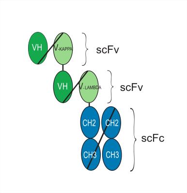
(scFv-heavy-kappa)-(scFv-heavy-lambda)-scFc
Species Reactivity
Human
IC50 & Target
CD3E & FLT3
In Vitro
Emirodatamab potently induces cytotoxicity in primary AML blasts with high FLT-3 expression under ex vivo conditions, and its activity is enhanced when combined with anti-PD-1 antibodies[2].
Emirodatamab mediates in vitro killing of primary AML cells in a manner dependent on FLT3 expression levels and effector-to-target cell ratio, and its activity is enhanced when combined with anti-PD-1 antibodies[4].
Emirodatamab induces potent, target-specific sub-picomolar TDCC effects in FLT3-expressing human AML cell lines, accompanied by T cell activation and cytokine secretion; it retains complete killing activity against target cells even in the presence of disease-relevant concentrations of sFLT3 or sFLT3L[5].
Emirodatamab exhibits lower potency against PD-L1-expressing MOLM-13 AML cells compared to parental MOLM-13 AML cells[5].
MedChemExpress (MCE) has not independently confirmed the accuracy of these methods. They are for reference only.
In Vivo
Emirodatamab (0.01-1 mg/kg; intravenous injection; once every 7 days; 6 doses total) significantly prolongs the survival of mice bearing orthotopic EOL-1 AML xenografts[5].
Emirodatamab (Cmax=1-10 nM; intravenous injection; 3 administrations) dose-dependently eliminates FLT3-expressing hematopoietic progenitor cells in cynomolgus monkeys, reduces FLT3 transcript levels in bone marrow and blood by up to 97%, and induces T cell activation and cytokine release[5].
MedChemExpress (MCE) has not independently confirmed the accuracy of these methods. They are for reference only.
-
Animal Model:NOD/SCID mcie with Acute myeloid leukemia (female, 7 weeks old, orthotopic xenograft via intravenous injection of 107 MOLM-13_Luc cells, sublethally irradiated)[5]
-
Dosage:0.12 mg/kg; 0.6 mg/kg; 3 mg/kg
-
Administration:i.v.; every 5 days; 6 doses (days 7, 12, 17, 22, 27, 34)
-
Result:Prolonged survival significantly at all doses tested, with median survival times of 36.5 days (3 mg/kg), 37 days (0.6 mg/kg), and 42.5 days (0.12 mg/kg).
All control group mice died within 20 days with a median survival of 18 days.
Remained above the in vitro TDCC EC50 (1.9 pM) in serum for at least 9 days following the final administration.
-
Animal Model:NOD/SCID mice with Acute myeloid leukemia (female, 7 weeks old, orthotopic xenograft via intravenous injection of 5 × 10⁶ EOL-1 cells, sublethally irradiated)[5]
-
Dosage:0.01 mg/kg; 0.1 mg/kg; 1 mg/kg
-
Administration:i.v.; every 7 days; 6 doses (days 9, 16, 23, 30, 37, 44)
-
Result:Prolonged survival significantly at all doses tested; ≥50% of treated animals survived until the study end on day 108 (17/30 total treated animals), while control group mice had a median survival of 36-37 days and were all euthanized by day 52.
-
Animal Model:Cynomolgus monkeys[5]
-
Dosage:Doses intended to achieve maximal serum concentrations (Cₘₐₓ) of 1 nM; 5 nM; 10 nM
-
Administration:i.v.; 3 doses (days 1, 2, and 5)
-
Result:Reduced bone marrow FLT3 transcript levels by 85-95% on day 4 and 93-97% on day 8 across all dose groups.
Reduced blood FLT3 transcript levels to nearly undetectable levels (≥97% reduction) at the earliest tested time point and maintained across all exposures and time points.
Induced T cell activation (upregulation of CD69) and increases in serum IFNγ, IL-6, MCP-1, and TNFα in response to the first dose, with cytokine responses attenuated in subsequent doses.
Increased soluble FLT3L levels dose-dependently, reaching maximum levels of 12,000-23,000 pg/mL per group.
- Cell Cytotoxicity Assay
- Mammalian live/dead viability and cytotoxicity staining
- Patient-Derived Xenograft (PDX)
- Splenic/Portal-Vein Liver Metastasis Xenograft
- Subcutaneous Cell-Line-Derived Xenograft
- Research Protocol for Cancer Immunology
- Orthotopic Cell-Line Xenograft
- Cell Viability Determination by MTT Colorimetric Assay
- Intraperitoneal/Peritoneal Dissemination Xenograft
Clinical Trial
| NCT Number | Sponsor | Condition | Start Date |
Phase

|
|---|---|---|---|---|
| NCT01329991 | 2011-05 | PHASE1 |
Gene ID
Accession
Conjugated
Unconjugated
Reconsititution
The product can be reconstituted/diluted with sterile PBS or saline.
Format
-
(scFv-heavy-kappa)-(scFv-heavy-lambda)-scFc
Application
ELISA, FACS, Functional assay, Research in vivo
Verified Bioactivity
-
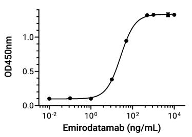 Immobilized CD3 epsilon Protein, Human (HEK293, His, HY-P70506) can bind Emirodatamab. The EC50 for this effect is 26.57 ng/mL.
 -
 Immobilized FLT3 Protein, Human (T227M, HEK293, His, HY-P75183) can bind Emirodatamab. The EC50 for this effect is 11.97 ng/mL.
 -
 Flow cytometric analysis of 1×10⁶ Jurkat cells labelling CD3E (red) with Emirodatamab (HY-P99916). Cells were fixed with 4% paraformaldehyde and permeabilised with 90% methanol. Then stained with the primary antibody at 1/200 dilution for an hour at 4℃. Alexa Goat Anti-Human IgG H&L (AF488) (HY-P83776) was used as the secondary antibody at 1/1,000 dilution for 30 minutes at 4℃. Human IgG1 kappa Isotype Control (HY-P99001, blue) was used as the isotype control, cells without incubation with primary antibody were used as the unlabeled control (black).
 -
 Flow cytometric analysis of 1X10⁶ THP-1 cells labeling FLT3 with Emirodatamab (HY-P99916, red). Cells were stained with the primary antibody at 1/200 dilution for an hour at 4℃. AF488-conjugated Goat Anti-Human IgG H&L (AF488) (HY-P83776) was used as the secondary antibody at 1/1,000 dilution for 30 minutes at 4℃. Human IgG1 kappa Isotype Control (HY-P99001, blue) was used as the isotype control, cells without incubation with primary antibody were used as the unlabeled control (black).


Chemical Information
-
CAS No. 2449199-61-3
-
Appearance Liquid
-
Molecular Weight 105.892 kDa
-
Color Colorless to light yellow
-
SMILES
[Emirodatamab]
-
Synonyms
AMG 427
-
Shipping
Shipping with dry ice.
-
Formulation
Please refer to the lot-specific COA for specific buffer information.
-
Storage
Please store the product under the recommended conditions in the Certificate of Analysis.


Purity & Documentation
-
Data Sheet (282 KB)
-
SDS (251 KB)
- English - EN (251 KB)
- Français - FR (251 KB)
- Deutsch - DE (251 KB)
- Norwegian - NO (251 KB)
- Español - ES (251 KB)
- Swedish - SV (251 KB)
- Italian - IT (251 KB)
- Korean - KR (251 KB)
- Portuguese - PT (251 KB)
-
Inhibitory Antibodies User Guide (603 KB)
References
[1]. Obszański P, et al. Molecular-Targeted Therapy of Pediatric Acute Myeloid Leukemia. Molecules. 2022;27(12):3911. Published 2022 Jun 18. [Content Brief]
[2]. Morse JW, et al. Antibody therapies for the treatment of acute myeloid leukemia: exploring current and emerging therapeutic targets. Expert Opin Investig Drugs. 2023;32(2):107-125. [Content Brief]
[3]. Einsele H, et al. The BiTE (bispecific T-cell engager) platform: Development and future potential of a targeted immuno-oncology therapy across tumor types. Cancer. 2020;126(14):3192-3201. [Content Brief]
[4]. Daver N, et al. T-cell-based immunotherapy of acute myeloid leukemia: current concepts and future developments. Leukemia. 2021;35(7):1843-1863. [Content Brief]

Calculators
Concentration (start) × Volume (start) = Concentration (final) × Volume (final)


Emirodatamab Related Classifications
HY-P99916 Related Classifications
 Powered by Bioz