



Methyl cholate

Based on 1 Customer Validation
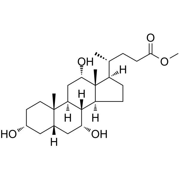
Methyl cholate is a bile acid analog and a specific inhibitor of TcdB toxin from Clostridioides difficile. Methyl cholate exerts a stronger selective inhibitory effect on TcdB than on TcdA. Methyl cholate induces conformational stabilization by binding to a unique site of TcdB, thereby blocking the binding of the toxin to host receptors and its self-processing process. Methyl cholate effectively protects human fibroblasts from TcdB-induced cytopathic effects. Methyl cholate exhibits dose-dependent anti-hepatic fibrosis activity in both cellular and zebrafish models, and significantly reduces the expression levels of α-SMA and COL-I. Methyl cholate is suitable for in-depth research in the fields of Clostridioides difficile infection and hepatic fibrosis.
For research use only. We do not sell to patients.
- Purity : 99.81%
- CAS No.: 1448-36-8
- Formula: C25H42O5
- Molecular Weight:422.60
-
Storage:Powder -20°C, 3 years , 4°C, 2 years ; In solvent -80°C, 2 years , -20°C, 1 year

To place orders, for customer services and technical support, please contact: MedChemExpress USA
Tel: 609-228-6898 E-mail: [email protected] [email protected]


-
 Biological Activity
-
Chemical Information
-
Solvent & Solubility
- Protocols
- Purity & Documentation
- References
-
 Help & FAQs


-
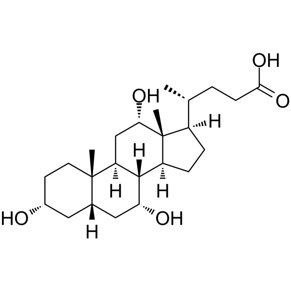
HY-N0324
Cholic acidEndogenous Metabolite -
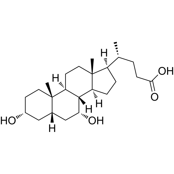
HY-76847
Chenodeoxycholic AcidEndogenous Metabolite -
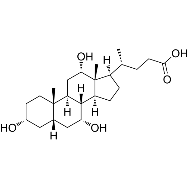
HY-N0324R
Cholic acid (Standard)Endogenous Metabolite -

HY-107830R
Methyl cholate (Standard)Methyl Ester Form of Cholic Acid -
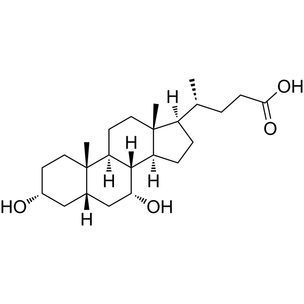
HY-76847R
Chenodeoxycholic Acid (Standard)Endogenous Metabolite -
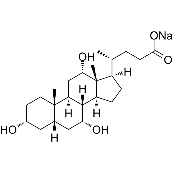
HY-N0324A
Cholic acid sodiumEndogenous Metabolite -
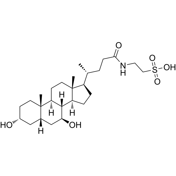
HY-19696
TauroursodeoxycholateEndoplasmic Reticulum Stress Inhibitor -
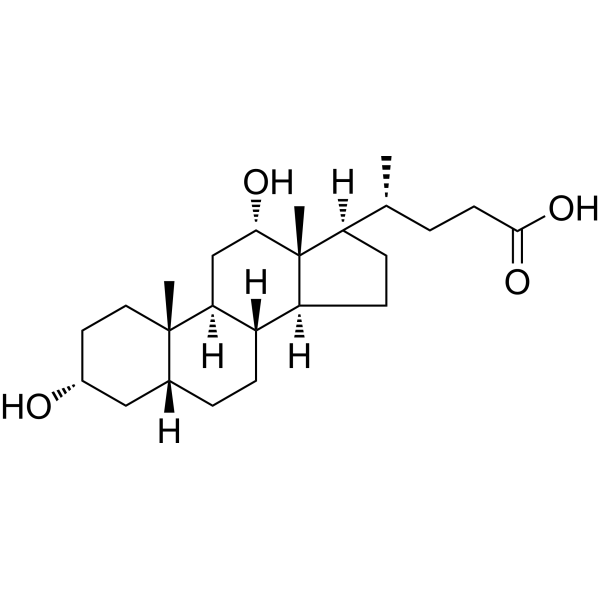
HY-N0593
Deoxycholic acidBile Acid Receptor Activator -
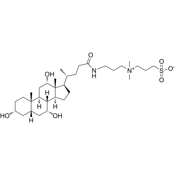
HY-15435
CHAPSIonic Detergent/Micelle Forming Agent -
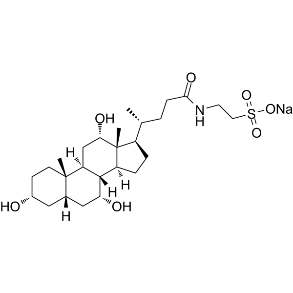
HY-N0545
Taurocholic acid sodiumImmunoregulation Agent -
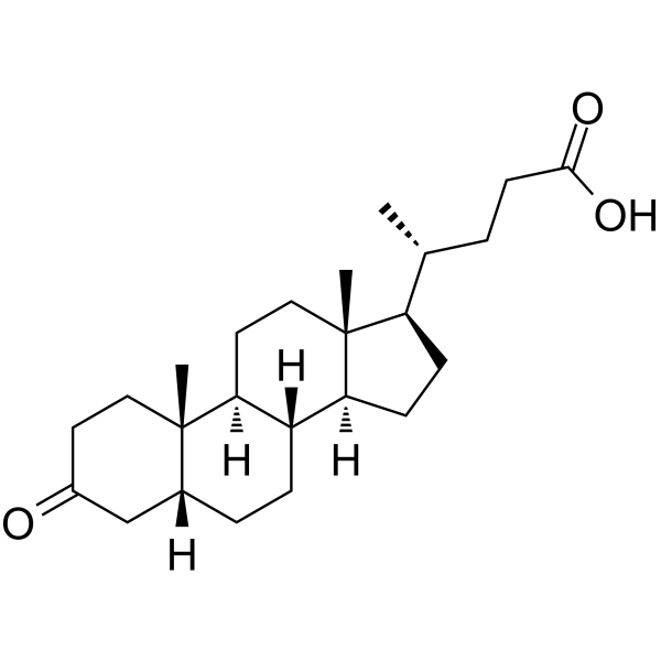
HY-125801
3-Oxo-5β-cholanoic acidTH17 Cell Differentiation Inhibitor -
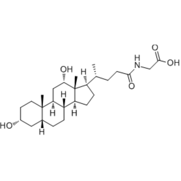
HY-125731
Glycodeoxycholic acidEndogenous Metabolite
All Endogenous Metabolite Isoforms
More

Biological Activity
Description
In Vitro
Methyl cholate (0.1-100 μM; 10 min pre-incubation, 3.5 hr combined exposure) dose-dependently protects human IMR-90 fibroblasts from TcdB-induced cell rounding with an IC50 of 11 μM, but does not protect against TcdA-induced cell rounding[1].
Methyl cholate (0.8-200 μM; 30 min pre-incubation, 3 hr reaction) dose-dependently inhibits IP6-induced autoprocessing of full-length Clostridium difficile TcdB, but does not affect autoprocessing of the isolated GTD-CPD truncated construct[1].
Methyl cholate (40 μM; 45 min pre-incubation with TcdB, 45 min incubation with cells) potently inhibits Clostridium difficile TcdB binding to Vero cells, reducing bound toxin to ~10% of vehicle control levels[1].
Methyl cholate (0.1-100 μM; 3 hr combined exposure) dose-dependently protects human IMR-90 fibroblasts from TcdB-induced necrosis with an IC50 of 4.3 μM[1].
Methyl cholate (80.64 μg/mL; 48 h) inhibits HSC-T6 hepatic stellate cell proliferation with an IC50 of 80.64 μg/mL after 48 h of incubation[2].
Methyl cholate (low to high dose range; duration sufficient to alter protein expression) dose-dependently reduces α-SMA protein expression, a marker of hepatic stellate cell activation, in HSC-T6 cells[2].
Methyl cholate (duration sufficient to alter protein expression) increases PIK3-α protein expression in HSC-T6 hepatic stellate cells[2].
MedChemExpress (MCE) has not independently confirmed the accuracy of these methods. They are for reference only. Further protocols information, click here.
-
Cell Line:Vero cells
-
Concentration:40 μM
-
Incubation Time:45 min pre-incubation with TcdB; 45 min incubation with cells
-
Result:Reduced TcdB binding to Vero cells to levels similar to the neutralizing monoclonal antibody bezlotoxumab, with only ~10% of TcdB remaining bound relative to vehicle controls.
-
Cell Line:HSC-T6 hepatic stellate cells
-
Concentration:80.64 μg/mL
-
Incubation Time:48 h
-
Result:Inhibited HSC-T6 cell proliferation in a dose-dependent manner, with a calculated half-maximal inhibitory concentration (IC50) of 80.64 μg/mL after 48 h of treatment.
In Vivo
MedChemExpress (MCE) has not independently confirmed the accuracy of these methods. They are for reference only.
-
Animal Model:Zebrafish larvae (2 days post-fertilization)[2]
-
Dosage:6.25 μM, 12.5 μM, 25 μM
-
Administration:aqueous immersion; post 3 days of TAA exposure
-
Result:Reduced liver intercellular gaps, cellular enlargement, and vacuolation in a dose-dependent manner relative to the TAA-induced hepatic fibrosis group.
Markedly decreased liver collagen-I levels compared to the fibrosis group (25 μM dose).


Chemical Information
-
CAS No. 1448-36-8
-
Appearance Solid
-
Molecular Weight 422.60
-
Formula C25H42O5
-
Color White to off-white
-
SMILES
O[C@H]1[C@@]2([H])[C@@]3([H])[C@@]([C@]([C@H](C)CCC(OC)=O)([H])CC3)([C@@H](O)C[C@]2([H])[C@]4(C)[C@](C[C@H](O)CC4)([H])C1)C
-
Structure Classification
-
Initial Source
Aspergillus niger
-
Shipping
Room temperature in continental US; may vary elsewhere.
-
Storage
Powder -20°C 3 years 4°C 2 years In solvent -80°C 2 years -20°C 1 year


Solvent & Solubility
In Vitro:
DMSO : 100 mg/mL (236.63 mM; Need ultrasonic; Hygroscopic DMSO has a significant impact on the solubility of product, please use newly opened DMSO)
Please refer to the solubility information to select the appropriate solvent. Once prepared, please aliquot and store the solution to prevent product inactivation from repeated freeze-thaw cycles.
Storage method and period of stock solution: -80°C, 2 years; -20°C, 1 year. When stored at -80°C, please use it within 2 years. When stored at -20°C, please use it within 1 year.
Please refer to the solubility information to select the appropriate solvent. Once prepared, please aliquot and store the solution to prevent product inactivation from repeated freeze-thaw cycles.
Storage method and period of stock solution: -80°C, 2 years; -20°C, 1 year. When stored at -80°C, please use it within 2 years. When stored at -20°C, please use it within 1 year.
Concentration (start) × Volume (start) = Concentration (final) × Volume (final)
In Vivo:
Select the appropriate dissolution method based on your experimental animal and administration route.
- For the following dissolution methods, please ensure to first prepare a clear stock solution using an In Vitro approach and then sequentially add co-solvents:
- To ensure reliable experimental results, the clarified stock solution can be appropriately stored based on storage conditions. As for the working solution for In Vivo experiments, it is recommended to prepare freshly and use it on the same day.
- The percentages shown for the solvents indicate their volumetric ratio in the final prepared solution. If precipitation or phase separation occurs during preparation, heat and/or sonication can be used to aid dissolution.
Add each solvent one by one: 10% DMSO 40% PEG300 5% Tween-80 45% Saline
Solubility: ≥ 2.5 mg/mL (5.92 mM); Clear solution
This protocol yields a clear solution of ≥ 2.5 mg/mL (saturation unknown).
Taking 1 mL working solution as an example, add 100 μL DMSO stock solution (25.0 mg/mL) to 400 μL PEG300, and mix evenly; then add 50 μL Tween-80 and mix evenly; then add 450 μL Saline to adjust the volume to 1 mL.
Preparation of Saline: Dissolve 0.9 g sodium chloride in ddH₂O and dilute to 100 mL to obtain a clear Saline solution.
Add each solvent one by one: 10% DMSO 90% (20% SBE-β-CD in Saline)
Solubility: ≥ 2.5 mg/mL (5.92 mM); Clear solution
This protocol yields a clear solution of ≥ 2.5 mg/mL (saturation unknown).
Taking 1 mL working solution as an example, add 100 μL DMSO stock solution (25.0 mg/mL) to 900 μL 20% SBE-β-CD in Saline, and mix evenly.
Preparation of 20% SBE-β-CD in Saline (4°C, storage for one week): 2 g SBE-β-CD powder is dissolved in 10 mL Saline, completely dissolve until clear.
In Vivo Dissolution Calculator
Please enter the basic information of animal experiments:
-
-
-
-
Recommended: Prepare an additional quantity of animals to account for potential losses during experiments.
Please enter your animal formula composition:
-
%DMSO +
Recommended: Keep the proportion of DMSO in working solution below 2% if your animal is weak.
-
%+
-
+%Tween-80 + +
-
%Saline +
The co-solvents required include: DMSO, . All of co-solvents are available by MedChemExpress (MCE). , Tween 80. All of co-solvents are available by MedChemExpress (MCE).
Working solution concentration: 0.22 mg/mL
Method for preparing stock solution: mg drug dissolved in μL DMSO. Stock solution concentration: mg/mL.
1. Take μL DMSO stock solution;
2. Add μL .
μL , mix evenly;
3. Then add μL Tween 80, mix evenly;
4. Then add μL
Please ensure that the stock solution in the first step is dissolved to a clear state, and add co-solvents in sequence. You can use ultrasonic heating (ultrasonic cleaner, recommended frequency 20-40 kHz), vortexing, etc. to assist dissolution.

Protocols
-
Fibrosis/Collagen Morphometry
Fibrosis and collagen morphometry is based on the quantitative visualization of fibrillar collagen deposition in tissue sections using histochemical stains such as Sirius Red (Picrosirius Red) or Masson's trichrome, followed by image-based or polarization-enhanced analysis to estimate collagen proportional area as a surrogate of extracellular matrix accumulation during fibrotic remodeling. Sirius Red combined with polarized light microscopy enhances detection of collagen fibers due to birefringence properties, enabling more specific visualization of collagen type I and III fibrils compared to conventional bright-field histology, while whole-section or region-restricted digital morphometry reduces field-selection bias in fibrosis assessment. Alternative quantitative approaches include second harmonic generation (SHG) and two-photon excited fluorescence microscopy, which enable label-free detection of fibrillar collagen and have been validated against histological staining and biochemica
-
Collagen: Sirius Red Staining
Sirius Red or picrosirius red staining is a histochemical method for visualizing collagen-rich extracellular matrix in tissue sections, and collagen fibers are detected as red-stained structures under bright-field microscopy with enhanced birefringence under polarized light. Picrosirius red is useful for assessing total collagen organization, distribution, and fibrosis burden, but polarized color should not be interpreted as a definitive collagen type I versus type III readout because color is affected by fiber orientation, thickness, and packing.
-
RNA extraction experimental
By lysing cells, releasing RNA, and removing impurities such as proteins and DNA, high-purity RNA products are finally obtained. The commonly used traditional method is the guanidine isothiocyanate/phenol/chloroform method (Trizol), which is suitable for a variety of animal materials including animal tissues, microorganisms, cultured cells, etc., and most plant materials.
-
Research Protocol for Infectious Diseases
Infectious-disease experiments test how pathogens interact with host barriers, innate immune receptors, inflammatory signaling, pathogen replication, and tissue injury; pattern-recognition receptors such as TLRs, RIG-I-like receptors, NOD-like receptors, and inflammasomes detect microbial molecules and activate NF-κB, interferon, and cytokine responses. The central hypothesis is that infection severity reflects the balance between pathogen burden and host response: protective inflammation restricts pathogen growth, whereas excessive or mislocalized inflammation contributes to tissue damage and disease phenotype. Unresolved questions include which host pathways are protective versus pathogenic, why some infection models fail to translate to human disease, and which combined readouts best predict clinically relevant infection outcomes.
-
Connective Tissue: Masson's Trichrome/Collagen Trichrome Staining
Masson’s Trichrome (collagen/trichrome staining) is a histological technique that differentially stains tissue compartments using sequential acidic dyes to distinguish collagen from muscle and cytoplasmic components based on dye affinity and tissue permeability differences, enabling visualization of fibrosis and connective tissue architecture in histological sections. The classical formulation typically uses Weigert's iron hematoxylin for nuclear staining, Biebrich scarlet-acid fuchsin for cytoplasm and muscle, and aniline blue (or light green variants) for collagen, producing a characteristic blue/green collagen signal contrasted against red cytoplasm and dark nuclei. The staining principle relies on selective displacement of smaller dye molecules by larger anionic dyes in collagen-rich regions under controlled acidified conditions, which enhances collagen-specific dye retention. This property makes the method widely used for fibrosis assessment in organs such as heart, liver, lung, a


Purity & Documentation
-
Data Sheet (288 KB)
-
SDS (393 KB)
- English - EN (393 KB)
- Français - FR (393 KB)
- Deutsch - DE (393 KB)
- Norwegian - NO (393 KB)
- Español - ES (393 KB)
- Swedish - SV (393 KB)
- Italian - IT (393 KB)
- Korean - KR (393 KB)
- Portuguese - PT (393 KB)
-
Handling Instructions (2659 KB)
References


Complete Stock Solution Preparation Table
Please refer to the solubility information to select the appropriate solvent. Once prepared, please aliquot and store the solution to prevent product inactivation from repeated freeze-thaw cycles.
Storage method and period of stock solution: -80°C, 2 years; -20°C, 1 year. When stored at -80°C, please use it within 2 years. When stored at -20°C, please use it within 1 year.
| Optional Solvent | Concentration Solvent Mass | 1 mg | 5 mg | 10 mg | 25 mg |
|---|---|---|---|---|---|
| DMSO | 1 mM | 2.3663 mL | 11.8315 mL | 23.6630 mL | 59.1576 mL |
| 5 mM | 0.4733 mL | 2.3663 mL | 4.7326 mL | 11.8315 mL | |
| 10 mM | 0.2366 mL | 1.1832 mL | 2.3663 mL | 5.9158 mL | |
| 15 mM | 0.1578 mL | 0.7888 mL | 1.5775 mL | 3.9438 mL | |
| 20 mM | 0.1183 mL | 0.5916 mL | 1.1832 mL | 2.9579 mL | |
| 25 mM | 0.0947 mL | 0.4733 mL | 0.9465 mL | 2.3663 mL | |
| 30 mM | 0.0789 mL | 0.3944 mL | 0.7888 mL | 1.9719 mL | |
| 40 mM | 0.0592 mL | 0.2958 mL | 0.5916 mL | 1.4789 mL | |
| 50 mM | 0.0473 mL | 0.2366 mL | 0.4733 mL | 1.1832 mL | |
| 60 mM | 0.0394 mL | 0.1972 mL | 0.3944 mL | 0.9860 mL | |
| 80 mM | 0.0296 mL | 0.1479 mL | 0.2958 mL | 0.7395 mL | |
| 100 mM | 0.0237 mL | 0.1183 mL | 0.2366 mL | 0.5916 mL |


Methyl cholate Related Classifications
HY-107830 Related Classifications
 Powered by Bioz