



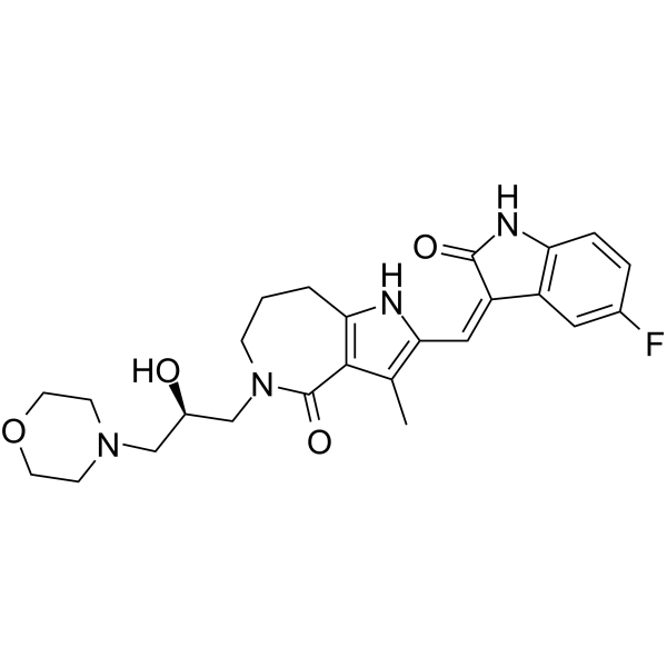
Henatinib
Henatinib is an orally active small-molecule multikinase inhibitor that has demonstrated broad and potent antitumor activities. Henatinib inhibits the activity of VEGFR-2, c-kit, PDGFR with IC50 values of 0.6 nM, 3.3 nM and 41.5 nM, respectively. Henatinib significantly inhibits VEGFR-2 phosphorylation and its downstream signal pathway in human umbilical vein endothelial cells (HUVECs).
연구목적의 판매만을 진행합니다. 환자를 대상으로 한 판매는 하지 않습니다.
- CAS No.: 1239269-51-2
- 화학식: C25H29FN4O4
- 분자량:468.52
-
보관:
Please store the product under the recommended conditions in the Certificate of Analysis.

To place orders, for customer services and technical support, please contact: MedChemExpress USA
Tel: 609-228-6898 E-mail: [email protected] [email protected]


-
 Biological Activity
-
Chemical Information
- Protocol
- 순도&문서
- References
-
 Help & FAQs


-
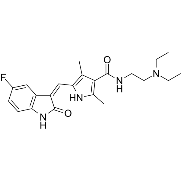
HY-10255A
SunitinibMulti-targeted RTK Inhibitor -
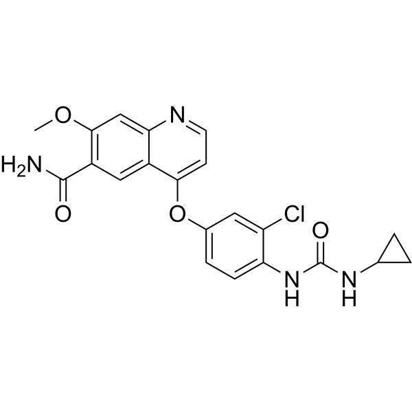
HY-10981
LenvatinibVEGFR/FGFR Inhibitor -

HY-10255AR
Sunitinib (Standard)Multi-targeted RTK Inhibitor -
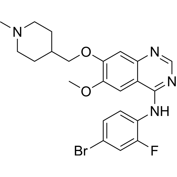
HY-10260
VandetanibVEGFR2/3 Inhibitor -
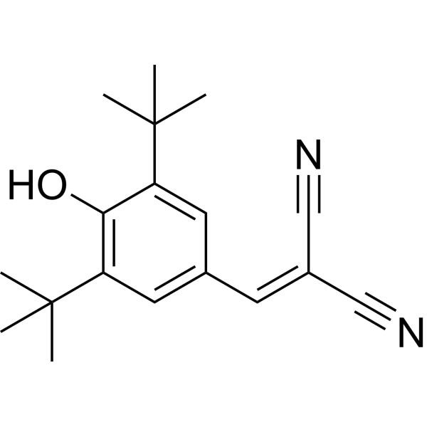
HY-15511
Tyrphostin A9PDGFR Inhibitor -
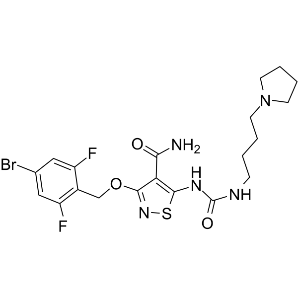
HY-13302
CP-547632VEGFR-2/FGF Inhibitor -
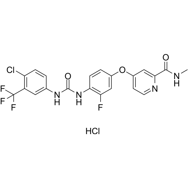
HY-13308
Regorafenib HydrochlorideAutophagy Inducer -
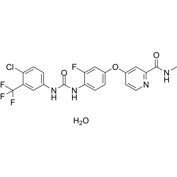
HY-10331A
Regorafenib monohydrateMulti-Kinase Inhibitor -
HY-15467A
ZM323881 hydrochlorideVEGFR2 Inhibitor -
HY-10337
BrivanibVEGFR2 Inhibitor -
HY-12038
Ki8751VEGFR Inhibitor
All VEGFR Isoforms
More

Biological Activity
제품 설명
IC50 & Target
|
VEGFR-2 0.6 nM (IC50) |
PDGFRα |
PDGFRβ |
In Vitro
Henatinib shows high binding affinities for VEGFRs, PDGFR and stem cell factor receptor[1].
Henatinib significantly inhibits VEGFR-2 phosphorylation and its downstream signal pathway in human umbilical vein endothelial cells (HUVECs), and consistently inhibited VEGF-stimulated HUVEC proliferation, migration and tubule formation[2].
MedChemExpress (MCE) has not independently confirmed the accuracy of these methods. They are for reference only.


Chemical Information
-
CAS No. 1239269-51-2
-
분자량 468.52
-
화학식 C25H29FN4O4
-
SMILES
O=C1N(C[C@H](O)CN2CCOCC2)CCCC3=C1C(C)=C(/C=C4C(NC5=C\4C=C(F)C=C5)=O)N3
-
선적
Room temperature in continental US; may vary elsewhere.
-
보관
Please store the product under the recommended conditions in the Certificate of Analysis.


순도&문서
References

Calculators
Concentration (start) × Volume (start) = Concentration (final) × Volume (final)


Henatinib Related Classifications
HY-13645 Related Classifications
 Powered by Bioz