



c-Jun Antibody (YA503)
(Synonyms: Transcription factor Jun, Activator protein 1, Proto-oncogene c-Jun, Transcription factor AP-1 subunit Jun, V-jun avian sarcoma virus 17 oncogene homolog, p39, AP1, JUN)Based on 2 publication(s) in Google Scholar
c-Jun Antibody (YA503) is a Rabbit-derived and non-conjugated IgG monoclonal antibody, targeting to c-Jun.
-
Host:
Rabbit
-
Isotype:
IgG
-
Application:
WB, IHC-P, ICC/IF, IP
-
Reactivity :
Human, Mouse, Rat
-
Formulation:
Supplied in 1*TBS (pH7.4), 0.05% BSA and 40% Glycerol. Preservative: 0.05% Sodium Azide.
-
Conjugation:
Non-conjugated


To place orders, for customer services and technical support, please contact: MedChemExpress USA
Tel: 609-228-6898 E-mail: [email protected] [email protected]



-

HY-P80801
Phospho-c-Jun (Ser63) Antibody (YA215)
-
HY-P80616
c-Fos Antibody
-
HY-P80802
Phospho-c-Jun (Ser73) Antibody
-
HY-P80938
p53 Antibody
-
HY-P80846
Phospho-PI3 Kinase p85/p55 (Tyr467/Tyr199) Antibody
-
HY-P80081
c-Fos Antibody (YA506)
-
HY-P80800
Phospho-c-Jun (Ser243) Antibody
-
HY-P80626
c-Myc Antibody (YA497)
-
HY-P80831
Phospho-JNK1 (Thr183/Tyr185) Antibody (YA180)
-
HY-P80456
Phospho-c-Jun(Ser63) Antibody (YA214)
Publications Citing Use of MedChemExpress (MCE) c-Jun Antibody (YA503)
More

Applications
| Application |
WB

WB: Western Blot
|
ICC/IF
ICC/IF: Immunocytochemistry/
Immunofluorescence |
IHC-P
IHC-P: Immunohistochemistry-Paraffin
|
IP
IP: Immunoprecipitation
|
|---|---|---|---|---|
| Dilution Ratio | 1:500-1:1000 | 1:100-1:500 | 1:50-1:800 | Use at an assay dependent concentration. |


Product Details
c-Jun Antibody (YA503) is a Rabbit-derived and non-conjugated IgG monoclonal antibody, targeting to c-Jun.
-
Host Rabbit

-
Clonality Recombinant,Monoclonal

-
Species ReactivityHuman, Mouse, Rat
-
Observed Molecular WeightObserved band size: 40 kDa
Note: Due to possible protein modifications or aggregation, the molecular weight should be confirmed by actual measurement, and the predicted value is for reference only.

-
Calculated Molecular Weight Predicted band size: 36 kDa

Entrez Gene: 3725 Human ; 16476 Mouse ; 24516 Rat
SwissProt: P05412 Human ; P05627 Mouse ; P17325 Rat
OMIM: 165160 Human
Synthetic peptide within N-terminal human c-Jun.
Endogenous
Protein A affinity purified.
Non-conjugated
Unmodified
IgG


Product Properties
-
Appearance
Solution
-
Formulation
Supplied in 1*TBS (pH7.4), 0.05% BSA and 40% Glycerol. Preservative: 0.05% Sodium Azide.
-
Concentration
Batch-dependent, Please check the COA for the concentration of each lot. Check Lot Concentration
-
Storage & Stability
Stored at -20°C for 1 year. Avoid repeated freeze / thaw cycles.
-
Shipping
Shipping with blue ice.


Publications (2)
-
Journal Impact Factor
-
Most Recent
-
J Inflamm Res
CCL20 Expression via AKT-ERK1/2-AP1 Pathway in Mycoplasma Pneumoniae Infection: Implications for EMT and Cell Migration. [Abstract]2025 Apr 28:18:5727-5739. PMID: 40322529


Verification Images
-
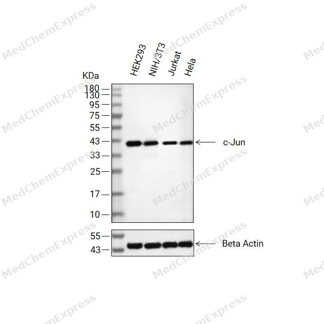 Western blot analysis of extracts from HEK293(lane 2(20μg) , NIH/3T3 (lane 3(20μg) ,Jurkat(lane 4(20μg)and Hela( lane 5(20μg) using c-Jun Antibody (HY-P80084). Proteins were transferred to a PVDF membrane and blocked with 5% non-fat milk in TBST for 2 hour at room temperature. The primary antibody ( 1/1000) and Loading control antibody (Beta Actin, HY-P80993,1/10000) was used in 5% non-fat milk in TBST at 4°C overnight. Goat Anti-Rabbit IgG-HRP Secondary Antibody (HY-P8001,1/10,000) was used for 1 hour at room temperature.
 -
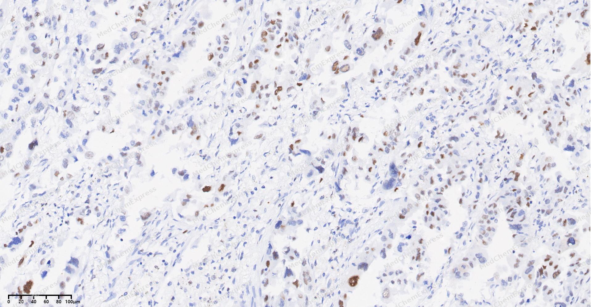 Immunohistochemical analysis of paraffin-embedded human Lung Adenocarcinoma tissue using c-Jun antibody was performed. The section was pretreated using high-temperature and high-pressure mediated EDTA antigen retrieval buffer (pH 9.0), for 5 minutes. The tissues were incubated with primary antibody (HY-P80084, 1:100 dilution) at room temperature for 60 minutes. Detection was performed using an HRP conjugated compact polymer system. DAB was used as the chromogen. The tissues were counterstained with hematoxylin and mounted with neutral balsam mounting medium.
 -
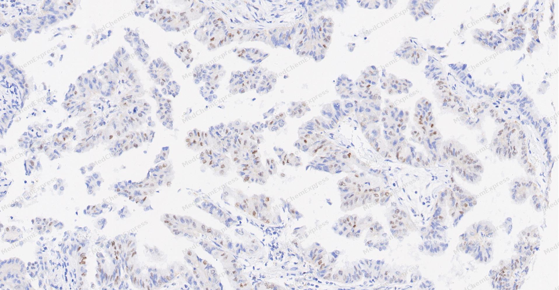 Immunohistochemical analysis of paraffin-embedded human Lung Adenocarcinoma tissue using c-Jun antibody was performed. The section was pretreated using high-temperature and high-pressure mediated EDTA antigen retrieval buffer (pH 9.0), for 5 minutes. The tissues were incubated with primary antibody (HY-P80084, 1:100 dilution) at room temperature for 60 minutes. Detection was performed using an HRP conjugated compact polymer system. DAB was used as the chromogen. The tissues were counterstained with hematoxylin and mounted with neutral balsam mounting medium.
 -
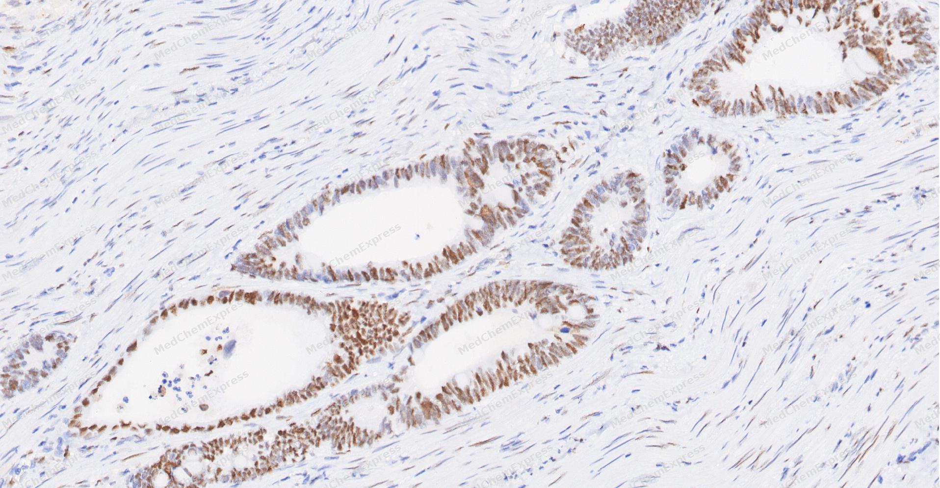 Immunohistochemical analysis of paraffin-embedded human Colon cancer tissue using c-Jun antibody was performed. The section was pretreated using high-temperature and high-pressure mediated EDTA antigen retrieval buffer (pH 9.0), for 5 minutes. The tissues were incubated with primary antibody (HY-P80084, 1:100 dilution) at room temperature for 60 minutes. Detection was performed using an HRP conjugated compact polymer system. DAB was used as the chromogen. The tissues were counterstained with hematoxylin and mounted with neutral balsam mounting medium.
 -
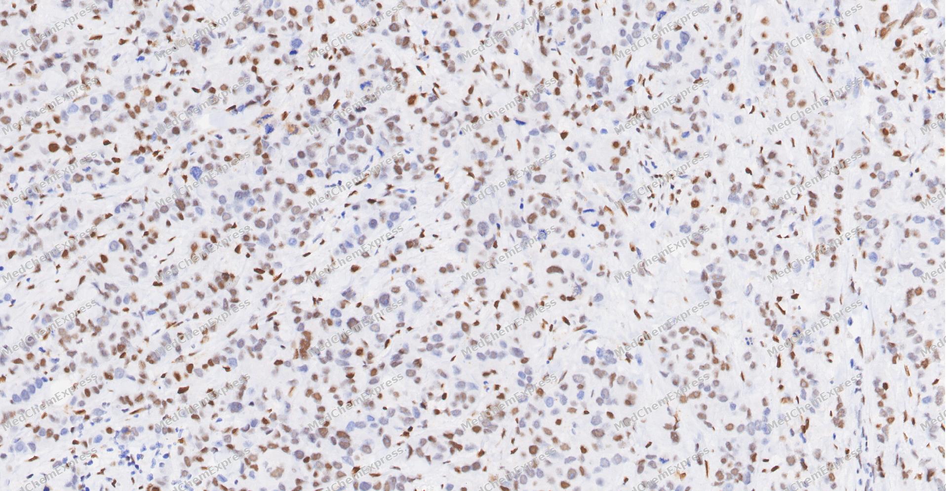 Immunohistochemical analysis of paraffin-embedded human Breast Cancer tissue using c-Jun antibody was performed. The section was pretreated using high-temperature and high-pressure mediated EDTA antigen retrieval buffer (pH 9.0), for 5 minutes. The tissues were incubated with primary antibody (HY-P80084, 1:100 dilution) at room temperature for 60 minutes. Detection was performed using an HRP conjugated compact polymer system. DAB was used as the chromogen. The tissues were counterstained with hematoxylin and mounted with neutral balsam mounting medium.
 -
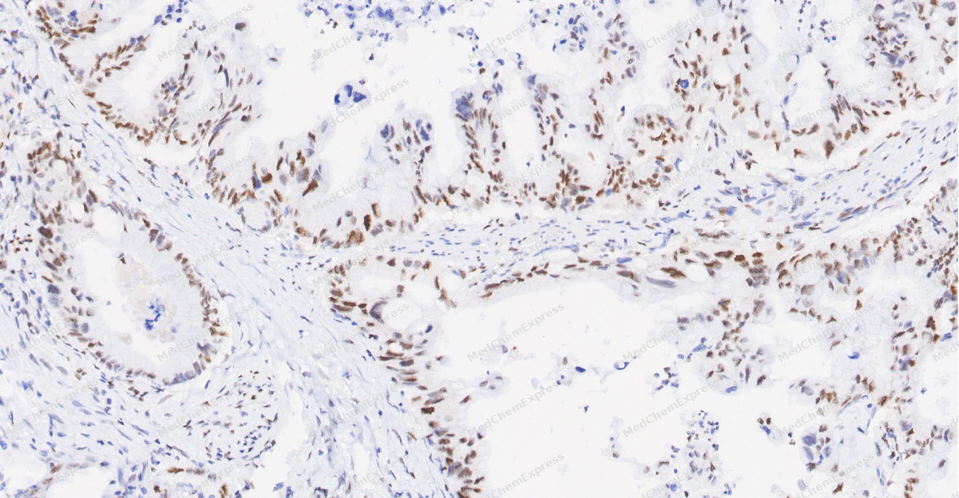 Immunohistochemical analysis of paraffin-embedded human Gastric Cancer tissue using c-Jun antibody was performed. The section was pretreated using high-temperature and high-pressure mediated EDTA antigen retrieval buffer (pH 9.0), for 5 minutes. The tissues were incubated with primary antibody (HY-P80084, 1:100 dilution) at room temperature for 60 minutes. Detection was performed using an HRP conjugated compact polymer system. DAB was used as the chromogen. The tissues were counterstained with hematoxylin and mounted with neutral balsam mounting medium.
 -
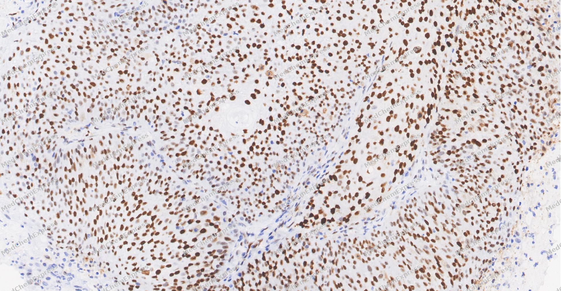 Immunohistochemical analysis of paraffin-embedded human Cervical Cancer tissue using c-Jun antibody was performed. The section was pretreated using high-temperature and high-pressure mediated EDTA antigen retrieval buffer (pH 9.0), for 5 minutes. The tissues were incubated with primary antibody (HY-P80084, 1:100 dilution) at room temperature for 60 minutes. Detection was performed using an HRP conjugated compact polymer system. DAB was used as the chromogen. The tissues were counterstained with hematoxylin and mounted with neutral balsam mounting medium.
 -
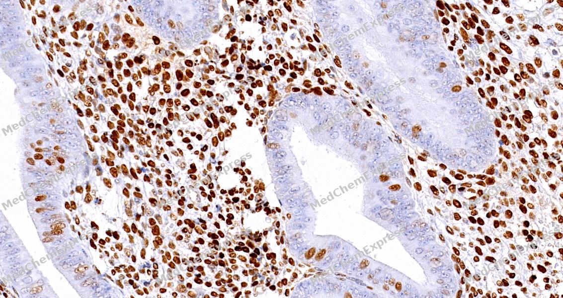 Immunohistochemical analysis of paraffin-embedded human endometrium tissue using c-Jun Antibody (HY-P80084, 1/500) . The section was pretreated using heat mediated antigen retrieval with sodium citrate buffer (PH 6.0)for 8 minutes. The tissues were blocked with quick block buffer for 0.5 hours at room temperature, washed with PBS and PBST, and then incubated with the primary antibody overnight at 6℃. The detection was performed using an HRP conjugated compact polymer system. DAB was used as the chromogen. Tissues were counterstained with hematoxylin and mounted with DPX.
 -
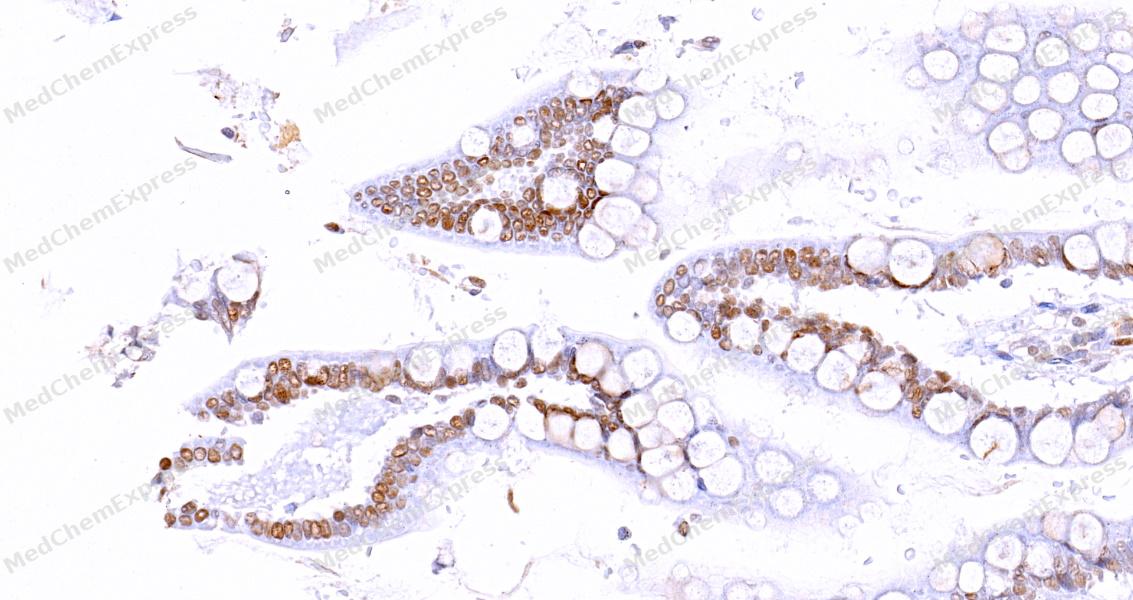 Immunohistochemical analysis of paraffin-embedded human small intestine tissue using c-Jun Antibody (HY-P80084, 1/500) . The section was pretreated using heat mediated antigen retrieval with sodium citrate buffer (PH 6.0)for 8 minutes. The tissues were blocked with quick block buffer for 0.5 hours at room temperature, washed with PBS and PBST, and then incubated with the primary antibody overnight at 6℃. The detection was performed using an HRP conjugated compact polymer system. DAB was used as the chromogen. Tissues were counterstained with hematoxylin and mounted with DPX.
 -
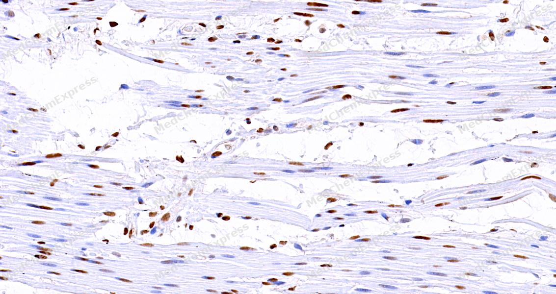 Immunohistochemical analysis of paraffin-embedded human esophagus tissue using c-Jun Antibody (HY-P80084, 1/500) . The section was pretreated using heat mediated antigen retrieval with sodium citrate buffer (PH 6.0)for 8 minutes. The tissues were blocked with quick block buffer for 0.5 hours at room temperature, washed with PBS and PBST, and then incubated with the primary antibody overnight at 6℃. The detection was performed using an HRP conjugated compact polymer system. DAB was used as the chromogen. Tissues were counterstained with hematoxylin and mounted with DPX.
 -
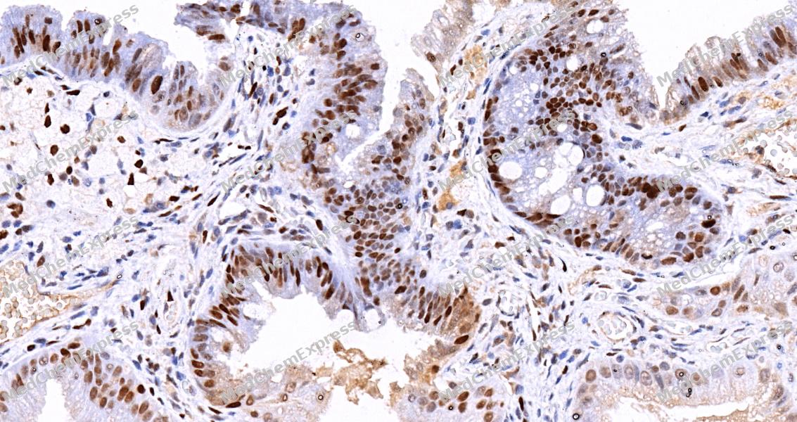 Immunohistochemical analysis of paraffin-embedded human gallbladder tissue using c-Jun Antibody (HY-P80084, 1/500) . The section was pretreated using heat mediated antigen retrieval with sodium citrate buffer (PH 6.0)for 8 minutes. The tissues were blocked with quick block buffer for 0.5 hours at room temperature, washed with PBS and PBST, and then incubated with the primary antibody overnight at 6℃. The detection was performed using an HRP conjugated compact polymer system. DAB was used as the chromogen. Tissues were counterstained with hematoxylin and mounted with DPX.
 -
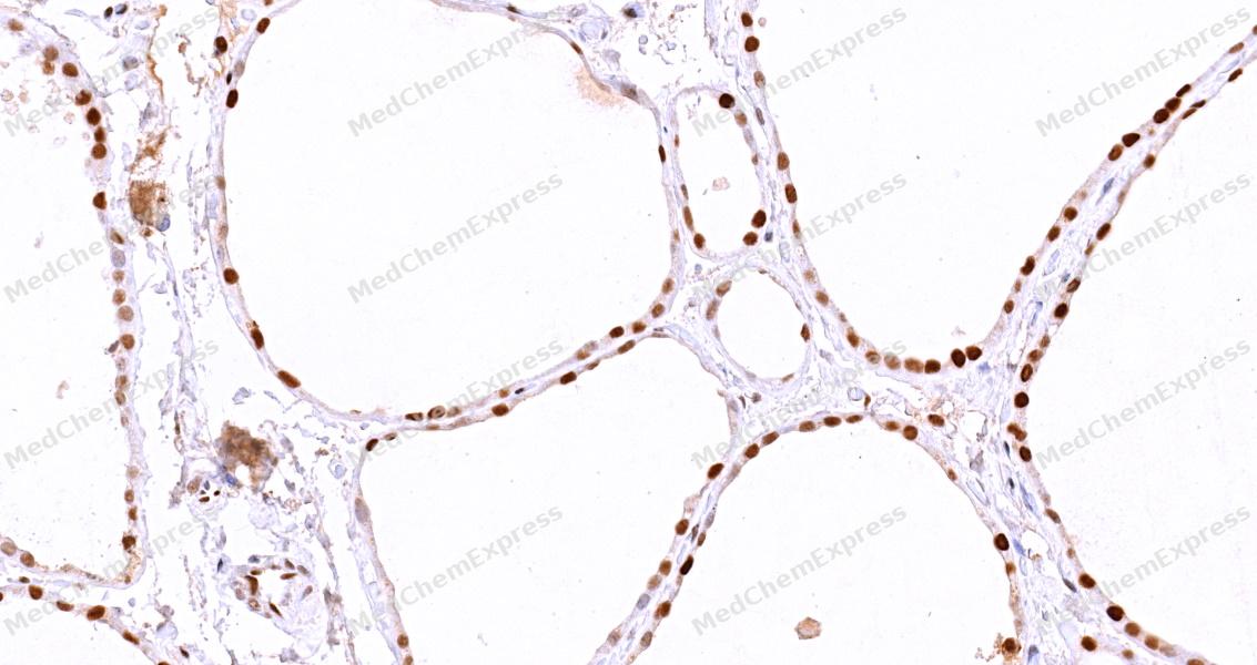 Immunohistochemical analysis of paraffin-embedded human thyroid gland tissue using c-Jun Antibody (HY-P80084, 1/500) . The section was pretreated using heat mediated antigen retrieval with sodium citrate buffer (PH 6.0)for 8 minutes. The tissues were blocked with quick block buffer for 0.5 hours at room temperature, washed with PBS and PBST, and then incubated with the primary antibody overnight at 6℃. The detection was performed using an HRP conjugated compact polymer system. DAB was used as the chromogen. Tissues were counterstained with hematoxylin and mounted with DPX.
 -
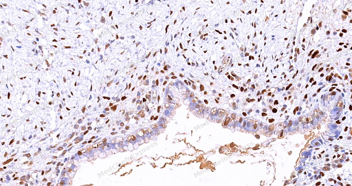 Immunohistochemical analysis of paraffin-embedded human cervix tissue using c-Jun Antibody (HY-P80084, 1/500) . The section was pretreated using heat mediated antigen retrieval with sodium citrate buffer (PH 6.0)for 8 minutes. The tissues were blocked with quick block buffer for 0.5 hours at room temperature, washed with PBS and PBST, and then incubated with the primary antibody overnight at 6℃. The detection was performed using an HRP conjugated compact polymer system. DAB was used as the chromogen. Tissues were counterstained with hematoxylin and mounted with DPX.
 -
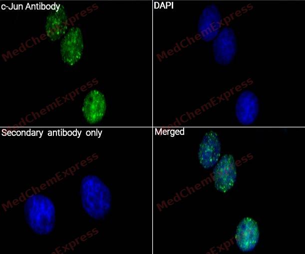 Immunocytochemistry analysis of NIH/3T3 cells labeling c-Jun with c-Jun Antibody (HY-P80084) at 1/200 dilution. Cells were fixed in 4% paraformaldehyde for 15 minutes at room temperature, permeabilized with 0.1% Triton X-100 in PBS for 15 minutes at room temperature, then blocked with quick block buffer for 10 minutes at room temperature. Cells were then incubated with c-Jun Antibody (HY-P80084) at 1/200 dilution in quick block buffer overnight at 4 ℃. AF488-conjugated Goat Anti-Rabbit IgG H&L(HY-P8002, Green) was used as the secondary antibody at 1/1,000 dilution. PBS instead of the primary antibody was used as the secondary antibody only control. The Nuclear counterstain was DAPI (Blue).
 -
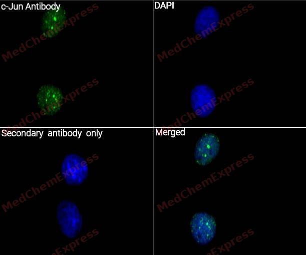 Immunocytochemistry analysis of NIH/3T3 cells labeling c-Jun with c-Jun Antibody (HY-P80084) at 1/400 dilution. Cells were fixed in 4% paraformaldehyde for 15 minutes at room temperature, permeabilized with 0.1% Triton X-100 in PBS for 15 minutes at room temperature, then blocked with quick block buffer for 10 minutes at room temperature. Cells were then incubated with c-Jun Antibody (HY-P80084) at 1/400 dilution in quick block buffer overnight at 4 ℃. AF488-conjugated Goat Anti-Rabbit IgG H&L(HY-P8002, Green) was used as the secondary antibody at 1/1,000 dilution. PBS instead of the primary antibody was used as the secondary antibody only control. The Nuclear counterstain was DAPI (Blue).


Background
-
Function
c-Jun, a basic leucine zipper transcription factor, regulates gene expression in response to multiple extracellular signals[1]. It is a central component of the activator protein-1 (AP-1) complex, mediating cellular proliferation, differentiation, and stress responses[2][3]. Mechanistically, c-Jun is primarily activated by phosphorylation through the c-Jun N-terminal kinase (JNK) pathway, linking stress signals to transcriptional outputs[3][4]. Isoform-specific functions of JNK1, JNK2, and JNK3 modulate c-Jun activity in tissue- and cell-specific contexts, influencing apoptosis, inflammation, and neuroplasticity[1][5][6]. In disease models, c-Jun contributes to pathological angiogenesis, rheumatoid arthritis, and neurodegeneration, where its inhibition reduces endothelial proliferation, metalloproteinase expression, and neuronal apoptosis[7][8][9][10][11][6]. Compared with related Jun-family members, c-Jun exhibits distinct phosphorylation patterns and transcriptional targets, enabling isoform-specific experimental interventions[6][12]. Pharmacological inhibitors targeting JNK or c-Jun itself, such as SP600125 or DNAzymes, demonstrate utility in blocking pro-apoptotic signaling, reducing inflammation, and modulating angiogenic pathways, supporting their research and therapeutic application[7][8][10][6]. In cancer and viral models, c-Jun interacts with oncogenic pathways, including Bcl-2/Bcl-xL upregulation and FGF-2-mediated proliferation, highlighting its role in cell survival and transcriptional regulation[2][13]. Collectively, c-Jun integrates stress, mitogenic, and inflammatory signals, serving as a critical target for isoform-specific modulation in experimental and disease contexts[1][3][6][7].
-
Subcellular Localization
Nucleus
-
Expression
Tissue_specificity:It is expressed in developing and adult prostate cells as well as prostate cancer cells. -
Subunit
Heterodimer with either BATF3 or ATF7 (PubMed:10376527, PubMed:12087103, PubMed:15467742). Heterodimer with FOS (By similarity). Heterodimer with FOSB isoform 1 and 2 (By similarity). Component of an AP-1 transcription factor complex composed of JUN-FOS heterodimers (By similarity). As part of the AP-1 transcription factor complex, forms heterodimers with FOSB, thereby binding to the AP-1 consensus sequence and stimulating transcription (By similarity). Interacts with FOS and FOSB isoform 1 and 2 (By similarity). The ATF7/JUN heterodimer is essential for ATF7 transactivation activity (PubMed:10376527). Interacts with TSC22D3 (via N-terminus); the interaction inhibits the binding of active AP1 to its target DNA (By similarity). Interacts with HIVEP3 and MYBBP1A (By similarity). Interacts with SP1, SPIB and TCF20 (PubMed:10196196, PubMed:16478997, PubMed:8663478). Interacts with COPS5; the interaction leads indirectly to its phosphorylation (PubMed:8837781). Component of the SMAD3/SMAD4/JUN/FOS/complex which forms at the AP1 promoter site (PubMed:10995748). The SMAD3/SMAD4 heterodimer acts synergistically with the JUN/FOS heterodimer to activate transcription in response to TGF-beta (PubMed:9732876). Interacts (via its basic DNA binding and leucine zipper domains) with SMAD3 (via an N-terminal domain); the interaction is required for TGF-beta-mediated transactivation of the SMAD3/SMAD4/JUN/FOS/complex (PubMed:10995748). Interacts with methylated RNF187 (PubMed:20852630, PubMed:23624934). Binds to HIPK3. Interacts (when phosphorylated) with FBXW7 (PubMed:14739463). Found in a complex with PRR7 and FBXW7 (PubMed:27458189). Interacts with PRR7 and FBXW7; the interaction inhibits ubiquitination-mediated JUN degradation promoting its phosphorylation and transcriptional activity (PubMed:27458189). Interacts with RBM39 (By similarity). Interacts with PAGE4 (PubMed:24263171, PubMed:24559171, PubMed:26242913). Interacts with ARK2N and CSNK2B; the interaction with ARK2N is mediated by CSNK2B (PubMed:31341047)
-
SwissProt ID
-
Synonyms
Transcription factor Jun, Activator protein 1, Proto-oncogene c-Jun, Transcription factor AP-1 subunit Jun, V-jun avian sarcoma virus 17 oncogene homolog, p39, AP1, JUN
-
Research Field
Epigenetics and Nuclear Signaling
Documentation
-
Data Sheet (264 KB)
-
SDS (252 KB)
- English - EN (252 KB)
- Français - FR (252 KB)
- Deutsch - DE (252 KB)
- Norwegian - NO (252 KB)
- Español - ES (252 KB)
- Swedish - SV (252 KB)
- Italian - IT (252 KB)
- Korean - KR (252 KB)
- Portuguese - PT (252 KB)
-
User Guide for Antibodies (1077 KB)
[1]. Bogoyevitch MA. The isoform-specific functions of the c-Jun N-terminal Kinases (JNKs): differences revealed by gene targeting. Bioessays. 2006 Sep;28(9):923-34. doi: 10.1002/bies.20458. PMID: 16937364. et al. The isoform-specific functions of the c-Jun N-terminal Kinases (JNKs): differences revealed by gene targeting. Bioessays. 2006 Sep;28(9):923-34. [Content Brief]
[2]. Hortala M, et al. Identification of c-Jun as a critical mediator for the intracrine 24 kDa FGF-2 isoform-induced cell proliferation. Int J Cancer. 2005 May 10;114(6):863-9. [Content Brief]
[3]. Shashikanth N, et al. Role of C-Jun N-Terminal Kinases on a Stressed Epithelium: Time for Testing Isoform Specificity. Biology (Basel). 2025 Jun 3;14(6):649. [Content Brief]
[4]. Yao R, et al. Specific activation of a c-Jun NH2-terminal kinase isoform and induction of neurite outgrowth in PC-12 cells by staurosporine. J Biol Chem. 1997 Jul 18;272(29):18261-6. [Content Brief]
[5]. Raivich G. c-Jun expression, et al. c-Jun expression, activation and function in neural cell death, inflammation and repair. J Neurochem. 2008 Nov;107(4):898-906. [Content Brief]
[6]. Tan J, et al. Isoform-specific functions of c-Jun N-terminal kinase 1 and 2 in lung ischemia-reperfusion injury through the c-Jun/activator protein-1 pathway. J Thorac Cardiovasc Surg. 2021 Aug;162(2):e143-e156. [Content Brief]
[7]. Folkman J. Angiogenesis and c-Jun. J Natl Cancer Inst. 2004 May 5;96(9):644. doi: 10.1093/jnci/djh148. PMID: 15126593. et al. Angiogenesis and c-Jun. J Natl Cancer Inst. 2004 May 5;96(9):644. [Content Brief]
[8]. Zhang GY, et al. Agents targeting c-Jun N-terminal kinase pathway as potential neuroprotectants. Expert Opin Investig Drugs. 2005 Nov;14(11):1373-83. [Content Brief]
[9]. Ouyang W, et al. Anthrax lethal toxin rapidly reduces c-Jun levels by inhibiting c-Jun gene transcription and promoting c-Jun protein degradation. J Biol Chem. 2017 Oct 27;292(43):17919-17927. [Content Brief]
[10]. Han Z, et al. c-Jun N-terminal kinase is required for metalloproteinase expression and joint destruction in inflammatory arthritis. J Clin Invest. 2001 Jul;108(1):73-81. [Content Brief]
[11]. Hepp Rehfeldt SC, et al. c-Jun N-Terminal Kinase Inhibitors as Potential Leads for New Therapeutics for Alzheimer's Diseases. Int J Mol Sci. 2020 Dec 18;21(24):9677. [Content Brief]
[12]. Zhang Y, et al. MAPK/c-Jun signaling pathway contributes to the upregulation of the anti-apoptotic proteins Bcl-2 and Bcl-xL induced by Epstein-Barr virus-encoded BARF1 in gastric carcinoma cells. Oncol Lett. 2018 May;15(5):7537-7544. [Content Brief]
[13]. Zoukhri D, et al. c-Jun NH2-terminal kinase mediates interleukin-1beta-induced inhibition of lacrimal gland secretion. J Neurochem. 2006 Jan;96(1):126-35. [Content Brief]

 Powered by Bioz
