



c-Myc Antibody (YA497)
(Synonyms: MYC; BHLHE39; Myc proto-oncogene protein; Class E basic helix-loop-helix protein 39; bHLHe39; Proto-oncogene c-Myc; Transcription factor p64)Based on 7 publication(s) in Google Scholar
c-Myc Antibody (YA497) is a Rabbit-derived and non-conjugated IgG monoclonal antibody, targeting to c-Myc.
-
Host:
Rabbit
-
Isotype:
IgG
-
Application:
WB, ICC/IF, IP
-
Reactivity :
Human, Mouse, Rat
-
Formulation:
Supplied in 50 mM Tris-Glycine (pH 7.4), 0.15 M NaCl, 40% Glycerol and 0.05% BSA. Preservative: 0.01% Sodium azide
-
Conjugation:
Non-conjugated


To place orders, for customer services and technical support, please contact: MedChemExpress USA
Tel: 609-228-6898 E-mail: [email protected] [email protected]



-

HY-P80488
beta Catenin Antibody (YA584) -
HY-P8001
HRP-conjugated Goat Anti-Rabbit IgG H&L -
HY-P8003
AF594-conjugated Goat Anti-Rabbit IgG H&L -
HY-P8004
HRP-conjugated Goat Anti-Mouse IgG H&L -
HY-15534
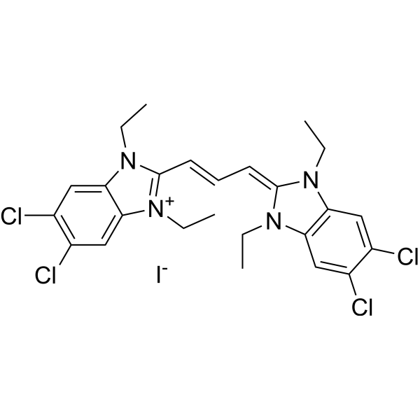
JC-1Mitochondrial Membrane Potential Probe -
HY-P80098
Cyclin D1/CCND1 Antibody (YA485) -
HY-K0207
Anti-Flag Magnetic Beads -
HY-P80535
AKT1+AKT2+AKT3 Antibody (YA635) -
HY-P80344
STAT3 Antibody (YA056) -

HY-K2015
Lentivirus Transfection Reagent -
HY-P80438
Beta Actin Antibody (YA823) -
HY-P80141
GFP Tag Antibody (YA415)
Publications Citing Use of MedChemExpress (MCE) c-Myc Antibody (YA497)
More- Signal Transduct Target Ther. 2025 Jul 9;10(1):217. [Abstract]
- Leukemia. 2025 Aug 1. [Abstract]
- Int J Biol Macromol. 2025 Aug 8;322(Pt 1):146678. [Abstract]
- Biochim Biophys Acta Mol Basis Dis. 2024 Apr 22;1870(5):167183. [Abstract]
- Funct Integr Genomics. 2025 Nov 4;25(1):230. [Abstract]
- Discov Oncol. 2024 Dec 18;15(1):768. [Abstract]
- Med Drug Discov. 2025 Sep 19;28:100229.

 7 Publications Citing Use of MCE c-Myc Antibody (YA497) (6)
-
IHC

-
WB

-
WB

-
WB

-
WB


Applications
| Application |
WB

WB: Western Blot
|
ICC/IF
ICC/IF: Immunocytochemistry/
Immunofluorescence |
IP
IP: Immunoprecipitation
|
|---|---|---|---|
| Dilution Ratio | 1:500-1:1000 | 1:50-1:200 | 1:20 |


Product Details
c-Myc Antibody (YA497) is a Rabbit-derived and non-conjugated IgG monoclonal antibody, targeting to c-Myc.
-
Host Rabbit

-
Clonality Recombinant,Monoclonal

-
Species ReactivityHuman, Mouse, Rat
-
Observed Molecular WeightObserved band size: 45/57-65 kDa
Note: Due to possible protein modifications or aggregation, the molecular weight should be confirmed by actual measurement, and the predicted value is for reference only.

-
Calculated Molecular Weight Predicted band size: 49 kDa

Entrez Gene: 4609 Human ; 17869 Mouse ; 24577 Rat
SwissProt: P01106 Human ; P01108 Mouse ; P09416 Rat
OMIM: 113970 Human
Synthetic peptide corresponding to Human c-Myc aa1-50.
Endogenous
affinity purified
Non-conjugated
Unmodified
IgG


Product Properties
-
Appearance
Solution
-
Formulation
Supplied in 50 mM Tris-Glycine (pH 7.4), 0.15 M NaCl, 40% Glycerol and 0.05% BSA. Preservative: 0.01% Sodium azide
-
Concentration
Batch-dependent, Please check the COA for the concentration of each lot. Check Lot Concentration
-
Storage & Stability
Stored at -20°C for 1 year. Avoid repeated freeze / thaw cycles.
-
Shipping
Shipping with blue ice.


Publications (7)
-
Journal Impact Factor
-
Most Recent
-
Signal Transduct Target Ther
A novel peptide 66CTG stabilizes Myc proto-oncogene protein to promote triple-negative breast cancer growth. [Abstract]2025 Jul 9;10(1):217. PMID: 40628713
c-Myc Antibody (YA497) purchased from MedChemExpress. Usage Cited in: Signal Transduct Target Ther. 2025 Jul 9;10(1):217. [Abstract]
Images of 66CTG, c-Myc, and Cyclin D1 expression in clinical TNBC paraffin-embedded continuous slicing samples using IHC. Scale bar: 200 μm. Primary antibodies against 66CTG (AB012233, 1.6 μg/mL), c-Myc (c-Myc Antibody (YA497), HY-P80626, 0.6 μg/mL), and Cyclin D1 (1 μg/mL) were used.
-
Leukemia
The bone marrow immune ecosystem shapes daratumumab acquired resistance in plasma cell myeloma. [Abstract]2025 Aug 1. PMID: 40750676
c-Myc Antibody (YA497) purchased from MedChemExpress. Usage Cited in: Leukemia. 2025 Aug 1. [Abstract]
Detection of MYC and phospho-MYC (p-MYC) induction by IFN-γ (10 ng/mL) in RPMI 8226 and MM.1S cells using Western blot assays. Following a 48-h incubation with IFN-γ recombinant protein, phospho-MYC (p-MYC) and total MYC protein levels in PCM cell lines were assessed by WB using an MYC antibody (c-Myc Antibody (YA497), HY-P80626) and phospho-c-Myc (Ser62) antibody (Phospho-c-Myc(Ser62) Antibody (YA211), HY-P80511).
-
Int J Biol Macromol
C-Arg9-APCR3-VHL promotes β-catenin degradation via the VHL-mediated ubiquitin-proteasome system in APC-mutant colorectal cancer. [Abstract]2025 Aug 8;322(Pt 1):146678. PMID: 40782852 -
Biochim Biophys Acta Mol Basis Dis
Ubiquitin ligase TRIM15 promotes the progression of pancreatic cancer via the upregulation of the IGF2BP2-TLR4 axis. [Abstract]2024 Apr 22;1870(5):167183. PMID: 38657551 -
Funct Integr Genomics
DDX1 facilitates lenvatinib resistance in hepatocellular carcinoma through regulating ephrin-A3 and activating the Wnt/β-catenin signaling pathway. [Abstract]2025 Nov 4;25(1):230. PMID: 41186842
c-Myc Antibody (YA497) purchased from MedChemExpress. Usage Cited in: Funct Integr Genomics. 2025 Nov 4;25(1):230. [Abstract]
Western blotting analysis showed that DDX1-induced increases in Wnt/β-catenin pathway related protein expression were reversed by Ephrin-A3 silencing in HCC cells. Antibodies used in this study were as follows: anti-DDX1 (1:4000), anti-Ephrin-A3 (1:4000), anti-cyclin D1 (1:5000), anti-β-catenin (1:5000), anti-c-MYC (1:1000, c-Myc Antibody (YA497), HY-P80626), anti-GSK-3β (1:1000), and anti-β-tubulin (1:4000).
-
Discov Oncol
Network pharmacology and anticancer mechanism study of Dendrobium nobile dendrobine in the treatment of colorectal cancer. [Abstract]2024 Dec 18;15(1):768. PMID: 39692955
c-Myc Antibody (YA497) purchased from MedChemExpress. Usage Cited in: Discov Oncol. 2024 Dec 18;15(1):768. [Abstract]
The impact of Dendrobium nobile dendrobine on the expression of key proteins in the Wnt/β-catenin signaling pathway in colorectal cancer cells. HT29 cells: Western blot image of protein expression levels. The following primary antibodies were used in the study: β-catenin, c-Myc (c-Myc Antibody (YA497), HY-P80626), and Cyclin D1.

c-Myc Antibody (YA497) purchased from MedChemExpress. Usage Cited in: Discov Oncol. 2024 Dec 18;15(1):768. [Abstract]
The impact of Dendrobium nobile dendrobine on the expression of key proteins in the Wnt/β-catenin signaling pathway in colorectal cancer cells. SW480 cells: Western blot image of protein expression levels. The following primary antibodies were used in the study: β-catenin, c-Myc (c-Myc Antibody (YA497), HY-P80626), and Cyclin D1.
-
c-Myc Antibody (YA497) purchased from MedChemExpress. Usage Cited in: Med Drug Discov. 2025 Sep 19;28:100229.
Western blot analysis of p-ERK1/2, ERK1/2, c-JUN, c-FOS and c-MYC in AML12 cells (n = 3). Primary antibodiy: c-Myc Antibody (YA497) (Incubated overnight at 4°C, HY-P80626).


Verification Images
-
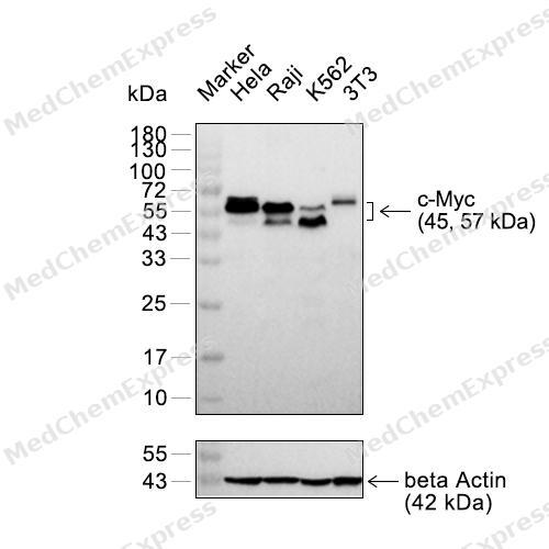 Western blot analysis of extracts from Hela (lane 2(20μg)), Raji (lane 3(20μg)), K562 (lane 4(20μg)) and 3T3 (lane 5(20μg)) using c-Myc Rabbit mAb. Proteins were transferred to a PVDF membrane and blocked with 5% non-fat milk in TBST for 2 hour at room temperature. The primary antibody (1/1000) and Loading control antibody (beta Actin, HY-P83730, 1/5000) was used in 5% non-fat milk in TBST at 4°C overnight. Goat Anti-Rabbit/Mouse IgG-HRP Secondary Antibody (1/10000) was used for 1 hour at room temperature.
 -
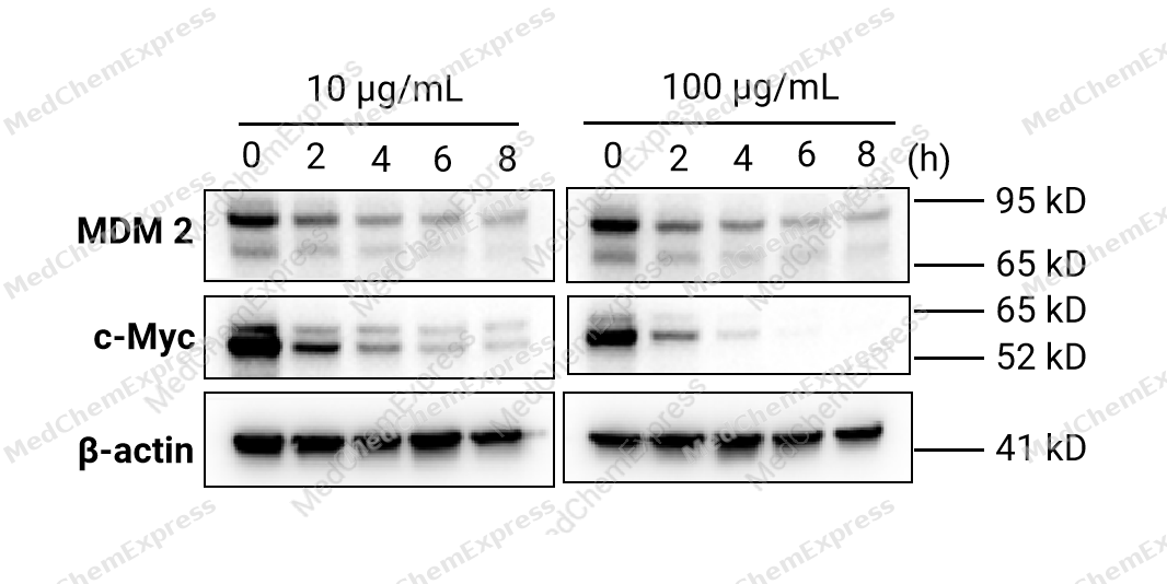 HCT 116 cells were seeded at 4*105 cells/well in a 6-well plate and were cultured overnight. Then, cells were treated with 10 and 100 μg/mL Cycloheximide (HY-12320) for 0-8 hours to assess the expression of MDM2 and c-Myc via Western blotting. The results demonstrated that Cycloheximide inhibited the expression of MDM2 and c-Myc in a time-dependent manner. Primary antibody: MDM2 antibody (HY-P83705), c-Myc antibody (HY-P80626), β-actin antibody (HY-P80438).
 -
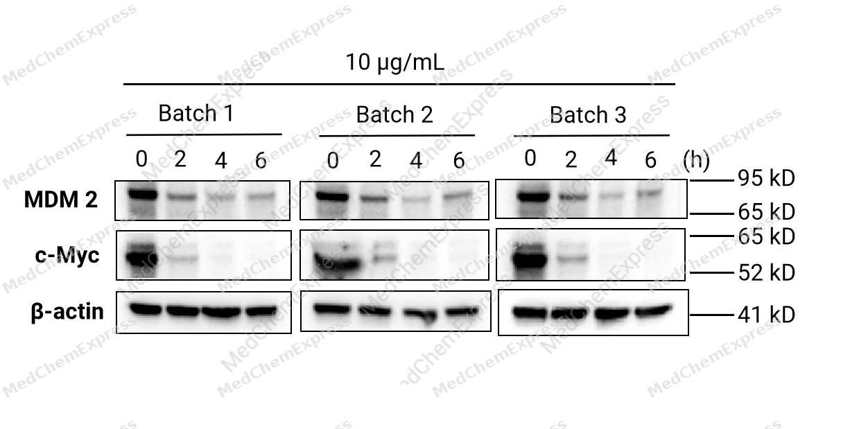 HCT 116 cells were seeded at 4*105 cells/well in a 6-well plate and were cultured overnight. Then, cells were treated with 10 μg/mL Cycloheximide (HY-12320) for 0-6 hours to assess the expression of MDM2 and c-Myc via Western blotting. The results demonstrated that Cycloheximide inhibited the expression of MDM2 and c-Myc proteins and showed lot-to-lot consistency. Primary antibody: MDM2 antibody (HY-P83705), c-Myc antibody (HY-P80626), β-actin antibody (HY-P80438).
 -
 Immunocytochemistry analysis of Hela cells labeling c-Myc with c-Myc Antibody (HY-P80626) at 1/100 dilution. Cells were fixed in 4% paraformaldehyde for 15 minutes at room temperature, permeabilized with 0.1% Triton X-100 in PBS for 15 minutes at room temperature, then blocked with quick block buffer for 10 minutes at room temperature. Cells were then incubated with c-Myc Antibody (HY-P80626) at 1/100 dilution in quick block buffer overnight at 4 ℃. AF488-conjugated Goat Anti-Rabbit IgG H&L(HY-P8002, Green) was used as the secondary antibody at 1/1,000 dilution. PBS instead of the primary antibody was used as the secondary antibody only control. The Nuclear counterstain was DAPI (Blue).
 -
 Immunocytochemistry analysis of NIH-3T3 cells labeling c-Myc with c-Myc Antibody (HY-P80626) at 1/100 dilution. Cells were fixed in 4% paraformaldehyde for 15 minutes at room temperature, permeabilized with 0.1% Triton X-100 in PBS for 15 minutes at room temperature, then blocked with quick block buffer for 10 minutes at room temperature. Cells were then incubated with c-Myc Antibody (HY-P80626) at 1/100 dilution in quick block buffer overnight at 4 ℃. AF488-conjugated Goat Anti-Rabbit IgG H&L(HY-P8002, Green) was used as the secondary antibody at 1/1,000 dilution. PBS instead of the primary antibody was used as the secondary antibody only control. The Nuclear counterstain was DAPI (Blue).


Background
-
Function
The transcription factor c-Myc is a member of the basic helix-loop-helix leucinezipper (bHLHZip) protein family. The target genes of the c-MYC protein participate in different cellular functions, including cell cycle, survival, protein synthesis, cell adhesion, and micro-RNA expression. c-Myc is also one of the four factors used in reprogramming somatic cells to induce pluripotent stem (iPS) cells and is implicated in maintaining cancer stem-like cells (CSCs) . Most biological functions of c-Myc require heterodimerization with its activation partner Max.
c-Myc is also part of a dynamic network whose members interact selectively with one another and with various transcriptional coregulators and histone-modifying enzymes. Deregulated expression of c-MYC caused by gene amplification, retroviral insertion, or chromosomal translocation is associated with tumorigenesis. c-Myc has been identified as a highly promising target for cancer therapy.
-
Subcellular Localization
Nucleus, nucleoplasm; Nucleus, nucleolus; Nucleus; Cytoplasm; Chromosome
-
Isoforms & Post-Translational Modification
P01106 has 3 isomers: P01106-2: 50565 Da (predicted); P01106-1: 48804 Da (predicted); P01106-3: 50437 Da (predicted).
Phosphorylated by PRKDC (PubMed:1597196). Phosphorylation at Ser-344 by PIM2 leads to the stabilization of MYC (By similarity). Phosphorylation at Ser-77 by CDK2 prevents Ras-induced senescence (PubMed:19966300, PubMed:20713526). Phosphorylated at Ser-77 by DYRK2; this primes the protein for subsequent phosphorylation by GSK3B at Thr-73 (PubMed:22307329). Phosphorylation at Thr-73 and Ser-77 by GSK3 is required for ubiquitination and degradation by the proteasome (PubMed:15103331, PubMed:17558397, PubMed:8386367). Dephosphorylation at multiple sites by the PNUTS-PP1 complex promotes MYC stability by preventing ubiquitination by the SCF(FBXW7) complex (PubMed:30158517). Dephosphorylation at Ser-77 by protein phosphatase 2A (PPP2CA) promotes its degradation; interaction with PPP2CA is enhanced by AMBRA1 (PubMed:25438055, PubMed:25803737);Ubiquitinated by the SCF(FBXW7) complex when phosphorylated at Thr-73 and Ser-77, leading to its degradation by the proteasome (PubMed:15103331, PubMed:17558397, PubMed:25775507, PubMed:30158517). In the nucleoplasm, ubiquitination is counteracted by USP28, which interacts with isoform 1 of FBXW7 (FBW7alpha), leading to its deubiquitination and preventing degradation (PubMed:17558397, PubMed:17873522). In the nucleolus, however, ubiquitination is not counteracted by USP28 but by USP36, due to the lack of interaction between isoform 3 of FBXW7 (FBW7gamma) and USP28, explaining the selective MYC degradation in the nucleolus (PubMed:17558397, PubMed:25775507). Also polyubiquitinated by the DCX(TRPC4AP) complex (PubMed:20551172, PubMed:29779948). Ubiquitinated by UBR5 when not forming a heterodimer with another bHLH protein, leading to its degradation: UBR5 recognizes and binds a degron that is only available upon heterodimer dissociation (PubMed:33208877, PubMed:37478862). Ubiquitinated by TRIM6 in a phosphorylation-independent manner (By similarity) -
Subunit
Efficient DNA binding requires dimerization with another bHLH protein. Binds DNA as a heterodimer with MAX (PubMed:9680483). Interacts with TAF1C and SPAG9. Interacts with PARP10. Interacts with KDM5A and KDM5B. Interacts (when phosphorylated at Thr-73 and Ser-77) with FBXW7 (PubMed:17558397, PubMed:25775507). Interacts with PIM2. Interacts with RIOX1. The heterodimer MYC:MAX interacts with ABI1; the interaction may enhance MYC:MAX transcriptional activity. Interacts with TRIM6 (By similarity). Interacts with NPM1; the binary complex is recruited to the promoter of MYC target genes and enhances their transcription (PubMed:25956029). Interacts with CIP2A; leading to the stabilization of MYC (PubMed:17632056). Interacts with NUP205 (PubMed:22719065). Interacts with HEATR1; the interaction is required for localization of MYC to the nucleolus (PubMed:38225354)
-
SwissProt ID
-
Synonyms
MYC; BHLHE39; Myc proto-oncogene protein; Class E basic helix-loop-helix protein 39; bHLHe39; Proto-oncogene c-Myc; Transcription factor p64
-
Research Field
Epigenetics and Nuclear Signaling
Documentation
-
Data Sheet (234 KB)
-
SDS (252 KB)
- English - EN (252 KB)
- Français - FR (252 KB)
- Deutsch - DE (252 KB)
- Norwegian - NO (252 KB)
- Español - ES (252 KB)
- Swedish - SV (252 KB)
- Italian - IT (252 KB)
- Korean - KR (252 KB)
- Portuguese - PT (252 KB)
-
User Guide for Antibodies (1077 KB)

 Powered by Bioz
