



MDM2 Antibody (YA3442)
(Synonyms: ACTFS antibody; Double minute 2 protein antibody; E3 ubiquitin-protein ligase Mdm2 antibody; Hdm 2 antibody; Hdm2 antibody; HDMX antibody; MDM 2 antibody; MDM2 antibody; MDM2 oncogene E3 ubiquitin protein ligase antibody; Mdm2 p53 E3 ubiquitin protein ligase homolog antibody; Mdm2 transformed 3T3 cell double minute 2 p53 binding protein (mouse) binding protein 104kDa antibody; MDM2_HUMAN antibody; MDM2BP antibody; Mouse Double Minute 2 antibody; MTBP antibody; Murine Double Minute Chromosome 2 antibody; Oncoprotein Mdm2 antibody; p53 Binding Protein Mdm2 antibody; p53-binding protein Mdm2 antibody; Ubiquitin protein ligase E3 Mdm2 antibody; Ubiquitin protein ligase E3 Mdm2 antibody)
Based on 1 Customer Validation
MDM2 Antibody (YA3442) is a Mouse-derived and non-conjugated IgG1 monoclonal antibody, targeting to MDM2.
-
Host:
Mouse
-
Isotype:
IgG
-
Application:
WB, IHC-P, ICC/IF
-
Reactivity :
Human, Monkey
-
Formulation:
Supplied in PBS (pH7.4), 0.1% BSA, 40% Glycerol. Preservative: 0.05% Sodium Azide.
-
Conjugation:
Non-conjugated


To place orders, for customer services and technical support, please contact: MedChemExpress USA
Tel: 609-228-6898 E-mail: [email protected] [email protected]





Applications
| Application |
WB

WB: Western Blot
|
ICC/IF
ICC/IF: Immunocytochemistry/
Immunofluorescence |
IHC-P
IHC-P: Immunohistochemistry-Paraffin
|
|---|---|---|---|
| Dilution Ratio | 1:1000 | 1:500 | 1:1000 |


Product Details
MDM2 Antibody (YA3442) is a Mouse-derived and non-conjugated IgG1 monoclonal antibody, targeting to MDM2.
-
Host Mouse

-
Clonality Monoclonal,Recombinant

-
Species ReactivityHuman, Monkey
-
Observed Molecular WeightObserved band size: 90 kDa
Note: Due to possible protein modifications or aggregation, the molecular weight should be confirmed by actual measurement, and the predicted value is for reference only.

-
Calculated Molecular Weight Predicted band size: 55 kDa

Entrez Gene: 4193 Human ; 17246 Mouse ; 314856 Rat
SwissProt: Q00987 Human ; P23804 Mouse ;
OMIM: 618681 Human
Recombinant protein within human MDM2 aa 1-491 / 491.
Affinity Purified
Non-conjugated
Unmodified
IgG


Product Properties
-
Appearance
Solution
-
Formulation
Supplied in PBS (pH7.4), 0.1% BSA, 40% Glycerol. Preservative: 0.05% Sodium Azide.
-
Concentration
Batch-dependent, Please check the COA for the concentration of each lot. Check Lot Concentration
-
Storage & Stability
Stored at -20°C for 1 year. Avoid repeated freeze / thaw cycles.
-
Shipping
Shipping with blue ice.


Verification Images
-
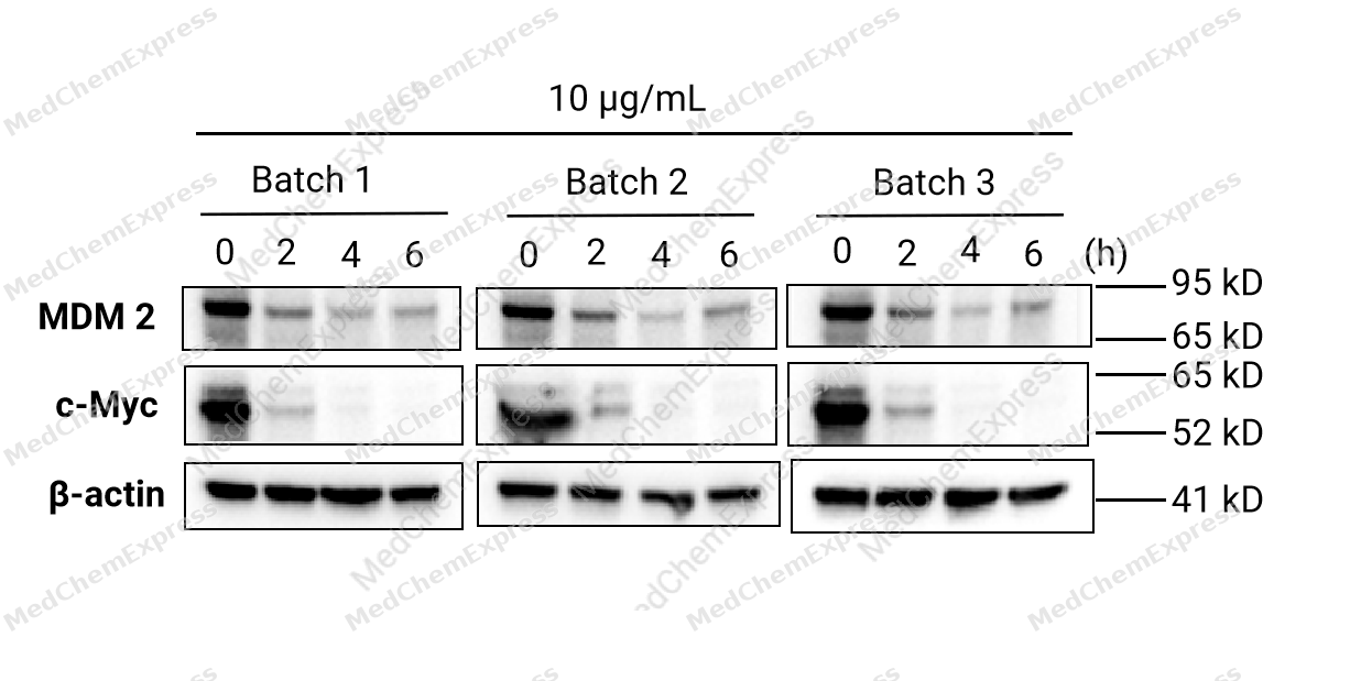 HCT 116 cells were seeded at 4*105 cells/well in a 6-well plate and were cultured overnight. Then, cells were treated with 10 μg/mL Cycloheximide (HY-12320) for 0-6 hours to assess the expression of MDM2 and c-Myc via Western blotting. The results demonstrated that Cycloheximide inhibited the expression of MDM2 and c-Myc proteins and showed lot-to-lot consistency. Primary antibody: MDM2 antibody (HY-P83705), c-Myc antibody (HY-P80626), β-actin antibody (HY-P80438).
 -
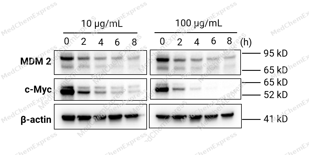 HCT 116 cells were seeded at 4*105 cells/well in a 6-well plate and were cultured overnight. Then, cells were treated with 10 and 100 μg/mL Cycloheximide (HY-12320) for 0-8 hours to assess the expression of MDM2 and c-Myc via Western blotting. The results demonstrated that Cycloheximide inhibited the expression of MDM2 and c-Myc in a time-dependent manner. Primary antibody: MDM2 antibody (HY-P83705), c-Myc antibody (HY-P80626), β-actin antibody (HY-P80438).
 -
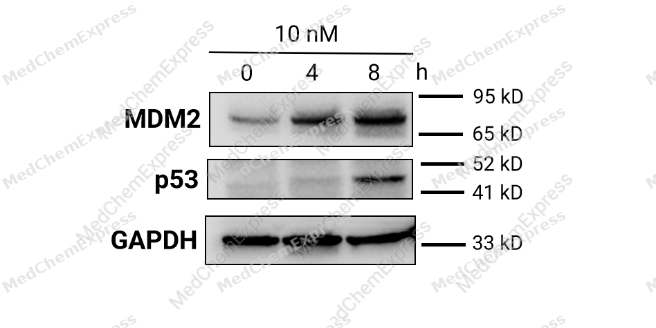 HCT-116 cells were seeded at 4*105 cells/well in a 6-well plate and were cultured overnight. Then, cells are treated with 10 nM Actinomycin D (HY-17559) for 0-8 hours to assess the expression of MDM2 and p53 via Western blotting. The results demonstrated that Actinomycin D upregulated the expression of MDM2 and p53 in a time-dependent manner. Primary antibody: MDM2 antibody (HY-P83705), p53 antibody (HY-P80938), GAPDH antibody (HY-P80137).
 -
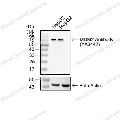 Western blot analysis of extracts from HepG2(lane2(20μg),HepG2(lane 3(40μg) , using MDM2 Antibody (HY-P80705) Mouse mAb. Proteins were transferred to a PVDF membrane and blocked with 5% non-fat milk in TBST for 2 hour at room temperature. The primary antibody (1/1000) and Loading control antibody (Beta Actin, HY-P80438, 1/10000) was used in 5% non-fat milk in TBST at 4°C overnight. Goat Anti-Mouse IgG-HRP Secondary Antibody (1/10000) was used for 1 hour at room temperature.
 -
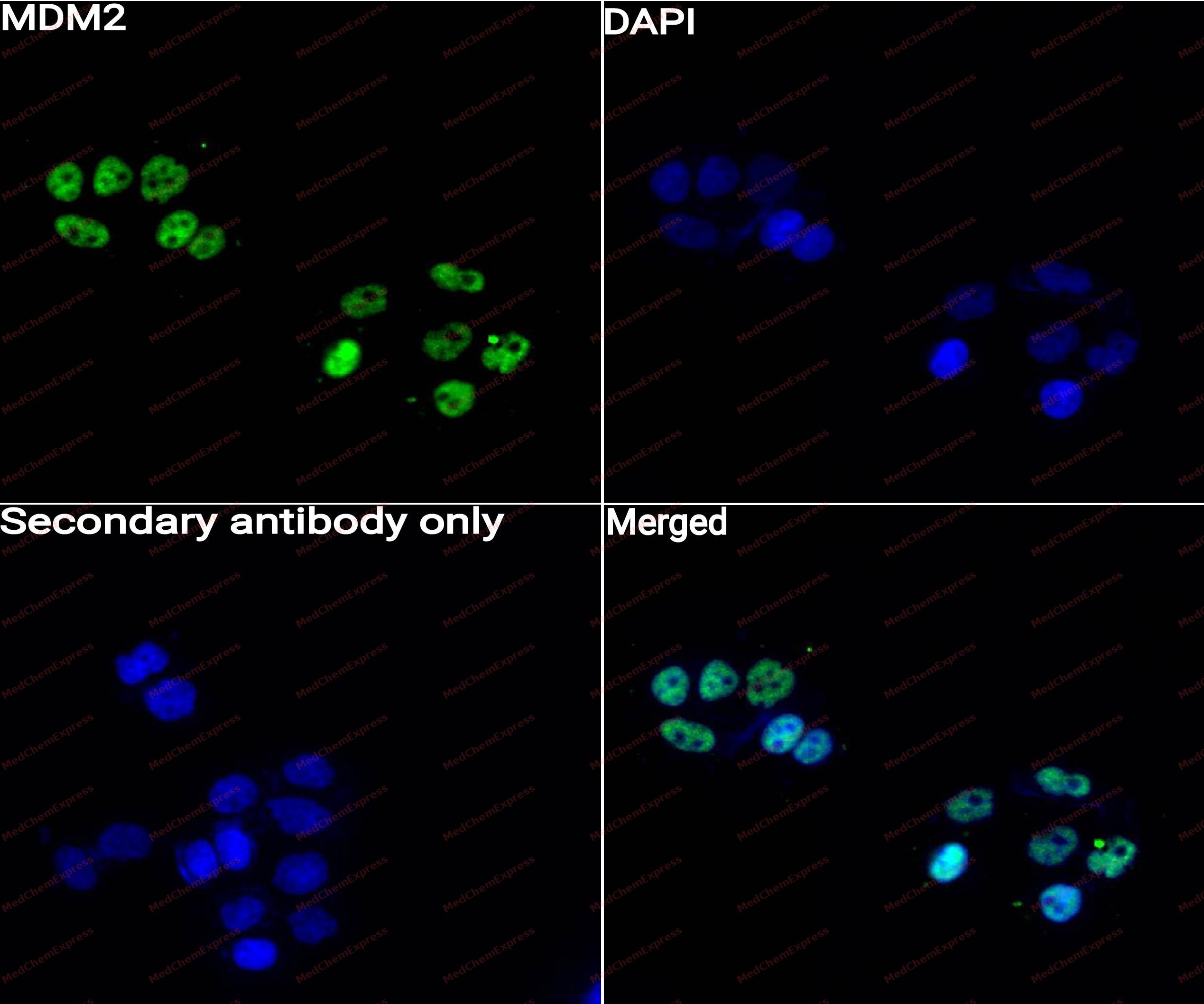 Immunocytochemistry analysis of MCF-7 cells labeling MDM2 with MDM2 Antibody (HY-P83705) at 1/50 dilution. Cells were fixed in 4% paraformaldehyde for 15 minutes at room temperature, permeabilized with 0.1% Triton X-100 for 10 minutes at room temperature, then blocked with QuickBlock™ Blocking Buffer for Immunol Staining for 10 min at room temperature. Cells were then incubated with MDM2 Antibody (HY-P83705) at 1/50 dilution in QuickBlock™ Blocking Buffer for Immunol Staining at 4 ℃. Alexa Fluor® 488-conjugated AffiniPure Goat Anti-Rabbit IgG H&L(HY-P8002, Green) was used as the secondary antibody at 1/1,000 dilution. PBS instead of the primary antibody was used as the secondary antibody only control. The Nuclear counterstain was DAPI (Blue).
 -
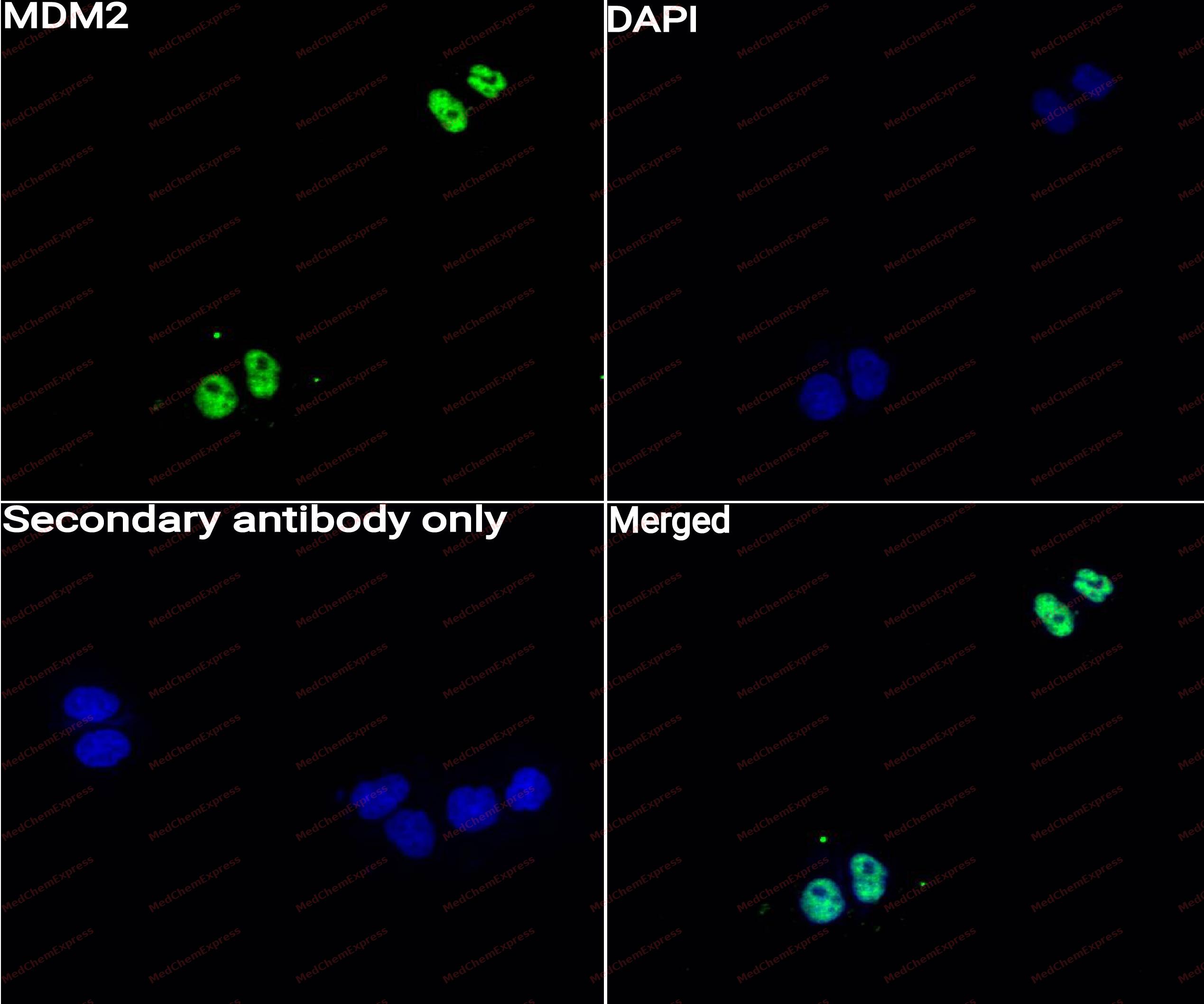 Immunocytochemistry analysis of Hela cells labeling MDM2 with MDM2 Antibody (HY-P83705) at 1/50 dilution. Cells were fixed in 4% paraformaldehyde for 15 minutes at room temperature, permeabilized with 0.1% Triton X-100 for 10 minutes at room temperature, then blocked with QuickBlock™ Blocking Buffer for Immunol Staining for 10 min at room temperature. Cells were then incubated with MDM2 Antibody (HY-P83705)at 1/50 dilution in QuickBlock™ Blocking Buffer for Immunol Staining at 4 ℃. Alexa Fluor® 488-conjugated AffiniPure Goat Anti-Rabbit IgG H&L(HY-P8002,Green) was used as the secondary antibody at 1/1,000 dilution. PBS instead of the primary antibody was used as the secondary antibody only control. The Nuclear counterstain was DAPI (Blue).


Background
-
Function
Murine double minute 2 (MDM2) is an E3 ubiquitin ligase that functions as a primary negative regulator of the tumor suppressor p53[1][2]. Mechanistically, MDM2 ubiquitinates p53, targeting it for proteasomal degradation, while also regulating additional transcription factors and post-transcriptional processes independently of p53[1][2][3]. MDM2 interacts with its homolog MDM4 (MDMX) through RING domain heterodimers, modulating protein stability and p53 activity[1][4]. Compared with MDM4, MDM2 possesses intrinsic E3 ligase activity and can degrade p53 even in the absence of MDM4, whereas MDM4 stabilizes the heterodimer complex without ubiquitin ligase function[1][4][5]. Dysregulation of MDM2 contributes to oncogenesis across multiple malignancies, including leukemia, neuroblastoma, breast cancer, and Theileria parva-induced lymphoproliferative disorders[4][6][7][8][9]. In experimental models, inhibition of MDM2 with small molecules such as RG7112, MI-63, CGM097, or XR-2 stabilizes p53, induces apoptosis, and can synergize with other pathway modulators to enhance anti-tumor activity[6][9][10][11][12]. Recent strategies exploit protein-protein interfaces, such as the MDM2-CK1α interaction, to induce targeted protein modifications and p53 activation independently of p53 status, providing tools for mechanistic studies and therapeutic design[13][14]. MDM2 inhibitors and dual MDM2/MDM4-targeting agents demonstrate isoform-specific effects, highlighting the importance of distinguishing functional contributions of MDM2 versus MDM4 in both experimental and therapeutic contexts[15][16][17].
-
Subcellular Localization
Nucleus, nucleoplasm; Cytoplasm; Nucleus, nucleolus; Nucleus
-
Expression
Tissue_specificity:They are ubiquitous. Mdm2-A, Mdm2-B, Mdm2-C, Mdm2-D, Mdm2-E, Mdm2-F, and Mdm2-G subtypes have been found in a variety of cancers, but are not present in normal tissues.
Induction:By DNA damage
Positive sample: U-2 OS treated with 10μM NUTLIN 3A for 24 hours cell lysate, HepG2 cell lysate, COS-1cell lysate, U-2 OS cells treated with 10μM NUTLIN 3A for 24 hours, human embryonal carcinoma tissue, -
Isoforms & Post-Translational Modification
Q00987 has 11 isomers: Q00987-1: 55233 Da (predicted); Q00987-2: 33140 Da (predicted); Q00987-3: 30265 Da (predicted); Q00987-4: 24467 Da (predicted); Q00987-5: 35980 Da (predicted); Q00987-6: 14689 Da (predicted); Q00987-7: 11587 Da (predicted); Q00987-8: 48488 Da (predicted); Q00987-9: 49899 Da (predicted); Q00987-10: 49249 Da (predicted); Q00987-11: 55991 Da (predicted).
Phosphorylation on Ser-166 by SGK1 activates ubiquitination of p53/TP53 (PubMed:19756449). Phosphorylated at multiple sites near the RING domain by ATM upon DNA damage; this promotes its ubiquitination and degradation, preventing p53/TP53 degradation (PubMed:10611322, PubMed:12167711, PubMed:18382127, PubMed:19816404, PubMed:26124108);Autoubiquitination leads to proteasomal degradation; resulting in p53/TP53 activation it may be regulated by SFN (PubMed:18382127, PubMed:30879903). Also ubiquitinated by TRIM13 (PubMed:21333377). ATM-phosphorylated MDM2 is ubiquitinated by the SCF(FBXO31) complex in response to genotoxic stress, promoting its degradation and p53/TP53-mediated DNA damage response (PubMed:26124108). Deubiquitinated by USP2 leads to its accumulation and increases deubiquitination and degradation of p53/TP53 (PubMed:17290220). Deubiquitinated by USP7 leading to its stabilization (PubMed:15053880) -
Subunit
Interacts with p53/TP53, TP73/p73, RBL5 and RP11. Binds specifically to RNA. Can interact with RB1, E1A-associated protein EP300 and the E2F1 transcription factor. Forms a ternary complex with p53/TP53 and WWOX. Interacts with CDKN2AIP, RFWD3, USP7, PYHIN1, and RBBP6. Interacts with ARRB1 and ARRB2. Interacts with PSMA3. Found in a trimeric complex with MDM2, MDM4 and USP2. Interacts with USP2 (via N-terminus and C-terminus). Interacts with MDM4. Part of a complex with MDM2, DAXX, RASSF1 and USP7. Part of a complex with DAXX, MDM2 and USP7. Interacts directly with DAXX and USP7. Interacts (via C-terminus) with RASSF1 isoform A (via N-terminus); the interaction is independent of TP53. Interacts with APEX1; leading to its ubiquitination and degradation. Interacts with RYBP; this inhibits ubiquitination of TP53. Identified in a complex with RYBP and p53/TP53. Also a component of the TRIM28/KAP1-MDM2-p53/TP53 complex involved in regulating p53/TP53 stabilization and activity. Binds directly both p53/TP53 and TRIM28. Component of the TRIM28/KAP1-ERBB4-MDM2 complex involved in connecting growth factor responses with DNA damage. Interacts directly with both TRIM28 and ERBB4 in the complex. Interacts with DYRK2. Interacts with IGF1R. Interacts with TRIM13; the interaction ubiquitinates MDM2 leading to its proteasomal degradation. Interacts with SNAI1; this interaction promotes SNAI1 ubiquitination. Interacts with NOTCH1 (via intracellular domain). Interacts with FHIT. Interacts with RFFL and RNF34; the interaction stabilizes MDM2. Interacts with CDK5RAP3 and CDKN2A/ARF; form a ternary complex involved in regulation of p53/TP53 (PubMed:16173922). Interacts with MTA1. Interacts with AARB2. Interacts with MTBP. Interacts with PML. Interacts with TBRG1. Interacts (via its RanBP2-type zinc finger domain) with RPL11 in the 5S RNP complex composed of 5S RNA, RPL5 and RPL11; this interaction occurs in the nucleoplasm and negatively regulates MDM2-mediated TP53 ubiquitination and degradation (PubMed:15195100, PubMed:24120868, PubMed:37291423). Interacts with ADGRB1; the interaction results in inhibition of MDM2-mediated ubiquitination and degradation of DLG4/PSD95, promoting DLG4 stability and regulating synaptic plasticity (By similarity). Interacts with RPL23A; this interaction may promote p53/TP53 polyubiquitination (PubMed:26203195). Interacts with NDUFS1 (PubMed:30879903). Interacts with MORN3; the interaction enhances the ubiquitination of p53/TP53 (PubMed:29681526)
-
SwissProt ID
-
Synonyms
ACTFS antibody; Double minute 2 protein antibody; E3 ubiquitin-protein ligase Mdm2 antibody; Hdm 2 antibody; Hdm2 antibody; HDMX antibody; MDM 2 antibody; MDM2 antibody; MDM2 oncogene E3 ubiquitin protein ligase antibody; Mdm2 p53 E3 ubiquitin protein ligase homolog antibody; Mdm2 transformed 3T3 cell double minute 2 p53 binding protein (mouse) binding protein 104kDa antibody; MDM2_HUMAN antibody; MDM2BP antibody; Mouse Double Minute 2 antibody; MTBP antibody; Murine Double Minute Chromosome 2 antibody; Oncoprotein Mdm2 antibody; p53 Binding Protein Mdm2 antibody; p53-binding protein Mdm2 antibody; Ubiquitin protein ligase E3 Mdm2 antibody; Ubiquitin protein ligase E3 Mdm2 antibody


Documentation
-
Data Sheet (265 KB)
-
SDS (254 KB)
- English - EN (254 KB)
- Français - FR (254 KB)
- Deutsch - DE (254 KB)
- Norwegian - NO (254 KB)
- Español - ES (254 KB)
- Swedish - SV (254 KB)
- Italian - IT (254 KB)
- Korean - KR (254 KB)
- Portuguese - PT (254 KB)
-
User Guide for Antibodies (1077 KB)
References
[1]. Zhu IY, et al. Structure and function of MDM2 and MDM4 in health and disease. Biochem J. 2025 Feb 17;482(4):BCJ20240757. [Content Brief]
[2]. Biderman L, et al. Mdm2 and MdmX as Regulators of Gene Expression. Genes Cancer. 2012 Mar;3(3-4):264-73. [Content Brief]
[3]. Thomasova D, et al. p53-independent roles of MDM2 in NF-κB signaling: implications for cancer therapy, wound healing, and autoimmune diseases. Neoplasia. 2012 Dec;14(12):1097-101. [Content Brief]
[4]. Lama R, et al. Small molecule MMRi62 targets MDM4 for degradation and induces leukemic cell apoptosis regardless of p53 status. Front Oncol. 2022 Aug 5;12:933446. [Content Brief]
[5]. Strachan GD, et al. A transcriptionally inactive E2F-1 targets the MDM family of proteins for proteolytic degradation. J Biol Chem. 2001 Dec 7;276(49):45677-85. [Content Brief]
[6]. Mitobe Y, et al. Antagonizing MDM2 Overexpression Induced by MDM4 Inhibitor CEP-1347 Effectively Reactivates Wild-Type p53 in Malignant Brain Tumor Cells. Cancers (Basel). 2023 Aug 30;15(17):4326. [Content Brief]
[7]. Hayashida K, et al. MDM2 regulates a novel form of incomplete neoplastic transformation of Theileria parva infected lymphocytes. Exp Mol Pathol. 2013 Feb;94(1):228-38. [Content Brief]
[8]. Qin JJ, et al. Experimental Therapy of Advanced Breast Cancer: Targeting NFAT1-MDM2-p53 Pathway. Prog Mol Biol Transl Sci. 2017;151:195-216. [Content Brief]
[9]. Maser T, et al. The MDM2 inhibitor CGM097 combined with the BET inhibitor OTX015 induces cell death and inhibits tumor growth in models of neuroblastoma. Cancer Med. 2020 Nov;9(21):8144-8158. [Content Brief]
[10]. Daniele S, et al. Long lasting inhibition of Mdm2-p53 interaction potentiates mesenchymal stem cell differentiation into osteoblasts. Biochim Biophys Acta Mol Cell Res. 2019 May;1866(5):737-749. [Content Brief]
[11]. Li Y, et al. Nonsense-mediated mRNA decay inhibition synergizes with MDM2 inhibition to suppress TP53 wild-type cancer cells in p53 isoform-dependent manner. Cell Death Discov. 2022;8(1):402. [Content Brief]
[12]. Gu D, et al. Inhibition of the MDM2 E3 Ligase induces apoptosis and autophagy in wild-type and mutant p53 models of multiple myeloma, and acts synergistically with ABT-737. PLoS One. 2014 Sep 2;9(9):e103015. [Content Brief]
[14]. Huart AS, et al. Exploiting the MDM2-CK1α protein-protein interface to develop novel biologics that induce UBL-kinase-modification and inhibit cell growth. PLoS One. 2012;7(8):e43391. [Content Brief]
[15]. Surmiak E, et al. A Unique Mdm2-Binding Mode of the 3-Pyrrolin-2-one- and 2-Furanone-Based Antagonists of the p53-Mdm2 Interaction. ACS Chem Biol. 2016 Dec 16;11(12):3310-3318. [Content Brief]
[17]. Jiang L, et al. Protoporphyrin IX is a dual inhibitor of p53/MDM2 and p53/MDM4 interactions and induces apoptosis in B-cell chronic lymphocytic leukemia cells. Cell Death Discov. 2019 Mar 11;5:77. [Content Brief]

 Powered by Bioz
