



KRAS inhibitor-9

Based on 1 Customer Validation
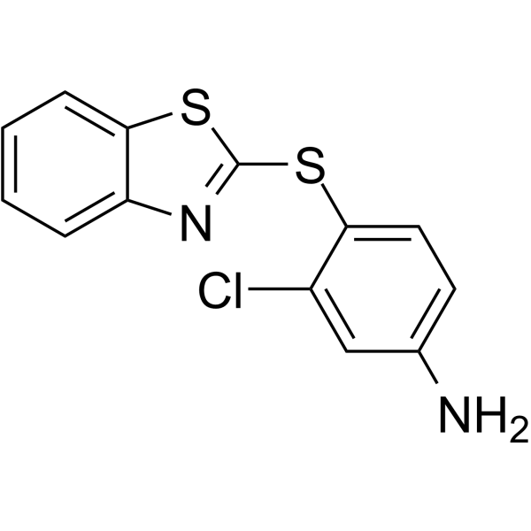
KRAS inhibitor-9, a potent KRAS inhibitor (Kd=92 μM), blocks the formation of GTP-KRAS and downstream activation of KRAS. KRAS inhibitor-9 binds to KRAS G12D, KRAS G12C and KRAS Q61H protein with a moderate binding affinity. KRAS inhibitor-9 causes G2/M cell cycle arrest and induces apoptosis. KRAS inhibitor-9 selectively inhibits the proliferation of NSCLC cells with KRAS mutation but not normal lung cells.
Nos produits utilisent uniquement pour la recherche. Nous ne vendons pas aux patients.
- Pureté : 99.85%
- CAS No.: 300809-71-6
- Formule: C13H9ClN2S2
- Masse moléculaire:292.81
-
Stockage:Powder -20°C, 3 years , 4°C, 2 years ; In solvent -80°C, 2 years , -20°C, 1 year

To place orders, for customer services and technical support, please contact: MedChemExpress USA
Tel: 609-228-6898 E-mail: [email protected] [email protected]


-
 Activité biologique
-
Chemical Information
-
Solvant et solubilité
- Protocole
- Pureté et documentation
- Références
-
 Help & FAQs


-
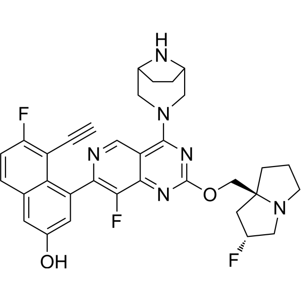
HY-134813
MRTX1133KRAS G12D Inhibitor -
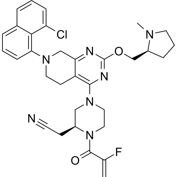
HY-130149
AdagrasibKRAS G12C Inhibitor -
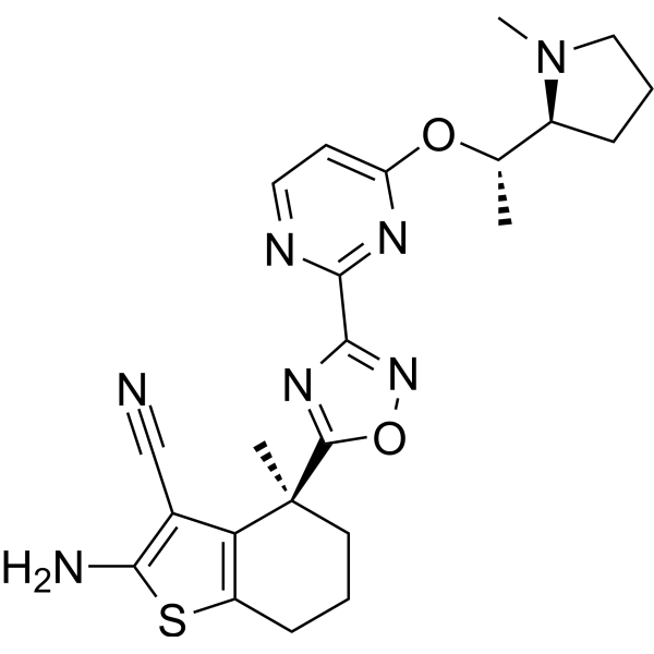
HY-153724
BI-2865Pan-KRAS Inhibitor -
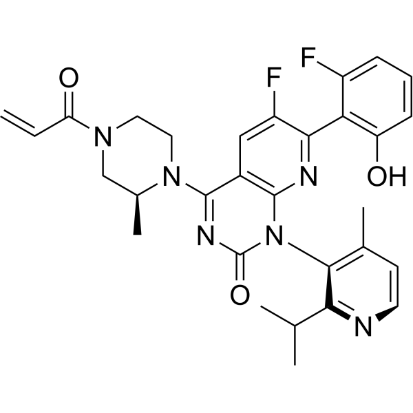
HY-114277
SotorasibKRAS G12C Inhibitor -
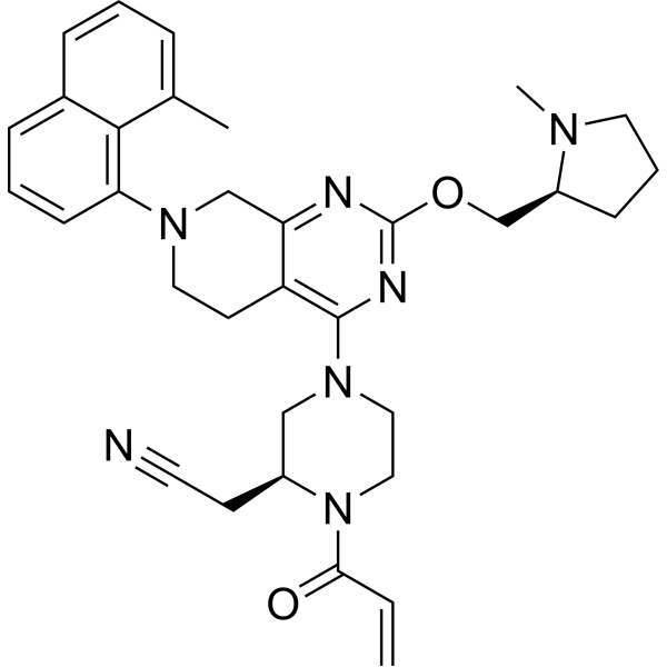
HY-114436
MRTX-1257KRAS G12C Inhibitor -
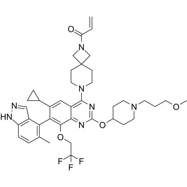
HY-132966
ASP2453Ras Inhibitor -
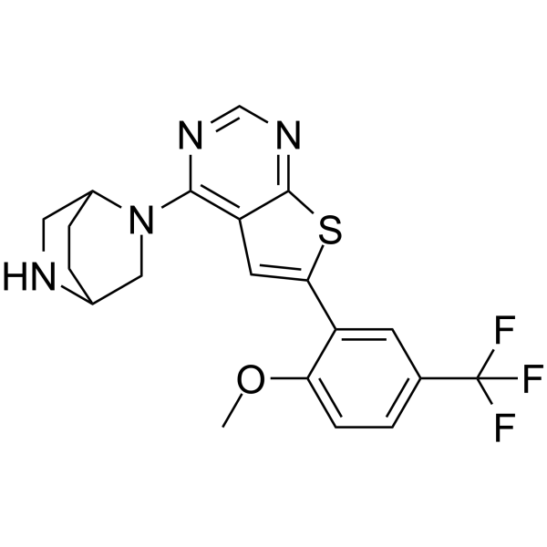
HY-144661
KRAS G12D inhibitor 14KRAS G12D Inhibitor -
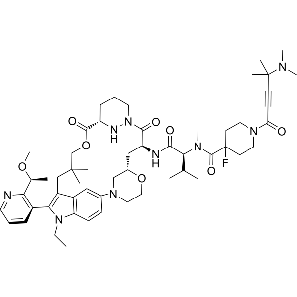
HY-153346
ElironrasibKRAS-G12C(ON) Inhibitor -
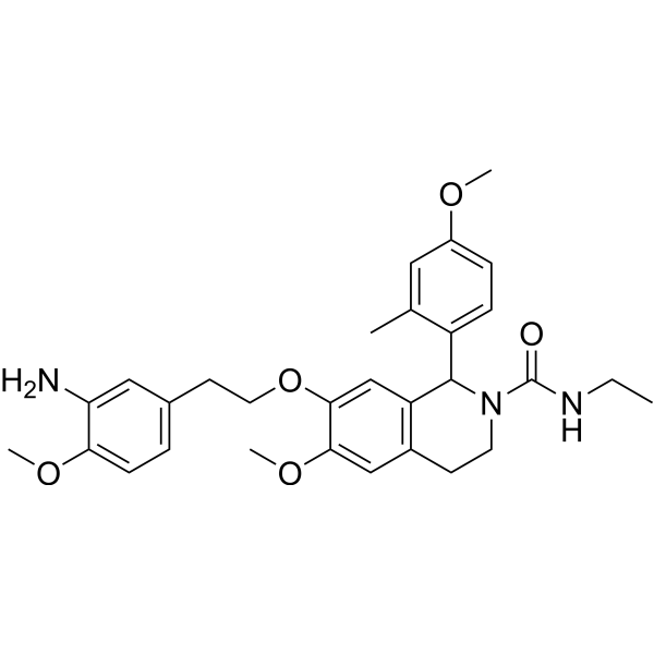
HY-138295
KRAS inhibitor-10KRAS Inhibitor -
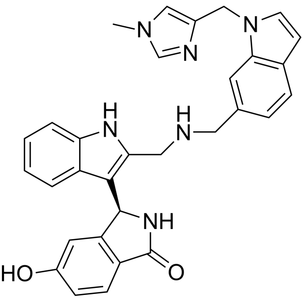
HY-126247
BI-2852KRAS Switch I/II Pocket Inhibitor -
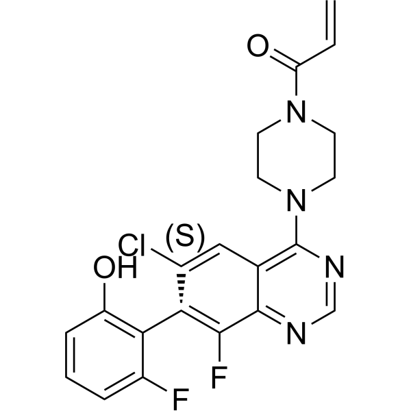
HY-U00418
ARS-1620 -
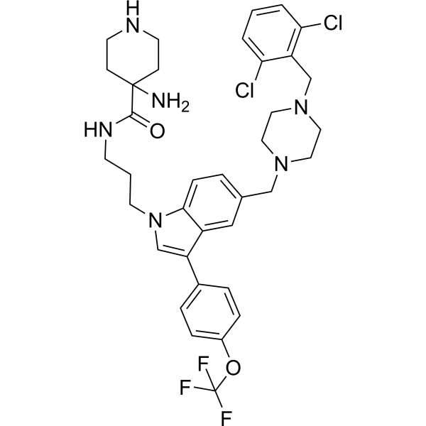
HY-101295
Pan-RAS-IN-1Ras Inhibitor


Activité biologique
Description
In Vitro
KRAS inhibitor-9 bound to KRASG12D, KRAS G12C and KRAS Q61H protein with a moderate binding affinity of -5.38, -5.41, and -3.97 kcal/mol, respectively[1].
KRAS inhibitor-9 (0-100 μM) shows strong inhibition selectivity in NSCLC cells with IC50s ranging from 39.56 to 66.02 μM for H2122, H358 and H460 cells (at 72 hours)[1].
KRAS inhibitor-9 (0-100 μM; 24 hours) blocks GTP-KRAS formation in H2122, H358 and H460 cells[1].
KRAS inhibitor-9 (25-100 μM; 48 hours) inhibits the activation of KRAS downstream signaling pathway[1].
KRAS inhibitor-9 (0-100 μM; 24-72 hours) induces cell cycle arrest and apoptosis in NSCLC[1].
MedChemExpress (MCE) has not independently confirmed the accuracy of these methods. They are for reference only.
-
Cell Line:H2122 (KRAS G12C), H358 (KRAS G12C) and H460 (KRAS Q61H) cell lines
-
Concentration:0, 25, 50, 100 μM
-
Incubation Time:24, 48, and 72 hours
-
Result:Inhibited three NSCLC cell lines in a dose- and time-dependent manner, but not in normal lung fibroblast cell line CCD-19Lu.
-
Cell Line:H2122, H358 and H460 cells
-
Concentration:0, 25, 50, 100 μM
-
Incubation Time:48 hours
-
Result:Reduces the levels of phosphorylation of CRAF and AKT in a dose-dependent manner in H2122, H358 and H460 cells.
-
Cell Line:H2122, H358, H460 cells
-
Concentration:0, 25, 50, 100 μM
-
Incubation Time:24-72 hours
-
Result:Significantly decreased in G0/G1 phase while remarkably increased in G2/M phase after 24 hours and induced a significantly increased apoptosis for 48h in NSCLC cell lines.
- Detection of Apoptosis
- TUNEL staining for apoptotic DNA fragmentation
- Flow cytometric DNA-content cell-cycle staining
- Annexin V plus membrane-impermeant dye apoptosis staining
- BrdU Incorporation Assay
- Apoptosis Solutions
- Protocol for Cell Cycle
- Genotoxicity/Mutagenicity Study
- Cell Viability Determination by MTT Colorimetric Assay


Chemical Information
-
CAS No. 300809-71-6
-
Appearance Solid
-
Masse moléculaire 292.81
-
Formule C13H9ClN2S2
-
Color White to off-white
-
SMILES
NC1=CC=C(SC2=NC3=CC=CC=C3S2)C(Cl)=C1
-
Livraison
Room temperature in continental US; may vary elsewhere.
-
Stockage
Powder -20°C 3 years 4°C 2 years In solvent -80°C 2 years -20°C 1 year


Solvant et solubilité
In Vitro:
DMSO : 250 mg/mL (853.80 mM; Need ultrasonic; Hygroscopic DMSO has a significant impact on the solubility of product, please use newly opened DMSO)
Please refer to the solubility information to select the appropriate solvent. Once prepared, please aliquot and store the solution to prevent product inactivation from repeated freeze-thaw cycles.
Storage method and period of stock solution: -80°C, 2 years; -20°C, 1 year. When stored at -80°C, please use it within 2 years. When stored at -20°C, please use it within 1 year.
Please refer to the solubility information to select the appropriate solvent. Once prepared, please aliquot and store the solution to prevent product inactivation from repeated freeze-thaw cycles.
Storage method and period of stock solution: -80°C, 2 years; -20°C, 1 year. When stored at -80°C, please use it within 2 years. When stored at -20°C, please use it within 1 year.
Concentration (start) × Volume (start) = Concentration (final) × Volume (final)
In Vivo:
Select the appropriate dissolution method based on your experimental animal and administration route.
- For the following dissolution methods, please ensure to first prepare a clear stock solution using an In Vitro approach and then sequentially add co-solvents:
- To ensure reliable experimental results, the clarified stock solution can be appropriately stored based on storage conditions. As for the working solution for In Vivo experiments, it is recommended to prepare freshly and use it on the same day.
- The percentages shown for the solvents indicate their volumetric ratio in the final prepared solution. If precipitation or phase separation occurs during preparation, heat and/or sonication can be used to aid dissolution.
Add each solvent one by one: 10% DMSO 90% (20% SBE-β-CD in Saline)
Solubility: ≥ 2.08 mg/mL (7.10 mM); Clear solution
This protocol yields a clear solution of ≥ 2.08 mg/mL (saturation unknown).
Taking 1 mL working solution as an example, add 100 μL DMSO stock solution (20.8 mg/mL) to 900 μL 20% SBE-β-CD in Saline, and mix evenly.
Preparation of 20% SBE-β-CD in Saline (4°C, storage for one week): 2 g SBE-β-CD powder is dissolved in 10 mL Saline, completely dissolve until clear.
In Vivo Dissolution Calculator
Please enter the basic information of animal experiments:
-
-
-
-
Recommended: Prepare an additional quantity of animals to account for potential losses during experiments.
Please enter your animal formula composition:
-
%DMSO +
Recommended: Keep the proportion of DMSO in working solution below 2% if your animal is weak.
-
%+
-
+%Tween-80 + +
-
%Saline +
The co-solvents required include: DMSO, . All of co-solvents are available by MedChemExpress (MCE). , Tween 80. All of co-solvents are available by MedChemExpress (MCE).
Working solution concentration: 0.22 mg/mL
Method for preparing stock solution: mg drug dissolved in μL DMSO. Stock solution concentration: mg/mL.
1. Take μL DMSO stock solution;
2. Add μL .
μL , mix evenly;
3. Then add μL Tween 80, mix evenly;
4. Then add μL
Please ensure that the stock solution in the first step is dissolved to a clear state, and add co-solvents in sequence. You can use ultrasonic heating (ultrasonic cleaner, recommended frequency 20-40 kHz), vortexing, etc. to assist dissolution.


Pureté et documentation
-
Fiche technique (275 KB)
-
SDS (393 KB)
- English - EN (393 KB)
- Français - FR (393 KB)
- Deutsch - DE (393 KB)
- Norwegian - NO (393 KB)
- Español - ES (393 KB)
- Swedish - SV (393 KB)
- Italian - IT (393 KB)
- Korean - KR (393 KB)
- Portuguese - PT (393 KB)
-
Instruction de manipulation (2659 KB)
Références


Complete Stock Solution Preparation Table
Please refer to the solubility information to select the appropriate solvent. Once prepared, please aliquot and store the solution to prevent product inactivation from repeated freeze-thaw cycles.
Storage method and period of stock solution: -80°C, 2 years; -20°C, 1 year. When stored at -80°C, please use it within 2 years. When stored at -20°C, please use it within 1 year.
| Optional Solvent | Concentration Solvent Mass | 1 mg | 5 mg | 10 mg | 25 mg |
|---|---|---|---|---|---|
| DMSO | 1 mM | 3.4152 mL | 17.0759 mL | 34.1518 mL | 85.3796 mL |
| 5 mM | 0.6830 mL | 3.4152 mL | 6.8304 mL | 17.0759 mL | |
| 10 mM | 0.3415 mL | 1.7076 mL | 3.4152 mL | 8.5380 mL | |
| 15 mM | 0.2277 mL | 1.1384 mL | 2.2768 mL | 5.6920 mL | |
| 20 mM | 0.1708 mL | 0.8538 mL | 1.7076 mL | 4.2690 mL | |
| 25 mM | 0.1366 mL | 0.6830 mL | 1.3661 mL | 3.4152 mL | |
| 30 mM | 0.1138 mL | 0.5692 mL | 1.1384 mL | 2.8460 mL | |
| 40 mM | 0.0854 mL | 0.4269 mL | 0.8538 mL | 2.1345 mL | |
| 50 mM | 0.0683 mL | 0.3415 mL | 0.6830 mL | 1.7076 mL | |
| 60 mM | 0.0569 mL | 0.2846 mL | 0.5692 mL | 1.4230 mL | |
| 80 mM | 0.0427 mL | 0.2134 mL | 0.4269 mL | 1.0672 mL | |
| 100 mM | 0.0342 mL | 0.1708 mL | 0.3415 mL | 0.8538 mL |


KRAS inhibitor-9 Related Classifications
HY-137497 Related Classifications
 Powered by Bioz