



LD4172

Based on 1 Customer Validation
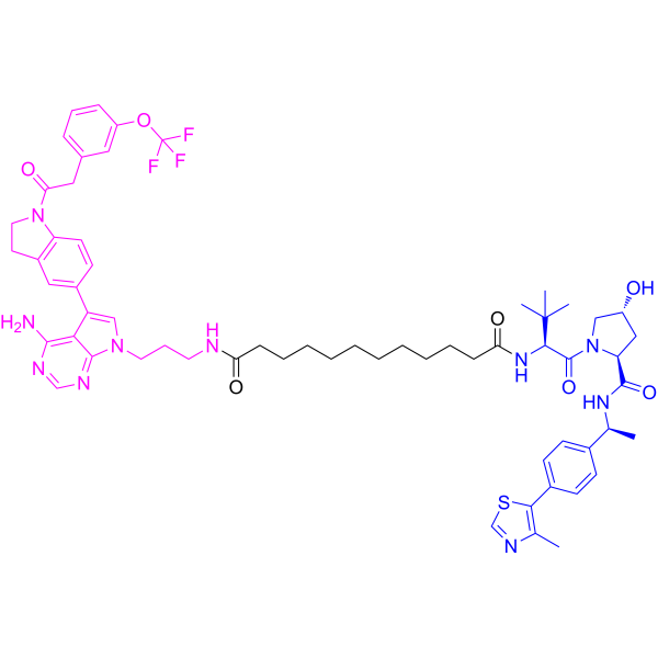
LD4172 is a selective RIPK1 PROTAC degrader with a Ki of 4.8 nM. LD4172 induces RIPK1 protein degradation via ternary complex formation with RIPK1 and VHL E3 ligase, driving ubiquitination and proteasomal breakdown. LD4172 abrogates TNF-induced classical NF-κB signaling in TRAF2-deficient cells, impairing IκBα phosphorylation and degradation, and reducing IL-8 production. LD4172 induces apoptosis and immunogenic cell death in tumor cells, enhances tumor-infiltrating lymphocyte responses, and sensitizes tumors to anti-PD1 therapy. LD4172 acts as a chemical probe for investigating RIPK1 scaffolding functions. LD4172 can be used for the research of melanoma, colon cancer.
(Pink: RIPK1 Target protein ligand; Blue: VHL ligand (HY-112078); Black: linker (HY-W012241)).
For research use only. We do not sell to patients.
- Purity : 98.32%
- CAS No.: 2782022-40-4
- Formula: C61H75F3N10O7S
- Molecular Weight:1149.37
-
Storage:Powder -20°C, 3 years , 4°C, 2 years ; In solvent -80°C, 6 months , -20°C, 1 month

To place orders, for customer services and technical support, please contact: MedChemExpress USA
Tel: 609-228-6898 E-mail: [email protected] [email protected]


-
 Biological Activity
-
Chemical Information
-
Solvent & Solubility
- Protocols
- Purity & Documentation
- References
-
 Help & FAQs


All PROTACs Isoforms
More

Biological Activity
Description
IC50 & Target
|
hRIP1 4.8 nM (Ki) |
VHL |
In Vitro
LD4172 (1 μM; 24 h) efficiently degrades RIPK1 in HCT116-PIK3CAmut, HeLa-RIPK3, and HT29 cells, and abrogates TNF-induced classical NFκB signaling in TRAF2-deficient variants of these cell lines, while leaving signaling intact in parental cells[1].
LD4172 potently binds to recombinant human RIPK1 protein with a biochemical Ki of 4.8 nM[2].
LD4172 engages intracellular RIPK1 in HEK293T cells with an apparent Ki of 1.9 μM[2].
LD4172 (10-100 nM; 4 h with 30 min pre-incubation with MG132) dose-dependently forms a ternary complex with RIPK1 and VHL in HEK293T cells[2].
LD4172 (0.016-10 μM; 24 h) potently degrades RIPK1 in a panel of human and mouse cell lines, with DC50 values of 4-400 nM and >90% degradation at 1 μM for 24 h in Jurkat and B16F10 cells[2].
LD4172 (200 nM; 6 h) induces highly specific degradation of RIPK1 in MDA-MB-231 cells, with no detectable degradation of other proteins including off-target kinases of the warhead[2].
LD4172 (1 μM; 24 h, co-treated with 100 ng/mL TNF-α) inhibits TNF-α-induced NF-κB activity in B16F10 mouse melanoma cells[2].
LD4172 (1 μM; 72 h, co-treated with 100 ng/mL TNF-α where indicated) sensitizes B16F10 mouse melanoma cells to TNF-α-induced apoptosis and immunogenic cell death[2].
MedChemExpress (MCE) has not independently confirmed the accuracy of these methods. They are for reference only.
-
Cell Line:HCT116-PIK3CAmut, HeLa-RIPK3, HT29 cells, and their TRAF2-deficient variants
-
Concentration:1 μM
-
Incubation Time:24 h
-
Result:Reduced RIPK1 expression efficiently in all tested cell lines and their TRAF2-deficient variants. Left TNF-induced NFκB signaling (measured by IκBα phosphorylation/degradation and IL-8 production) largely unaffected in parental cells. Abrogated TNF-induced NFκB signaling practically in TRAF2-deficient cells.
-
Cell Line:Jurkat, B16F10 cells
-
Concentration:1 μM
-
Incubation Time:0, 2, 4, 8, 12, 24, 48 h
-
Result:Induced >90% RIPK1 degradation within 2 h in Jurkat cells and 4 h in B16F10 cells. Initiated RIPK1 resynthesis 4 h post-washout, with resynthesis half-lives of ~48 h in Jurkat cells and ~24 h in B16F10 cells.
-
Cell Line:B16F10 mouse melanoma cells
-
Concentration:1 μM (co-treated with 100 ng/mL TNF-α where indicated)
-
Incubation Time:72 h
-
Result:Combined with TNF-α to induce significant apoptosis, evidenced by increased Annexin V/PI positivity, cleaved caspase 3/7 staining, PI uptake, and elevated levels of cleaved caspase-3, cleaved caspase-7, and cleaved PARP. Combined with TNF-α to trigger immunogenic cell death, marked by increased extracellular ATP, release of HMGB1, and downregulation of intracellular calreticulin.
Parmacokinetics
In Vivo
LD4172 (20 mg/kg; i.p.; daily) synergizes with anti-PD1 to improve antitumor efficacy in female C57BL/6J mice bearing the MC38 colon carcinoma model[2].
MedChemExpress (MCE) has not independently confirmed the accuracy of these methods. They are for reference only.
-
Animal Model:C57BL/6J (female, 7 weeks old) injected with B16F10 cells[2]
-
Dosage:20 mg/kg
-
Administration:i.p.; daily
-
Result:Induced a 60% reduction in RIPK1 levels in tumors; less than 50% RIPK1 degradation was observed in the spleen, and no significant degradation was seen in lymph nodes, PBMCs, lungs, or bone marrow.
-
Animal Model:C57BL/6J (female, 7 weeks old) injected with MC38 cells[2]
-
Dosage:20 mg/kg
-
Administration:i.p.; daily
-
Result:Sensitized MC38 tumors to anti-PD1 therapy, enhancing the antitumor response relative to single-agent treatments.
- Kinase activity and phosphorylation assays
- Western Blot
- Detection of Apoptosis
- TUNEL staining for apoptotic DNA fragmentation
- Cell Cytotoxicity Assay
- Mammalian live/dead viability and cytotoxicity staining
- Annexin V plus membrane-impermeant dye apoptosis staining
- Apoptosis Solutions
- Research Protocol for Inflammation-related Diseases
- Protocol for Kinase activity and phosphorylation assays


Chemical Information
-
CAS No. 2782022-40-4
-
Appearance Solid
-
Molecular Weight 1149.37
-
Formula C61H75F3N10O7S
-
Color White to off-white
-
SMILES
O=C(CC1=CC=CC(OC(F)(F)F)=C1)N2CCC3=C2C=CC(C4=CN(C5=NC=NC(N)=C45)CCCNC(CCCCCCCCCCC(N[C@H](C(N6C[C@@H](C[C@H]6C(N[C@H](C7=CC=C(C=C7)C8=C(N=CS8)C)C)=O)O)=O)C(C)(C)C)=O)=O)=C3
-
Shipping
Room temperature in continental US; may vary elsewhere.
-
Storage
Powder -20°C 3 years 4°C 2 years In solvent -80°C 6 months -20°C 1 month


Solvent & Solubility
In Vitro:
DMSO : ≥ 100 mg/mL (87.00 mM; Hygroscopic DMSO has a significant impact on the solubility of product, please use newly opened DMSO)
* "≥" means soluble, but saturation unknown.
Please refer to the solubility information to select the appropriate solvent. Once prepared, please aliquot and store the solution to prevent product inactivation from repeated freeze-thaw cycles.
Storage method and period of stock solution: -80°C, 6 months; -20°C, 1 month. When stored at -80°C, please use it within 6 months. When stored at -20°C, please use it within 1 month.
Please refer to the solubility information to select the appropriate solvent. Once prepared, please aliquot and store the solution to prevent product inactivation from repeated freeze-thaw cycles.
Storage method and period of stock solution: -80°C, 6 months; -20°C, 1 month. When stored at -80°C, please use it within 6 months. When stored at -20°C, please use it within 1 month.
Concentration (start) × Volume (start) = Concentration (final) × Volume (final)


Purity & Documentation
-
Data Sheet (281 KB)
-
SDS (254 KB)
- English - EN (254 KB)
- Français - FR (254 KB)
- Deutsch - DE (254 KB)
- Norwegian - NO (254 KB)
- Español - ES (254 KB)
- Swedish - SV (254 KB)
- Italian - IT (254 KB)
- Korean - KR (254 KB)
- Portuguese - PT (254 KB)
-
Handling Instructions (2659 KB)
References
[1]. Wagner J, et al. TRAF2 and RIPK1 redundantly mediate classical NFκB signaling by TNFR1 and CD95-type death receptors. Cell Death Dis. 2025;16(1):35. Published 2025 Jan 21. [Content Brief]
[2]. Yu X, et al. Development of a RIPK1 degrader to enhance antitumor immunity. Nat Commun. 2024;15(1):10683. Published 2024 Dec 16. [Content Brief]


Complete Stock Solution Preparation Table
Please refer to the solubility information to select the appropriate solvent. Once prepared, please aliquot and store the solution to prevent product inactivation from repeated freeze-thaw cycles.
Storage method and period of stock solution: -80°C, 6 months; -20°C, 1 month. When stored at -80°C, please use it within 6 months. When stored at -20°C, please use it within 1 month.
| Optional Solvent | Concentration Solvent Mass | 1 mg | 5 mg | 10 mg | 25 mg |
|---|---|---|---|---|---|
| DMSO | 1 mM | 0.8700 mL | 4.3502 mL | 8.7004 mL | 21.7510 mL |
| 5 mM | 0.1740 mL | 0.8700 mL | 1.7401 mL | 4.3502 mL | |
| 10 mM | 0.0870 mL | 0.4350 mL | 0.8700 mL | 2.1751 mL | |
| 15 mM | 0.0580 mL | 0.2900 mL | 0.5800 mL | 1.4501 mL | |
| 20 mM | 0.0435 mL | 0.2175 mL | 0.4350 mL | 1.0876 mL | |
| 25 mM | 0.0348 mL | 0.1740 mL | 0.3480 mL | 0.8700 mL | |
| 30 mM | 0.0290 mL | 0.1450 mL | 0.2900 mL | 0.7250 mL | |
| 40 mM | 0.0218 mL | 0.1088 mL | 0.2175 mL | 0.5438 mL | |
| 50 mM | 0.0174 mL | 0.0870 mL | 0.1740 mL | 0.4350 mL | |
| 60 mM | 0.0145 mL | 0.0725 mL | 0.1450 mL | 0.3625 mL | |
| 80 mM | 0.0109 mL | 0.0544 mL | 0.1088 mL | 0.2719 mL |


LD4172 Related Classifications
HY-153598 Related Classifications
 Powered by Bioz