
9 Results for "

cytoplasmic staining

" in MedChemExpress (MCE) Product Catalog:
Products (9)

9 Results for "cytoplasmic staining" in MCE Product Catalog:

2
2 Cited Publications
Cat. No.: HY-D0944
CAS No.: 51811-82-6
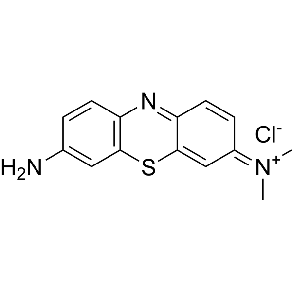
Giemsa stain is a composite dye composed of methylene azure, methylene blue, eosin and other components. Giemsa stain stains chromatin, nuclear membranes, specific cytoplasmic components and microorganisms. Giemsa stain is used in research on cytological and parasitological staining .
loading...
    loading...
1
1 Cited Publications
Cat. No.: HY-126367
CAS No.: 5141-20-8
Purity:  ≥75.0%
Synonyms: Acid Green 5
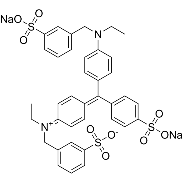
Light green SF yellowish (Acid Green 5) is a water-soluble triarylmethane dye. Light green SF yellowish serves as a histological stain that selectively labels mitochondria, collagen, and cartilage, while being an essential component of Papanicolaou staining. Light green SF yellowish is commonly used as a cytoplasmic counterstain for nuclear stains, and is applied in Masson's trichrome staining for collagen fibers, Pap staining, and cytological polychromatic staining in histopathology. Light green SF yellowish also induces growth inhibition and local fibrosarcomas in rats and exerts mild pulmonary tumorigenicity in mice .
loading...
    loading...
Cat. No.: HY-W088068
CAS No.: 68988-92-1
Wright's stain is a composite cell stain that mainly binds to intracellular nucleic acids, proteins and other components through thiazine dyes (such as methylene blue) and eosin. Wright's stain is pH-dependent (optimal pH 6.4-6.7) and achieves cell morphology resolution by differentially staining the cytoplasm and nucleus. Under alkaline conditions, thiazine dyes bind to nucleic acids to form purple, and acidic eosin binds to cytoplasmic proteins to form red, which can form contrasting cell morphological features. Wright's stain can clearly display the fine structures of blood cells and bone marrow cells (such as nuclear chromatin and granules) and quickly evaluate cell morphological abnormalities .
loading...
    loading...
Cat. No.: HY-W110793
CAS No.: 60-10-6
Synonyms: Diphenylthiocarbazone
Research Areas:  

Metabolic Disease

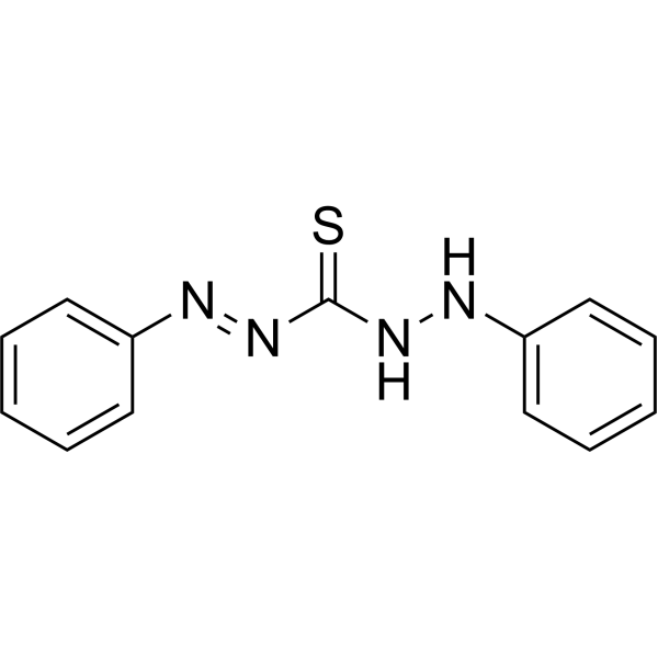
Dithizone can be toxic to pancreatic islet cells and cause experimental diabetes. Dithizone can be used for in vitro in vivo staining of transplanted islets and cytoplasmic granules of bone marrow cells. Dithizone can be used as a chelating agent and colorimetric indicator for the detection and extraction of various heavy metals, including lead and mercury, in analytical and biochemical applications. Dithizone forms stable colored complexes with metal ions and can be quantitatively analyzed by methods such as spectrophotometry .
loading...
    loading...
Cat. No.: HY-D0920
CAS No.: 166196-17-4
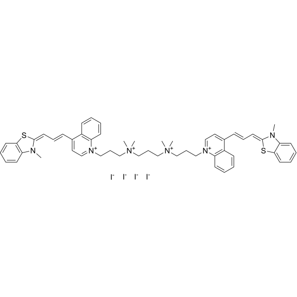
TOTO-3 is a cyanine nucleic acid intercalating fluorescent probe that binds to both RNA and DNA (Ex=642 nm, Em=660 nm). TOTO-3 cannot enter intact cells, but can penetrate cells with permeabilized plasma membranes, staining cytoplasmic RNA and nucleoli to distinguish intact cells from damaged ones. TOTO-3 is taken up by macrophages in an ultrasound-independent manner. TOTO-3 accumulates in tumor cells via ultrasound-mediated enhancement of plasma membrane permeability. TOTO-3 supports optical imaging for real-time assessment of plasma membrane integrity, nucleic acid detection, and monitoring of ultrasound-mediated intracellular delivery processes. TOTO-3 is applicable to studies related to rectal cancer .
loading...
    loading...
Cat. No.: HY-W342930
CAS No.: 5858-33-3
Synonyms: Acid Red 17
Bordeaux Red (Acid Red 17) is a redox indicator that can be used for cytoplasm staining, such as spleen, testis, and liver slice staining. Bordeaux Red is a kind of biological materials or organic compounds that are widely used in life science research .
loading...
    loading...
Cat. No.: HY-DY2020
Target:  

Fluorescent Dye

Research Areas:  

Others

Neutral Red Staining Solution (for Live Cells) is a vital stain used for vital staining of peripheral blood cells, and it can also be used for staining other living cells or tissues. Neutral Red Staining Solution (for Live Cells) accumulates in cytoplasmic vacuoles, lysosomes of living cells, as well as plant/fungal vacuoles. It binds to anionic sites, suberin, lignin, cell walls, DNA and phenolic substances, and can intercalate into DNA. Neutral Red Staining Solution (for Live Cells) stains the cytoplasm of specific phytoplankton and estuarine zooplankton, while dead cells are not stained, enabling viability determination of such organisms .
loading...
    loading...
Cat. No.: HY-DY2025
Target:  

Fluorescent Dye

Research Areas:  

Neurological Disease

Luxol Fast Blue Staining Solution is a copper phthalocyanine-based histological dye that specifically binds to lipid-rich structures such as myelin sheaths and mitochondria, staining them blue-green. Luxol Fast Blue Staining Solution is suitable for displaying myelin sheaths in neural tissues, staining mitochondria in tissues such as kidney/liver, and staining sections for phase contrast microscopy. It is often used in combination with PAS, Phloxine or Cresyl Violet to enhance structural contrast .
loading...
    loading...
Cat. No.: HY-DY2027
Target:  

Fluorescent Dye

Research Areas:  

Others

Nissl Staining Solution (Thionin Method) is a neural tissue staining solution with thionin as the core component, which stains Nissl bodies purplish blue and nuclear chromatin blue. Nissl Staining Solution (Thionin Method) relies on positively charged thionin molecules to specifically bind to the negatively charged nucleic acid components in the rough endoplasmic reticulum inside Nissl bodies, and achieves differential staining by regulating pH through a buffer system. Nissl Staining Solution (Thionin Method) has pH-dependent staining selectivity; it exhibits the highest staining specificity for neuronal Nissl bodies under pH 3.65 conditions, and can efficiently label glial processes under pH 4.6 conditions .
loading...
    loading...

