



Soquelitinib
Based on 3 publication(s) in Google Scholar
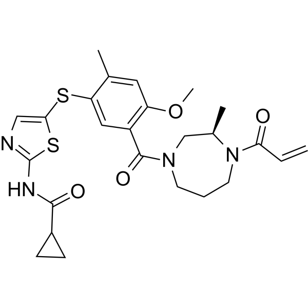
Soquelitinib (CPI-818) is an orally active and highly selective covalent interleukin-2-inducible kinase (ITK) inhibitor. Soquelitinib is active in six different models of T cell-mediated inflammatory and immune disease, including acute and chronic asthma, pulmonary fibrosis, systemic sclerosis (scleroderma), psoriasis, and acute graft versus host disease with Th2 cytokine product inhibition. Soquelitinib increases tumor infiltration of normal CD8+ cells that possess enhanced T effector function.
Nur für Forschungszwecke. Wir verkaufen nicht an Patienten.
- Reinheit : 99.98%
- CAS. Nr.: 2226636-04-8
- Formel: C25H30N4O4S2
- Molecular Weight:514.66
-
Speicherung:Powder -20°C, 3 years , 4°C, 2 years ; In solvent -80°C, 6 months , -20°C, 1 month

To place orders, for customer services and technical support, please contact: MedChemExpress USA
Tel: 609-228-6898 E-mail: [email protected] [email protected]


-
 Biologische Aktivität
-
Chemical Information
-
Lösungsmittel & Löslichkeit
- Protokoll
- Reinheit & Dokumentation
- Verweise
-
 Help & FAQs


-
HY-10997
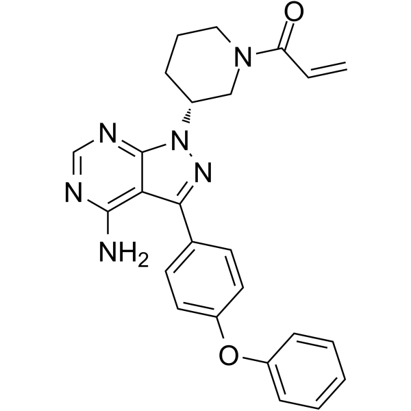
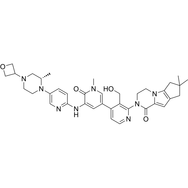
IbrutinibBtk Inhibitor -
HY-11092
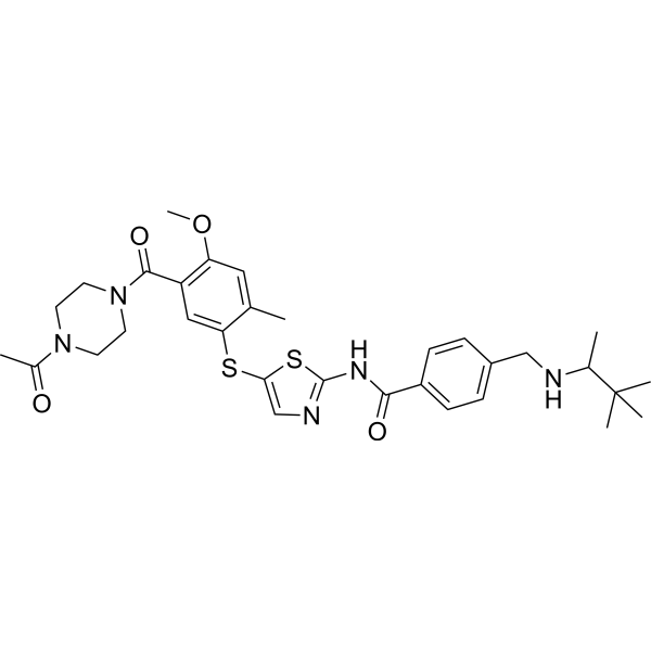
BMS-509744ITK Inhibitor -
HY-12680
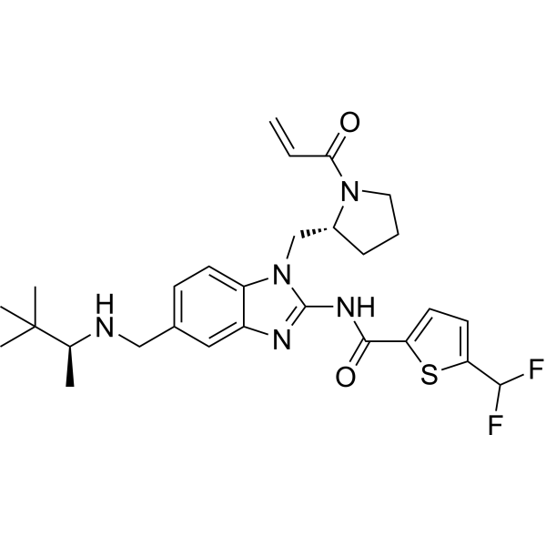
PRN694ITK/RLK Inhibitor -
HY-19834
FenebrutinibBtk Inhibitor -
HY-19569
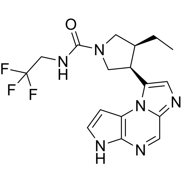
UpadacitinibJAK1 Inhibitor -
HY-100754
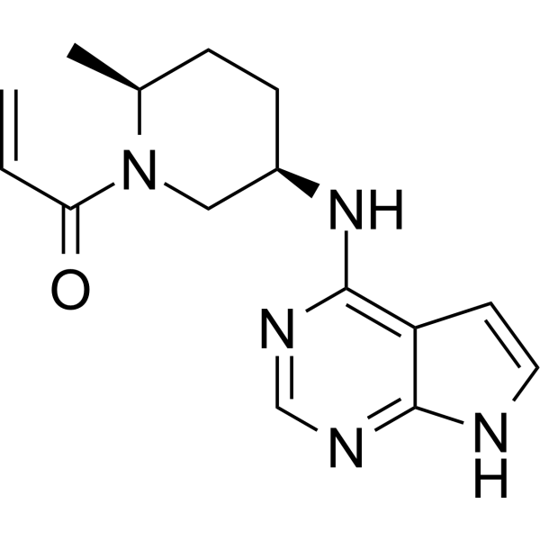
RitlecitinibJAK3 Inhibitor -
HY-13453
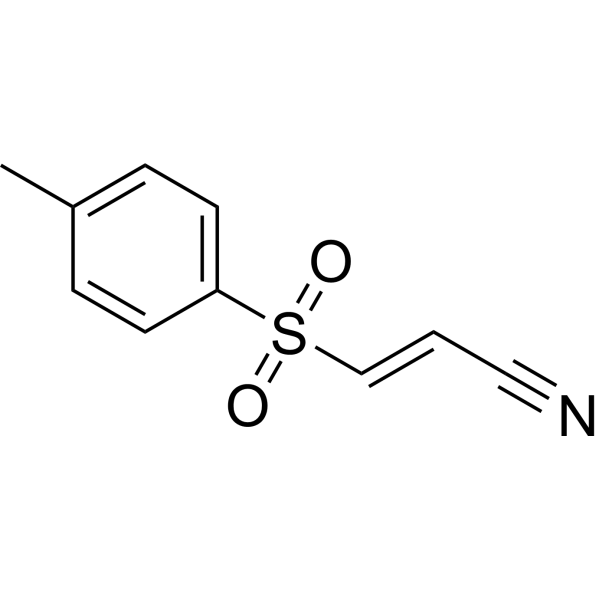
BAY 11-7082IκBα/NF-κB Inhibitor -
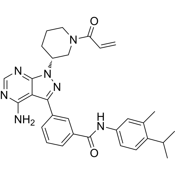
HY-108691
PF-06465469ITK Inhibitor -
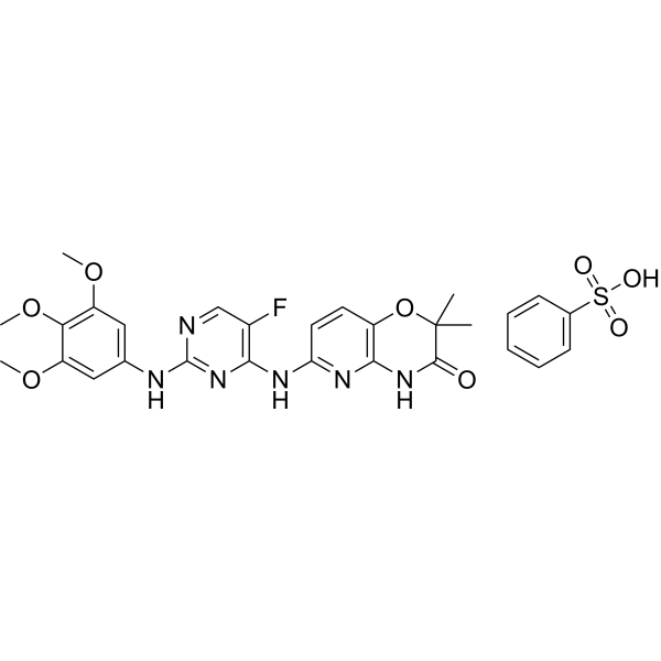
HY-12067
R406Syk/FLT3 Inhibitor -

HY-101474AR
Zanubrutinib (Standard)Btk Inhibitor
Publications Citing Use of MedChemExpress (MCE) Soquelitinib
More Customer Validation & Images

-
WB
-
WB


Biologische Aktivität
Beschreibung
In Vitro
Soquelitinib (10 μM, 2 h) inhibits TCR signaling downstream of ITK and blocks lL-2 production in Jurkat cells[1].
Soquelitinib (10 μM, 1 h) suppresses Th2 associated cytokines and induces Th1 skewing in CD4 T cells[1].
MedChemExpress (MCE) has not independently confirmed the accuracy of these methods. They are for reference only.
In Vivo
MedChemExpress (MCE) has not independently confirmed the accuracy of these methods. They are for reference only.
-
Animal Model:Murine syngeneictumor models[2]
-
Dosage:30 mg/kg
-
Administration:oral gavage, twice daily for 20 days
-
Result:Induced significantly higher frequency of intra-tumoral CD8 T cells in the CT26 model and a moderate increase in intra-tumoral CD8 infiltration.
Augmented the antitumor efficacy of immune checkpoint blockade, leading to durable antitumor response in the CT26 model.
- Fibrosis/Collagen Morphometry
- Collagen: Sirius Red Staining
- Imiquimod-Induced Psoriasiform Dermatitis
- Naïve CD4+ T-cell subset differentiation/polarization
- Connective Tissue: Masson's Trichrome/Collagen Trichrome Staining
- Research Protocol for Inflammation-related Diseases
- Research Protocol for Cancer Immunology
Clinical Trial
| NCT Number | Sponsor | Condition | Start Date |
Phase

|
|---|---|---|---|---|
| NCT01329991 | 2011-05 | PHASE1 |


Chemical Information
-
CAS. Nr. 2226636-04-8
-
Appearance Solid
-
Molecular Weight 514.66
-
Formel C25H30N4O4S2
-
Color White to off-white
-
SMILES
O=C(N1C[C@H](N(CCC1)C(C=C)=O)C)C2=C(C=C(C(SC3=CN=C(NC(C4CC4)=O)S3)=C2)C)OC
-
Synonyms
CPI-818
-
Versand
Room temperature in continental US; may vary elsewhere.
-
Speicherung
Powder -20°C 3 years 4°C 2 years In solvent -80°C 6 months -20°C 1 month


Publications (3)
-
Journal Impact Factor
-
Most Recent
-
Cell Signal
Phosphoproteomic analysis of successive Jurkat CD19-CAR generations reveals TCRζ-driven signalling. [Abstract]2026 Feb:138:112204. PMID: 41177417
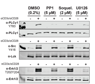
Soquelitinib purchased from MedChemExpress. Usage Cited in: Cell Signal. 2026 Feb:138:112204. [Abstract]
Western blot analysis of PLCγ1Y783, LckY394, and Erk1T202Y204/Erksup>2T185Y187, respectively, in samples treated with PP1, Soquelitinib (2 μM), or U0126. One sample is shown here for display.
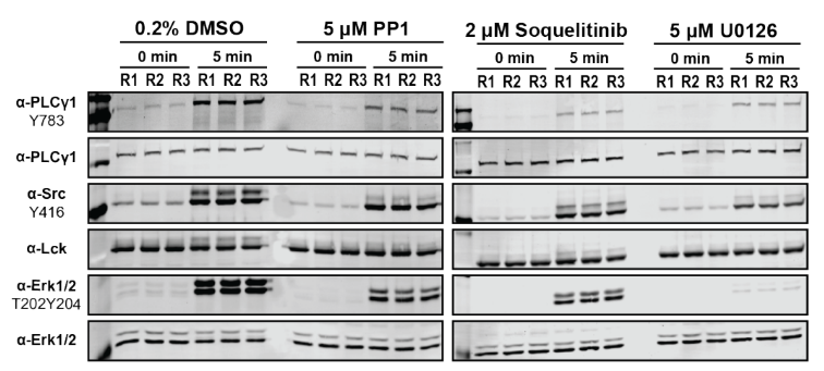
Soquelitinib purchased from MedChemExpress. Usage Cited in: Cell Signal. 2026 Feb:138:112204. [Abstract]
Western blot analysis of PLCγ1Y783, LckY394, and Erk1T202Y204/Erksup>2T185Y187 phosphorylation in the basal and stimulated states of untreated or kinase inhibitor treated Jurkat T cells with Soquelitinib (2 μM; 0-5 min).
-
-
bioRxiv
2025 Jul 7:2025.07.04.663030. PMID: 40672291


Lösungsmittel & Löslichkeit
In Vitro:
DMSO : ≥ 100 mg/mL (194.30 mM; Hygroscopic DMSO has a significant impact on the solubility of product, please use newly opened DMSO)
* "≥" means soluble, but saturation unknown.
Please refer to the solubility information to select the appropriate solvent. Once prepared, please aliquot and store the solution to prevent product inactivation from repeated freeze-thaw cycles.
Storage method and period of stock solution: -80°C, 6 months; -20°C, 1 month. When stored at -80°C, please use it within 6 months. When stored at -20°C, please use it within 1 month.
Please refer to the solubility information to select the appropriate solvent. Once prepared, please aliquot and store the solution to prevent product inactivation from repeated freeze-thaw cycles.
Storage method and period of stock solution: -80°C, 6 months; -20°C, 1 month. When stored at -80°C, please use it within 6 months. When stored at -20°C, please use it within 1 month.
Konzentration (Stammlösung) × Volumen (Stammlösung) = Konzentration (Ziellösung) × Volumen (Ziellösung)
In Vivo:
Select the appropriate dissolution method based on your experimental animal and administration route.
- For the following dissolution methods, please ensure to first prepare a clear stock solution using an In Vitro approach and then sequentially add co-solvents:
- To ensure reliable experimental results, the clarified stock solution can be appropriately stored based on storage conditions. As for the working solution for In Vivo experiments, it is recommended to prepare freshly and use it on the same day.
- The percentages shown for the solvents indicate their volumetric ratio in the final prepared solution. If precipitation or phase separation occurs during preparation, heat and/or sonication can be used to aid dissolution.
Add each solvent one by one: 10% DMSO 40% PEG300 5% Tween-80 45% Saline
Solubility: ≥ 10 mg/mL (19.43 mM); Clear solution
This protocol yields a clear solution of ≥ 10 mg/mL (saturation unknown).
Taking 1 mL working solution as an example, add 100 μL DMSO stock solution (100.0 mg/mL) to 400 μL PEG300, and mix evenly; then add 50 μL Tween-80 and mix evenly; then add 450 μL Saline to adjust the volume to 1 mL.
Preparation of Saline: Dissolve 0.9 g sodium chloride in ddH₂O and dilute to 100 mL to obtain a clear Saline solution.
Add each solvent one by one: 10% DMSO 90% (20% SBE-β-CD in Saline)
Solubility: ≥ 10 mg/mL (19.43 mM); Clear solution
This protocol yields a clear solution of ≥ 10 mg/mL (saturation unknown).
Taking 1 mL working solution as an example, add 100 μL DMSO stock solution (100.0 mg/mL) to 900 μL 20% SBE-β-CD in Saline, and mix evenly.
Preparation of 20% SBE-β-CD in Saline (4°C, storage for one week): 2 g SBE-β-CD powder is dissolved in 10 mL Saline, completely dissolve until clear.
In Vivo Dissolution Calculator
Please enter the basic information of animal experiments:
-
-
-
-
Recommended: Prepare an additional quantity of animals to account for potential losses during experiments.
Please enter your animal formula composition:
-
%DMSO +
Recommended: Keep the proportion of DMSO in working solution below 2% if your animal is weak.
-
%+
-
+%Tween-80 + +
-
%Saline +
The co-solvents required include: DMSO, . All of co-solvents are available by MedChemExpress (MCE). , Tween 80. All of co-solvents are available by MedChemExpress (MCE).
Working solution concentration: 0.22 mg/mL
Method for preparing stock solution: mg drug dissolved in μL DMSO. Stock solution concentration: mg/mL.
1. Take μL DMSO stock solution;
2. Add μL .
μL , mix evenly;
3. Then add μL Tween 80, mix evenly;
4. Then add μL
Please ensure that the stock solution in the first step is dissolved to a clear state, and add co-solvents in sequence. You can use ultrasonic heating (ultrasonic cleaner, recommended frequency 20-40 kHz), vortexing, etc. to assist dissolution.


Reinheit & Dokumentation
-
Data Sheet (280 KB)
-
SDS (254 KB)
- English - EN (254 KB)
- Français - FR (254 KB)
- Deutsch - DE (254 KB)
- Norwegian - NO (254 KB)
- Español - ES (254 KB)
- Swedish - SV (254 KB)
- Italian - IT (254 KB)
- Korean - KR (254 KB)
- Portuguese - PT (254 KB)
-
Handling Instructions (2659 KB)
Verweise


Complete Stock Solution Preparation Table
Please refer to the solubility information to select the appropriate solvent. Once prepared, please aliquot and store the solution to prevent product inactivation from repeated freeze-thaw cycles.
Storage method and period of stock solution: -80°C, 6 months; -20°C, 1 month. When stored at -80°C, please use it within 6 months. When stored at -20°C, please use it within 1 month.
| Optional Solvent | Concentration Solvent Mass | 1 mg | 5 mg | 10 mg | 25 mg |
|---|---|---|---|---|---|
| DMSO | 1 mM | 1.9430 mL | 9.7152 mL | 19.4303 mL | 48.5758 mL |
| 5 mM | 0.3886 mL | 1.9430 mL | 3.8861 mL | 9.7152 mL | |
| 10 mM | 0.1943 mL | 0.9715 mL | 1.9430 mL | 4.8576 mL | |
| 15 mM | 0.1295 mL | 0.6477 mL | 1.2954 mL | 3.2384 mL | |
| 20 mM | 0.0972 mL | 0.4858 mL | 0.9715 mL | 2.4288 mL | |
| 25 mM | 0.0777 mL | 0.3886 mL | 0.7772 mL | 1.9430 mL | |
| 30 mM | 0.0648 mL | 0.3238 mL | 0.6477 mL | 1.6192 mL | |
| 40 mM | 0.0486 mL | 0.2429 mL | 0.4858 mL | 1.2144 mL | |
| 50 mM | 0.0389 mL | 0.1943 mL | 0.3886 mL | 0.9715 mL | |
| 60 mM | 0.0324 mL | 0.1619 mL | 0.3238 mL | 0.8096 mL | |
| 80 mM | 0.0243 mL | 0.1214 mL | 0.2429 mL | 0.6072 mL | |
| 100 mM | 0.0194 mL | 0.0972 mL | 0.1943 mL | 0.4858 mL |


Soquelitinib Related Classifications
HY-150298 Related Classifications
 Powered by Bioz