



SU056
Based on 2 publication(s) in Google Scholar
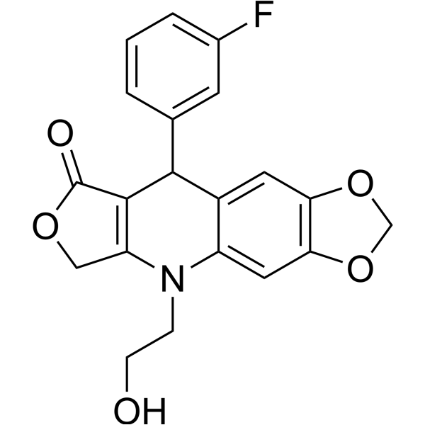
SU056 is a YB-1 inhibitor. SU056 induces cell-cycle arrest, apoptosis, and inhibits cell migration in ovarian cancer cells. SU056 interacts with YB-1 and inhibits and its associated downstream proteins and pathways. SU056 can enhance the cytotoxic effects of Paclitaxel (HY-B0015).
Nos produits utilisent uniquement pour la recherche. Nous ne vendons pas aux patients.
- Pureté : 99.77%
- CAS No.: 2376580-08-2
- Formule: C20H16FNO5
- Masse moléculaire:369.34
-
Stockage:Powder -20°C, 3 years , 4°C, 2 years ; In solvent -80°C, 6 months , -20°C, 1 month

To place orders, for customer services and technical support, please contact: MedChemExpress USA
Tel: 609-228-6898 E-mail: [email protected] [email protected]


-
 Activité biologique
-
Chemical Information
-
Solvant et solubilité
- Protocole
- Pureté et documentation
- Références
-
 Help & FAQs


-
HY-15531
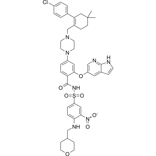
VenetoclaxBCL-2 Inhibitor -

HY-10219
RapamycinmTOR Inhibitor -

HY-15141
StaurosporinePKC/PKA Inhibitor -
HY-13966
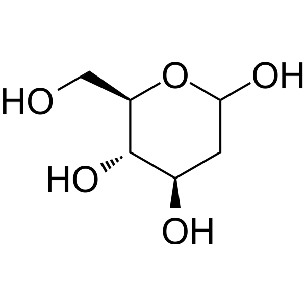
2-Deoxy-D-glucoseGlycolysis Inhibitor -

HY-13259
MG-132Proteasome Inhibitor -

HY-10182
LaduviglusibGSK-3 Inhibitor -
HY-Y0873
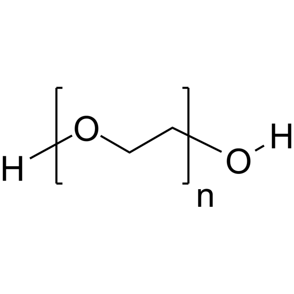
PEG300Neutral Polymer -
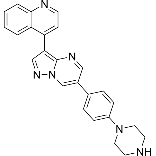
HY-12071
LDN193189ALK2/3 Inhibitor -

HY-14648
DexamethasoneGR Agonist -
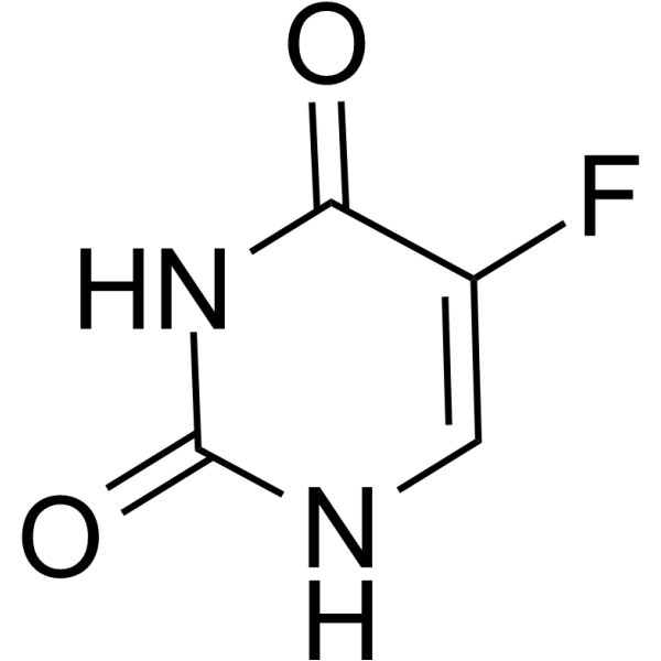
HY-90006
5-FluorouracilThymidylate Synthase Inhibitor -
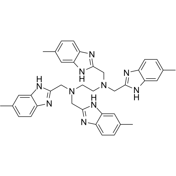
HY-13915
NSC348884NPM Inhibitor -
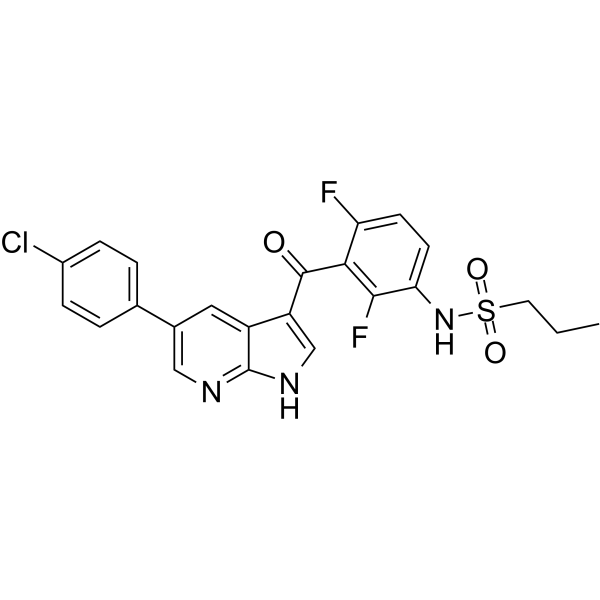
HY-12057
VemurafenibB-Raf Inhibitor
Publications Citing Use of MedChemExpress (MCE) SU056
More Customer Validation & Images

-
Cell Imaging/Staining


Activité biologique
Description
IC50 & Target
YB-1[1]
In Vitro
SU056 (0-10 μM approximately, 48 h) inhibits cell growth in OVCAR3/4/5/8, SKOV3 and ID8 cells[1].
SU056 (1 μM, 5-8 days) inhibits colony formation in a dose-dependent manner in OVCAR-8 and ID8 cells[1].
SU056 (1-5 μM, 6 h) arrests OVCAR8, SKOV3, and ID8 cells in the sub-G1 and G1 phases[1].
SU056 (0-1 μM, 12 h) inhibits cell migration in OVCAR8, SKOV3, and ID8 cells[1].
SU056 (0-5 μM, 24 h) induces apoptotic cell death in OVCAR8, SKOV3, and ID8 cells[1].
SU056 (1-5 μM, 12 h) inhibited the expression of YB-1, TMSB10, SUMO2, and PMSB2 proteins in OVCAR8 cells[1].
SU056 (0.1, 0.5, and 1 μM, 48 h) enhances the cytotoxic effects of Paclitaxel (HY-B0015) (0.1, 0.5, and 1 nM)[1].
MedChemExpress (MCE) has not independently confirmed the accuracy of these methods. They are for reference only.
-
Cell Line:OVCAR3/4/5/8, SKOV3 and ID8 cells
-
Concentration:0-10 μM approximately
-
Incubation Time:48 h
-
Result:Inhibited cell growth with IC50 values of 1.27, 6.8, 4.33, 3.18, 1.73, 3.75 μM.
-
Cell Line:OVCAR8, SKOV3, and ID8 cells
-
Concentration:0, 0.5, 1 μM
-
Incubation Time:12 h
-
Result:Dose-dependently inhibited cell migration.
-
Cell Line:OVCAR8 cell
-
Concentration:1-5 μM
-
Incubation Time:12 h
-
Result:Dose-dependently inhibited the YB-1, TMSB10, SUMO2, and PMSB2 proteins.
In Vivo
SU056 (10 mg/kg, i.p., daily) combined with Paclitaxel (HY-B0015) (5 mg/kg, weekly, i.p.) shows a much greater reduction in OC tumor growth in immunodeficiency mice implanted with OVCAR8 OC tumors[1].
MedChemExpress (MCE) has not independently confirmed the accuracy of these methods. They are for reference only.
-
Animal Model:C57BL/6 mice implanted (s.c.) with luciferase-expressing ID8 cells[1]
-
Dosage:20 mg/kg
-
Administration:Intraperitoneal injection (i.p.)
-
Result:Reduced the tumor weight by 2-fold, and is well-tolerated.
Reduced the lung metastases.
Inhibited YB-1 expression and downstream MDR1 (IHC assay of tumor sample).
- Scratch/Wound-Healing Migration Assay
- Annexin V plus membrane-impermeant dye apoptosis staining
- Cell migration
- Neural Crest/Neuronal Cell Migration Explant Assay
- Cell Viability Determination by MTT Colorimetric Assay
- Detection of Apoptosis
- TUNEL staining for apoptotic DNA fragmentation
- Zigmond Chamber Chemotaxis Assay
- Under-Agarose Cell Migration Assay
- Transwell/Boyden Chamber Migration Assay
- Apoptosis Solutions
- 3D Collagen/Hydrogel Matrix Migration Assay
- Cell-Exclusion Zone Migration Assay
- Dunn/Insall Chamber Chemotaxis Assay


Chemical Information
-
CAS No. 2376580-08-2
-
Appearance Solid
-
Masse moléculaire 369.34
-
Formule C20H16FNO5
-
Color White to off-white
-
SMILES
O=C1OCC2=C1C(C3=CC4=C(C=C3N2CCO)OCO4)C5=CC(F)=CC=C5
-
Livraison
Room temperature in continental US; may vary elsewhere.
-
Stockage
Powder -20°C 3 years 4°C 2 years In solvent -80°C 6 months -20°C 1 month


Publications (2)
-
Journal Impact Factor
-
Most Recent
-
Int J Mol Sci
Downregulation of IL-8 and IL-10 by LRRC8A Inhibition through the NOX2-Nrf2-CEBPB Transcriptional Axis in THP-1-Derived M2 Macrophages. [Abstract]2024 Sep 5;25(17):9612. PMID: 39273558 -
J Virol
YBX1 is required for assembly of viral replication complexes of chikungunya virus and replication of multiple alphaviruses. [Abstract]2025 Feb 25;99(2):e0201524. PMID: 39745458
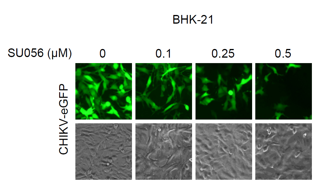
SU056 purchased from MedChemExpress. Usage Cited in: J Virol. 2025 Feb 25;99(2):e0201524. [Abstract]
SU056 (0.1-0.5 μM; 12 h) inhibited CHIKV replication in BHK-21 cells at a dose-dependent manner.


Solvant et solubilité
In Vitro:
DMSO : 50 mg/mL (135.38 mM; Need ultrasonic; Hygroscopic DMSO has a significant impact on the solubility of product, please use newly opened DMSO)
Please refer to the solubility information to select the appropriate solvent. Once prepared, please aliquot and store the solution to prevent product inactivation from repeated freeze-thaw cycles.
Storage method and period of stock solution: -80°C, 6 months; -20°C, 1 month. When stored at -80°C, please use it within 6 months. When stored at -20°C, please use it within 1 month.
Please refer to the solubility information to select the appropriate solvent. Once prepared, please aliquot and store the solution to prevent product inactivation from repeated freeze-thaw cycles.
Storage method and period of stock solution: -80°C, 6 months; -20°C, 1 month. When stored at -80°C, please use it within 6 months. When stored at -20°C, please use it within 1 month.
Concentration (start) × Volume (start) = Concentration (final) × Volume (final)
In Vivo:
Select the appropriate dissolution method based on your experimental animal and administration route.
- For the following dissolution methods, please ensure to first prepare a clear stock solution using an In Vitro approach and then sequentially add co-solvents:
- To ensure reliable experimental results, the clarified stock solution can be appropriately stored based on storage conditions. As for the working solution for In Vivo experiments, it is recommended to prepare freshly and use it on the same day.
- The percentages shown for the solvents indicate their volumetric ratio in the final prepared solution. If precipitation or phase separation occurs during preparation, heat and/or sonication can be used to aid dissolution.
Add each solvent one by one: 10% DMSO 40% PEG300 5% Tween-80 45% Saline
Solubility: 2.5 mg/mL (6.77 mM); Suspended solution; Need ultrasonic
This protocol yields a suspended solution of 2.5 mg/mL. Suspended solution can be used for oral and intraperitoneal injection.
Taking 1 mL working solution as an example, add 100 μL DMSO stock solution (25.0 mg/mL) to 400 μL PEG300, and mix evenly; then add 50 μL Tween-80 and mix evenly; then add 450 μL Saline to adjust the volume to 1 mL.
Preparation of Saline: Dissolve 0.9 g sodium chloride in ddH₂O and dilute to 100 mL to obtain a clear Saline solution.
Add each solvent one by one: 10% DMSO 90% (20% SBE-β-CD in Saline)
Solubility: ≥ 2.5 mg/mL (6.77 mM); Clear solution
This protocol yields a clear solution of ≥ 2.5 mg/mL (saturation unknown).
Taking 1 mL working solution as an example, add 100 μL DMSO stock solution (25.0 mg/mL) to 900 μL 20% SBE-β-CD in Saline, and mix evenly.
Preparation of 20% SBE-β-CD in Saline (4°C, storage for one week): 2 g SBE-β-CD powder is dissolved in 10 mL Saline, completely dissolve until clear.
For the following dissolution methods, please prepare the working solution directly:
It is recommended to prepare fresh solutions and use them promptly within a short period of time.
The percentages shown for the solvents indicate their volumetric ratio in the final prepared solution. If precipitation or phase separation occurs during preparation, heat and/or sonication can be used to aid dissolution.
Add each solvent one by one: 50% PEG300 50% Saline
Solubility: 5 mg/mL (13.54 mM); Suspended solution; Need ultrasonic
In Vivo Dissolution Calculator
Please enter the basic information of animal experiments:
-
-
-
-
Recommended: Prepare an additional quantity of animals to account for potential losses during experiments.
Please enter your animal formula composition:
-
%DMSO +
Recommended: Keep the proportion of DMSO in working solution below 2% if your animal is weak.
-
%+
-
+%Tween-80 + +
-
%Saline +
The co-solvents required include: DMSO, . All of co-solvents are available by MedChemExpress (MCE). , Tween 80. All of co-solvents are available by MedChemExpress (MCE).
Working solution concentration: 0.22 mg/mL
Method for preparing stock solution: mg drug dissolved in μL DMSO. Stock solution concentration: mg/mL.
1. Take μL DMSO stock solution;
2. Add μL .
μL , mix evenly;
3. Then add μL Tween 80, mix evenly;
4. Then add μL
Please ensure that the stock solution in the first step is dissolved to a clear state, and add co-solvents in sequence. You can use ultrasonic heating (ultrasonic cleaner, recommended frequency 20-40 kHz), vortexing, etc. to assist dissolution.


Pureté et documentation
-
Fiche technique (282 KB)
-
SDS (251 KB)
- English - EN (251 KB)
- Français - FR (251 KB)
- Deutsch - DE (251 KB)
- Norwegian - NO (251 KB)
- Español - ES (251 KB)
- Swedish - SV (251 KB)
- Italian - IT (251 KB)
- Korean - KR (251 KB)
- Portuguese - PT (251 KB)
-
Instruction de manipulation (2659 KB)
Références


Complete Stock Solution Preparation Table
Please refer to the solubility information to select the appropriate solvent. Once prepared, please aliquot and store the solution to prevent product inactivation from repeated freeze-thaw cycles.
Storage method and period of stock solution: -80°C, 6 months; -20°C, 1 month. When stored at -80°C, please use it within 6 months. When stored at -20°C, please use it within 1 month.
| Optional Solvent | Concentration Solvent Mass | 1 mg | 5 mg | 10 mg | 25 mg |
|---|---|---|---|---|---|
| DMSO | 1 mM | 2.7075 mL | 13.5377 mL | 27.0753 mL | 67.6883 mL |
| 5 mM | 0.5415 mL | 2.7075 mL | 5.4151 mL | 13.5377 mL | |
| 10 mM | 0.2708 mL | 1.3538 mL | 2.7075 mL | 6.7688 mL | |
| 15 mM | 0.1805 mL | 0.9025 mL | 1.8050 mL | 4.5126 mL | |
| 20 mM | 0.1354 mL | 0.6769 mL | 1.3538 mL | 3.3844 mL | |
| 25 mM | 0.1083 mL | 0.5415 mL | 1.0830 mL | 2.7075 mL | |
| 30 mM | 0.0903 mL | 0.4513 mL | 0.9025 mL | 2.2563 mL | |
| 40 mM | 0.0677 mL | 0.3384 mL | 0.6769 mL | 1.6922 mL | |
| 50 mM | 0.0542 mL | 0.2708 mL | 0.5415 mL | 1.3538 mL | |
| 60 mM | 0.0451 mL | 0.2256 mL | 0.4513 mL | 1.1281 mL | |
| 80 mM | 0.0338 mL | 0.1692 mL | 0.3384 mL | 0.8461 mL | |
| 100 mM | 0.0271 mL | 0.1354 mL | 0.2708 mL | 0.6769 mL |


SU056 Related Classifications
HY-150231 Related Classifications
 Powered by Bioz