



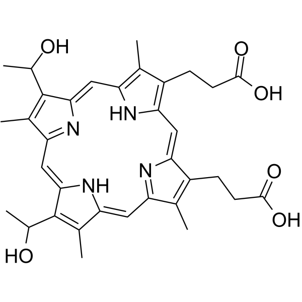
Hematoporphyrin
Based on 15 publication(s) in Google Scholar
Hematoporphyrin (Hematoporphyrin IX), a photosensitizer, is a substrate for affinity chromatography of heme-binding proteins. Hematoporphyrin can induce apoptosis in U87 glioma cells and decrease tumor growth in vivo when exposed to red light.
For research use only. We do not sell to patients.
- Purity : 95.81%
- CAS No.: 14459-29-1
- Formula: C34H38N4O6
- Molecular Weight:598.69
-
Storage:
-20°C, protect from light
* The compound is unstable in solutions, freshly prepared is recommended.

To place orders, for customer services and technical support, please contact: MedChemExpress USA
Tel: 609-228-6898 E-mail: [email protected] [email protected]


-
 Biological Activity
-
Chemical Information
-
Solvent & Solubility
- Protocols
- Purity & Documentation
- References
-
 Help & FAQs


Publications Citing Use of MedChemExpress (MCE) Hematoporphyrin
More- Nat Biomed Eng. 2023 Mar;7(3):298-312. [Abstract]
- Nat Commun. 2022 Jun 16;13(1):3468. [Abstract]
- ACS Nano. 2023 Dec 12;17(23):23535-23544. [Abstract]
- Chem Eng J. 390 (2020) 124521.
- Acta Biomater. 2025 Jan 24:193:231-241. [Abstract]
- Free Radic Biol Med. 2023 Oct:207:239-246. [Abstract]
- Drug Deliv. 2022 Dec;29(1):3358-3369. [Abstract]
- Pharmaceutics. 2023 Aug 3;15(8):2076. [Abstract]
- Int J Mol Sci. 2022 Apr 11;23(8):4218. [Abstract]
- J Biol Chem. 2022 Oct;298(10):102417. [Abstract]
- FEBS Lett. 2021 Jan;595(2):206-219. [Abstract]
- PLoS One. 2019 Sep 25;14(9):e0222331. [Abstract]
- Lasers Med Sci. 2020 Feb;35(1):71-78. [Abstract]
- Nanyang Technological University. 2024.
- Cancer Adv. 2024;7:e24021.
All Endogenous Metabolite Isoforms
More

Biological Activity
Description
IC50 & Target
|
Human Endogenous Metabolite |
Cellular Effect
|
Cell Line

|
Type | Value | Description | References |
|---|---|---|---|---|
| Caco-2 | CC50 |
160.08 μM
Compound: HEMATOPORPHYRIN
|
Toxicity against Caco-2 cells determined at 48 hours by intracellular ATP concentration using the CellTiter-Glo Luminescent Cell Viability Assay
Toxicity against Caco-2 cells determined at 48 hours by intracellular ATP concentration using the CellTiter-Glo Luminescent Cell Viability Assay
|
10.21203/rs.3.rs-23951/v1 |
| Caco-2 | IC50 |
1.85 μM
Compound: HEMATOPORPHYRIN
|
Determination of IC50 values for inhibition of SARS-CoV-2 induced cytotoxicity of Caco-2 cells after 48 hours by high content imaging
Determination of IC50 values for inhibition of SARS-CoV-2 induced cytotoxicity of Caco-2 cells after 48 hours by high content imaging
|
10.21203/rs.3.rs-23951/v1 |
| Daudi | IC50 |
15 μM
Compound: 3, HPIX, hematoporphyrin
|
Cytotoxicity against human Daudi cells under deem light after 96 hrs by MTT assay
Cytotoxicity against human Daudi cells under deem light after 96 hrs by MTT assay
|
[PMID: 17993275] |
| HeLa | IC50 |
71 μM
Compound: 3, HPIX, hematoporphyrin
|
Cytotoxicity against human HeLa cells under deem light after 96 hrs by MTT assay
Cytotoxicity against human HeLa cells under deem light after 96 hrs by MTT assay
|
[PMID: 17993275] |
| K562 | IC50 |
44 μM
Compound: 3, HPIX, hematoporphyrin
|
Cytotoxicity against human K562 cells under deem light after 96 hrs by MTT assay
Cytotoxicity against human K562 cells under deem light after 96 hrs by MTT assay
|
[PMID: 17993275] |
| NCI-H69 | IC50 |
70 μM
Compound: 3, HPIX, hematoporphyrin
|
Cytotoxicity against human H69 cells under deem light after 96 hrs by MTT assay
Cytotoxicity against human H69 cells under deem light after 96 hrs by MTT assay
|
[PMID: 17993275] |
| Raji | IC50 |
57 μM
Compound: 3, HPIX, hematoporphyrin
|
Cytotoxicity against human Raji cells under deem light after 96 hrs by MTT assay
Cytotoxicity against human Raji cells under deem light after 96 hrs by MTT assay
|
[PMID: 17993275] |
| SiHa | IC50 |
56 μM
Compound: 3, HPIX, hematoporphyrin
|
Cytotoxicity against human SIHA cells under deem light after 96 hrs by MTT assay
Cytotoxicity against human SIHA cells under deem light after 96 hrs by MTT assay
|
[PMID: 17993275] |
In Vitro
Hematoporphyrin (20-120 nM; 60 min) dose-dependently inhibits cell viability in U87 and U251 glioma cells, with IC50s of 85 and 166 nM, respectively[2].
Hematoporphyrin (85 nM; 60 min) induces cell apoptosis via induction of ROS in U87 cells[2].
Hematoporphyrin (85 nM; 60 min) induces morphological changes of U87 cells under the red light, including shrinking, fragmentation[2].
MedChemExpress (MCE) has not independently confirmed the accuracy of these methods. They are for reference only. Further protocols information, click here.
-
Cell Line:U87 and U251 cells
-
Concentration:20, 40, 60, 80, 100, 120 nM
-
Incubation Time:60 min
-
Result:Inhibited cell viability in a dose-dependent manner.
Was more effective under the red light than white light.
-
Cell Line:U87 cells
-
Concentration:85 nM
-
Incubation Time:60 min
-
Result:Induced apoptotic nuclei in U87 cells with low cell density.
Induced the ROS and decreased the mitochondrial membrane potential.
In Vivo
MedChemExpress (MCE) has not independently confirmed the accuracy of these methods. They are for reference only.
-
Animal Model:Wistar albino rats of both sexes (20 d; 60-80 g) bearing a subcutaneous solid Yoshida hepatoma AH-130[3]
-
Dosage:5, 10 mg/kg
-
Administration:I.p. daily during the initial 10 days and biweekly for the next 2 months
-
Result:No tumor could be palpated a few days after exposure of the rats to light.
The skin healed completely and regrowth of the hair occurred.
Massive coagulation necrosis of the tumor 24 h after phototreatment (×40).
Clinical Trial
| NCT Number | Sponsor | Condition | Start Date |
Phase
|
|---|---|---|---|---|
| NCT01329991 | Plexxikon| | 2011-05 | PHASE1 |


Chemical Information
-
CAS No. 14459-29-1
-
Appearance Solid
-
Molecular Weight 598.69
-
Formula C34H38N4O6
-
Color Pale purple to purple
-
SMILES
O=C(O)CCC1=C2/C=C3C(CCC(O)=O)=C(C)C(/C=C(N/4)/C(C)=C(C(O)C)C4=C\C5=N/C(C(C(O)C)=C5C)=C\C(N2)=C1C)=N/3
-
Synonyms
Hematoporphyrin IX
-
Structure Classification
-
Initial Source
-
Shipping
Room temperature in continental US; may vary elsewhere.
-
Storage
-20°C, protect from light
* The compound is unstable in solutions, freshly prepared is recommended.


Publications (15)
-
Journal Impact Factor
-
Most Recent
-
Nat Biomed Eng
Nanoparticles with ultrasound-induced afterglow luminescence for tumour-specific theranostics. [Abstract]2023 Mar;7(3):298-312. PMID: 36550302 -
Nat Commun
Catalytical nano-immunocomplexes for remote-controlled sono-metabolic checkpoint trimodal cancer therapy. [Abstract]2022 Jun 16;13(1):3468. PMID: 35710545 -
ACS Nano
A Side-Effect-Free Interventional Therapy for Precisely Eliminating Unresectable Cancer Pain. [Abstract]2023 Dec 12;17(23):23535-23544. PMID: 38084419 -
-
Acta Biomater
Regulation of antigen presentation and interleukin 10 production in murine dendritic cells via the oxidative stimulation of cell membrane using a polycation-porphyrin-conjugate-immobilized cell culture dish. [Abstract]2025 Jan 24:193:231-241. PMID: 39788307 -
Free Radic Biol Med
Singlet oxygen-generating cell-adhesive glass surfaces for the fundamental investigation of plasma membrane-targeted photodynamic therapy. [Abstract]2023 Oct:207:239-246. PMID: 37499887 -
Drug Deliv
Docetaxel prodrug and hematoporphyrin co-assembled nanoparticles for anti-tumor combination of chemotherapy and photodynamic therapy. [Abstract]2022 Dec;29(1):3358-3369. PMID: 36397301 -
Pharmaceutics
Locally Administered Photodynamic Therapy for Cancer Using Nano-Adhesive Photosensitizer. [Abstract]2023 Aug 3;15(8):2076. PMID: 37631290 -
Int J Mol Sci
Long-Term Fluorescent Tissue Marking Using Tissue-Adhesive Porphyrin with Polycations Consisting of Quaternary Ammonium Salt Groups. [Abstract]2022 Apr 11;23(8):4218. PMID: 35457034 -
J Biol Chem
α-Crystallin chaperone mimetic drugs inhibit lens γ-crystallin aggregation: Potential role for cataract prevention. [Abstract]2022 Oct;298(10):102417. PMID: 36037967 -
FEBS Lett
2021 Jan;595(2):206-219. PMID: 33210733 -
PLoS One
A pull-down and slot blot-based screening system for inhibitor compounds of the podoplanin-CLEC-2 interaction. [Abstract]2019 Sep 25;14(9):e0222331. PMID: 31553741 -
Lasers Med Sci
The study of killing effect and inducing apoptosis of 630-nm laser on lung adenocarcinoma A549 cells mediated by hematoporphyrin derivatives in vitro. [Abstract]2020 Feb;35(1):71-78. PMID: 31049741 -
-


Solvent & Solubility
In Vitro:
DMSO : 150 mg/mL (250.55 mM; Need ultrasonic; Hygroscopic DMSO has a significant impact on the solubility of product, please use newly opened DMSO)
Please refer to the solubility information to select the appropriate solvent. The compound is unstable in solutions, freshly prepared is recommended.
Please refer to the solubility information to select the appropriate solvent. The compound is unstable in solutions, freshly prepared is recommended.
Concentration (start) × Volume (start) = Concentration (final) × Volume (final)
In Vivo:
Select the appropriate dissolution method based on your experimental animal and administration route.
- For the following dissolution methods, please ensure to first prepare a clear stock solution using an In Vitro approach and then sequentially add co-solvents:
- To ensure reliable experimental results, the clarified stock solution can be appropriately stored based on storage conditions. As for the working solution for In Vivo experiments, it is recommended to prepare freshly and use it on the same day.
- The percentages shown for the solvents indicate their volumetric ratio in the final prepared solution. If precipitation or phase separation occurs during preparation, heat and/or sonication can be used to aid dissolution.
Add each solvent one by one: 10% DMSO 40% PEG300 5% Tween-80 45% Saline
Solubility: ≥ 2.5 mg/mL (4.18 mM); Clear solution
This protocol yields a clear solution of ≥ 2.5 mg/mL (saturation unknown).
Taking 1 mL working solution as an example, add 100 μL DMSO stock solution (25.0 mg/mL) to 400 μL PEG300, and mix evenly; then add 50 μL Tween-80 and mix evenly; then add 450 μL Saline to adjust the volume to 1 mL.
Preparation of Saline: Dissolve 0.9 g sodium chloride in ddH₂O and dilute to 100 mL to obtain a clear Saline solution.
Add each solvent one by one: 10% DMSO 90% (20% SBE-β-CD in Saline)
Solubility: 2.5 mg/mL (4.18 mM); Suspended solution; Need ultrasonic
This protocol yields a suspended solution of 2.5 mg/mL. Suspended solution can be used for oral and intraperitoneal injection.
Taking 1 mL working solution as an example, add 100 μL DMSO stock solution (25.0 mg/mL) to 900 μL 20% SBE-β-CD in Saline, and mix evenly.
Preparation of 20% SBE-β-CD in Saline (4°C, storage for one week): 2 g SBE-β-CD powder is dissolved in 10 mL Saline, completely dissolve until clear.
In Vivo Dissolution Calculator
Please enter the basic information of animal experiments:
-
-
-
-
Recommended: Prepare an additional quantity of animals to account for potential losses during experiments.
Please enter your animal formula composition:
-
%DMSO +
Recommended: Keep the proportion of DMSO in working solution below 2% if your animal is weak.
-
%+
-
+%Tween-80 + +
-
%Saline +
The co-solvents required include: DMSO, . All of co-solvents are available by MedChemExpress (MCE). , Tween 80. All of co-solvents are available by MedChemExpress (MCE).
Working solution concentration: 0.22 mg/mL
Method for preparing stock solution: mg drug dissolved in μL DMSO. Stock solution concentration: mg/mL. * The compound is unstable in solutions, freshly prepared is recommended.
1. Take μL DMSO stock solution;
2. Add μL .
μL , mix evenly;
3. Then add μL Tween 80, mix evenly;
4. Then add μL
Please ensure that the stock solution in the first step is dissolved to a clear state, and add co-solvents in sequence. You can use ultrasonic heating (ultrasonic cleaner, recommended frequency 20-40 kHz), vortexing, etc. to assist dissolution.

Protocols
-
Detection of Apoptosis
Apoptosis, also called programmed cell death, is generally characterized by distinct morphological characteristics.
-
TUNEL staining for apoptotic DNA fragmentation
TUNEL staining detects DNA strand breaks by using terminal deoxynucleotidyl transferase to add labeled nucleotides to exposed 3′-OH DNA termini, generating either microscopic staining in fixed cells or tissue sections, or fluorescence/cytometric signal in cell suspensions. TUNEL positivity reflects DNA fragmentation but should not be interpreted alone as definitive apoptosis, because TUNEL can also label necrotic, autolytic, mechanically damaged, or DNA-repair-associated DNA breaks.
-
Annexin V plus membrane-impermeant dye apoptosis staining
Annexin V-based apoptosis assays rely on the detection of phosphatidylserine (PS) externalization from the inner leaflet of the plasma membrane to the outer leaflet, an early biochemical hallmark of apoptosis. Fluorescently labeled Annexin V binds PS in a calcium-dependent manner, enabling identification of early apoptotic cells by flow cytometry or fluorescence microscopy. When combined with a membrane-impermeant DNA-binding dye (e. g. , propidium iodide), this approach allows discrimination between viable (Annexin V−/dye−), early apoptotic (Annexin V+/dye−), and late apoptotic or necrotic (Annexin V+/dye+) cell populations by assessing membrane integrity and PS exposure.
-
Patient-Derived Xenograft (PDX)
Patient-derived xenograft (PDX) models are generated by engrafting primary human tumor tissue directly into immunodeficient mice, allowing in vivo propagation of patient tumor biology without initial in vitro adaptation. These models are used to preserve key histopathological and molecular characteristics of the original tumor and enable assessment of tumor growth dynamics and therapeutic response in a living organism. The biological readout is tumor engraftment and subsequent growth in the murine host, which reflects the ability of human tumor cells to survive, vascularize, and expand in an immunocompromised microenvironment.
-
Apoptosis Solutions
Apoptosis is a regulated, generally non-lytic cell-death pathway that removes unwanted, damaged, infected, or abnormal cells through coordinated morphological changes, caspase activation, DNA fragmentation, and membrane remodeling. The intrinsic apoptosis pathway is controlled mainly by mitochondrial outer membrane permeabilization, BCL-2 family proteins, cytochrome c release, apoptosome formation, caspase-9 activation, and downstream executioner caspase-3/7 activation. The extrinsic apoptosis pathway is initiated by death receptors such as Fas, TNFR, and TRAIL receptors, which recruit adaptor proteins and activate caspase-8 before engaging executioner caspases or mitochondrial amplification through BID cleavage. Apoptosis is linked to many phenotypes, including cancer cell killing, tissue homeostasis, immune regulation, neurodegeneration, infection response, and treatment-induced cytotoxicity; unresolved questions include how apoptosis interacts with necroptosis, pyroptosis, ferroptos
-
Patient-Derived Orthotopic Xenograft (PDOX)
Patient-derived orthotopic xenograft (PDOX) modeling implants fresh patient tumor tissue or patient-derived tumor cells into the anatomically corresponding organ or tissue site of immunodeficient mice, usually by surgical orthotopic implantation, to preserve patient tumor histology, local microenvironmental context, invasion, metastatic behavior, and treatment-response features better than subcutaneous implantation. PDOX readouts include tumor engraftment, orthotopic tumor growth, local invasion, metastasis, recurrence after resection, histologic similarity to the donor tumor, biomarker retention, molecular concordance, survival, and response or resistance to therapy. PDOX models are used for preclinical drug testing and individualized therapy evaluation, but engraftment success varies by tumor type and specimen quality.
-
Subcutaneous Cell-Line-Derived Xenograft
Subcutaneous cell-line-derived xenograft (CDX) models are established by implanting cultured human cancer cell lines into immunodeficient mice, where the injected cells form localized tumors that can be monitored in vivo as a measure of tumorigenic potential, growth kinetics, and treatment response. These models are widely used in oncology research because they allow reproducible tumor formation and enable comparative assessment of tumor growth between different cell lines or genetic manipulations in a controlled in vivo microenvironment. Subcutaneous implantation of cancer cells in immunodeficient mice is a standard approach for evaluating tumor growth behavior and therapeutic response across multiple cancer types, including prostate, esophageal, pancreatic, and colon cancer models.
-
Orthotopic Cell-Line Xenograft
Orthotopic cell-line xenograft models involve implantation of human cancer cell lines into the anatomically corresponding organ of immunodeficient mice to reproduce tumor growth within a native microenvironment, enabling more clinically relevant tumor behavior compared with subcutaneous models. These models are widely used because orthotopic placement better recapitulates tumor progression, including invasion and metastatic spread, which are often underrepresented in heterotopic implantation systems. Compared with conventional xenografts, orthotopic implantation is described as more technically complex but provides improved simulation of tumor-microenvironment interactions and metastatic behavior, making it particularly valuable for translational oncology research. Surgical orthotopic implantation approaches have been emphasized as enabling faithful reproduction of clinical cancer features, including metastasis and disease progression patterns that align with the tumor’s organ of origi


Purity & Documentation
-
Data Sheet (288 KB)
-
SDS (252 KB)
- English - EN (252 KB)
- Français - FR (252 KB)
- Deutsch - DE (252 KB)
- Norwegian - NO (252 KB)
- Español - ES (252 KB)
- Swedish - SV (252 KB)
- Italian - IT (252 KB)
- Korean - KR (252 KB)
- Portuguese - PT (252 KB)
-
Handling Instructions (2659 KB)
References
[1]. Olsen, K.W., Affinity chromatography of heme-binding proteins: synthesis and characterization of hematin- and hematoporphyrin-agarose. Methods Enzymol, 1986. 123: p. 324-31. [Content Brief]
[2]. Yuan SX, et, al. Underlying mechanism of the photodynamic activity of hematoporphyrin‑induced apoptosis in U87 glioma cells. Int J Mol Med. 2018 Apr;41(4):2288-2296. [Content Brief]
[3]. Tomio L, et, al. Effect of hematoporphyrin and red light on AH-130 solid tumors in rats. Acta Radiol Oncol. 1983;22(1):49-53. [Content Brief]


Complete Stock Solution Preparation Table
Please refer to the solubility information to select the appropriate solvent. The compound is unstable in solutions, freshly prepared is recommended.
| Optional Solvent | Concentration Solvent Mass | 1 mg | 5 mg | 10 mg | 25 mg |
|---|---|---|---|---|---|
| DMSO | 1 mM | 1.6703 mL | 8.3516 mL | 16.7031 mL | 41.7578 mL |
| 5 mM | 0.3341 mL | 1.6703 mL | 3.3406 mL | 8.3516 mL | |
| 10 mM | 0.1670 mL | 0.8352 mL | 1.6703 mL | 4.1758 mL | |
| 15 mM | 0.1114 mL | 0.5568 mL | 1.1135 mL | 2.7839 mL | |
| 20 mM | 0.0835 mL | 0.4176 mL | 0.8352 mL | 2.0879 mL | |
| 25 mM | 0.0668 mL | 0.3341 mL | 0.6681 mL | 1.6703 mL | |
| 30 mM | 0.0557 mL | 0.2784 mL | 0.5568 mL | 1.3919 mL | |
| 40 mM | 0.0418 mL | 0.2088 mL | 0.4176 mL | 1.0439 mL | |
| 50 mM | 0.0334 mL | 0.1670 mL | 0.3341 mL | 0.8352 mL | |
| 60 mM | 0.0278 mL | 0.1392 mL | 0.2784 mL | 0.6960 mL | |
| 80 mM | 0.0209 mL | 0.1044 mL | 0.2088 mL | 0.5220 mL | |
| 100 mM | 0.0167 mL | 0.0835 mL | 0.1670 mL | 0.4176 mL |


Hematoporphyrin Related Classifications
HY-B0754 Related Classifications
 Powered by Bioz