



Evogliptin tartrate
Based on 2 publication(s) in Google Scholar
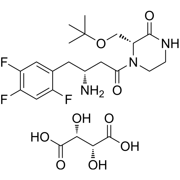
Evogliptin (DA-1229) tartrate is an orally active DPP4 inhibitor with significant and sustained hypoglycaemic effects in mouse models. Evogliptin tartrate also inhibits the production of inflammatory and fibrotic signals in hepatocytes by inducing autophagy. Evogliptin tartrate can be used in studies of type 2 diabetes, osteoporosis, renal impairment and chronic liver inflammation.
For research use only. We do not sell to patients.
- Purity : 98.11%
- CAS No.: 1222102-51-3
- Formula: C23H32F3N3O9
- Molecular Weight:551.51
-
Storage:
4°C, sealed storage, away from moisture
* In solvent : -80°C, 6 months; -20°C, 1 month (sealed storage, away from moisture)

To place orders, for customer services and technical support, please contact: MedChemExpress USA
Tel: 609-228-6898 E-mail: [email protected] [email protected]


-
 Biological Activity
-
Chemical Information
-
Solvent & Solubility
- Protocols
- Purity & Documentation
- References
-
 Help & FAQs


-
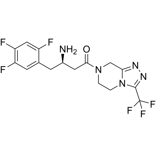
HY-13749
SitagliptinDPP4 Inhibitor -
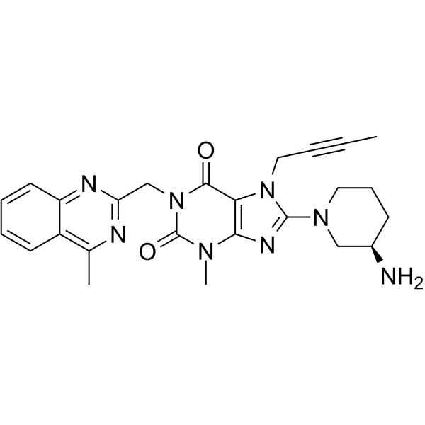
HY-10284
LinagliptinDPP-4 Inhibitor -
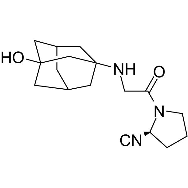
HY-14291
VildagliptinDPP-IV Inhibitor -
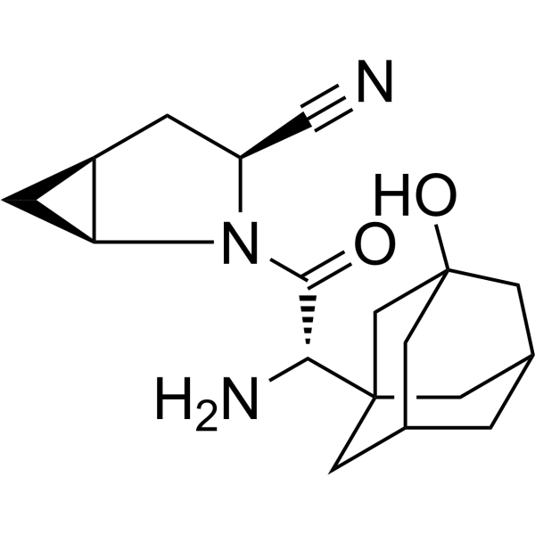
HY-10285
SaxagliptinDPP-4 Inhibitor -
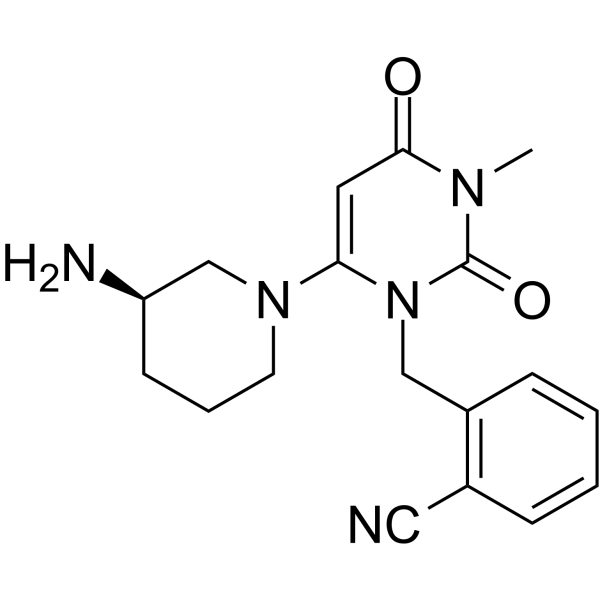
HY-A0023A
AlogliptinDPP-4 Inhibitor -
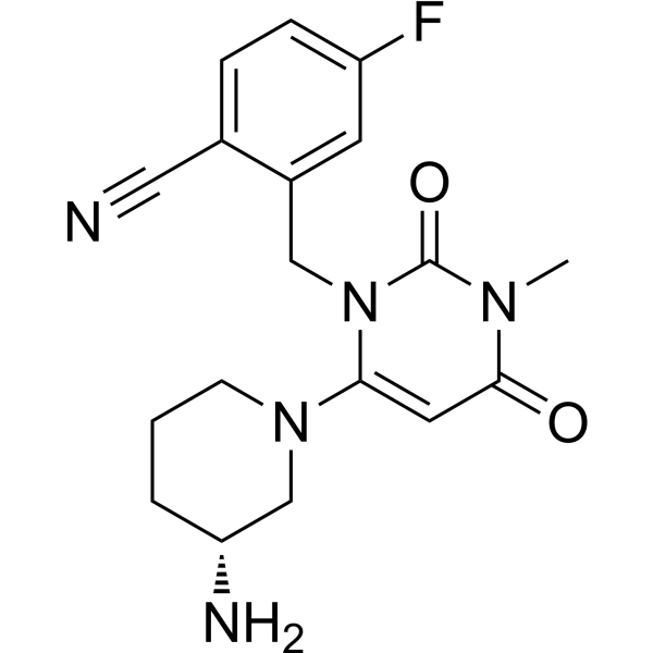
HY-15408
TrelagliptinDPP-4 Inhibitor -
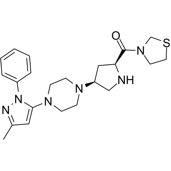
HY-14806
TeneligliptinDPP-4 Inhibitor -
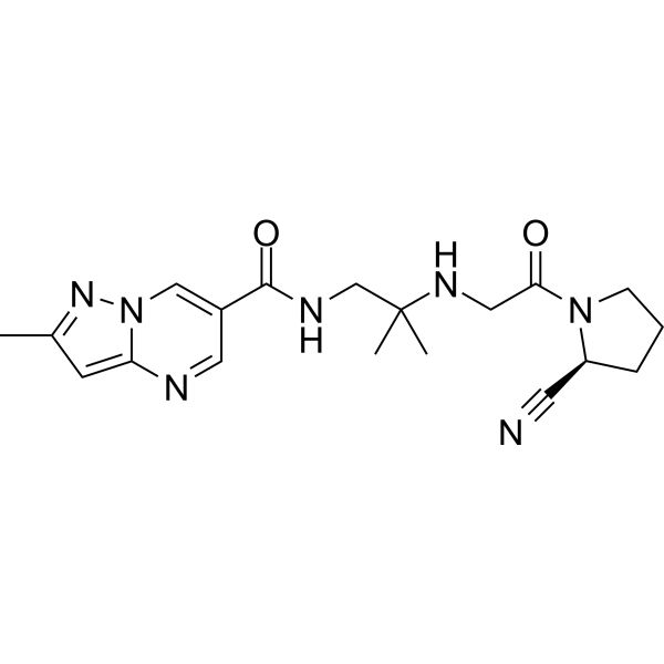
HY-14877
AnagliptinDPP-4 Inhibitor -
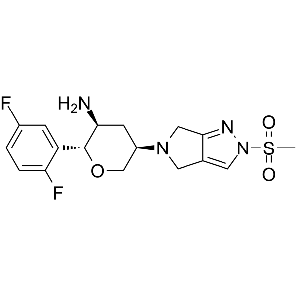
HY-15981
OmarigliptinDPP-4 Inhibitor -
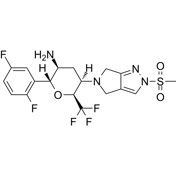
HY-147257
CofrogliptinDPP-4 Inhibitor -
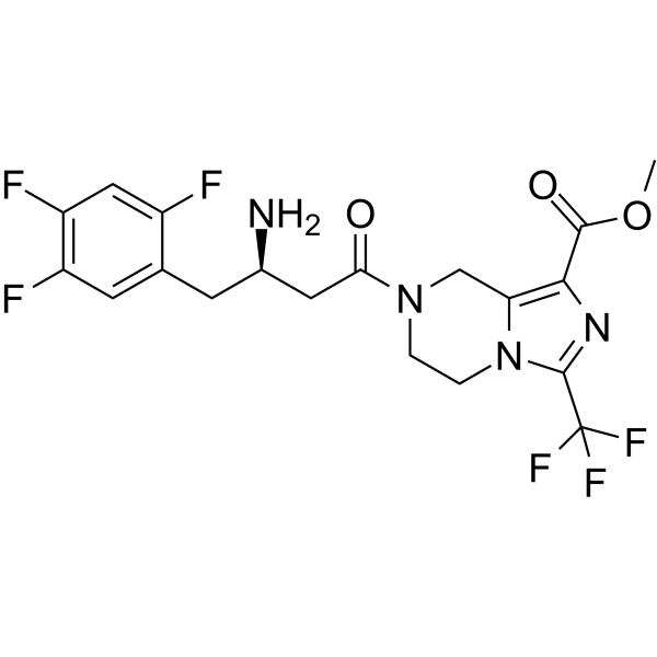
HY-112668A
RetagliptinDPP-4 Inhibitor -
HY-10287
GosogliptinDPP-IV Inhibitor
Publications Citing Use of MedChemExpress (MCE) Evogliptin tartrate
More

Biological Activity
Description
IC50 & Target
|
DPP-4 |
In Vitro
Evogliptin tartrate (2.49 mM; 12 h) efficiently inhibits mDPP4 (membrane DPP4) enzymatic activity in PWM-induced H9 Th1 cells[1].
Evogliptin tartrate prevents inflammatory and fibrotic signaling through autophagy induction in primary hepatocytes of ATG7f/f-Cre+ mice[2].
MedChemExpress (MCE) has not independently confirmed the accuracy of these methods. They are for reference only.
-
Cell Line:H9 Th1 cells (pokeweed mitogen (PWM)-induced)
-
Concentration:2.49 mM (1 µg/mL)
-
Incubation Time:12 h
-
Result:Potently inhibited mDPP4 (membrane DPP4) activity in a dose-dependent manner but did not affect either the cytokine profile or cell viability in PWM-activated CD4+CD26+ H9 Th1 cells.
In Vivo
MedChemExpress (MCE) has not independently confirmed the accuracy of these methods. They are for reference only.
-
Animal Model:Male ICR mice (four-week-old; HFD/STZ mice model)[3].
-
Dosage:100, 300 mg/kg
-
Administration:In animal feedings; single daily for 10 weeks
-
Result:Decreased the blood glucose level from the second weekand persisted through the 10-week treatment, when at 300 mg/kg.
Significantly reduced HbA1c level when dosage at 300 mg/kg.
Significantly decreased 6 h-fasted blood glucose levels in a dose-dependent manner.


Chemical Information
-
CAS No. 1222102-51-3
-
Appearance Solid
-
Molecular Weight 551.51
-
Formula C23H32F3N3O9
-
Color White to off-white
-
SMILES
O=C1NCCN(C(C[C@H](N)CC2=CC(F)=C(F)C=C2F)=O)[C@@H]1COC(C)(C)C.O=C(O)[C@H](O)[C@@H](O)C(O)=O
-
Synonyms
DA-1229 tartrate
-
Shipping
Room temperature in continental US; may vary elsewhere.
-
Storage
4°C, sealed storage, away from moisture
* In solvent : -80°C, 6 months; -20°C, 1 month (sealed storage, away from moisture)


Publications (2)
-
Journal Impact Factor
-
Most Recent
-
Cell Rep
Aortic valve stenosis serum IGFBP2 mediates female-specific Evogliptin resistance in valve myofibroblasts. [Abstract]2026 Jul 13;45(7):117678. PMID: 42441401 -
bioRxiv
Circulating biomarkers in serum from aortic valve stenosis patients predict sex-specific drug responses in valve myofibroblasts. [Abstract]2026 Jan 20:2026.01.16.700010. PMID: 41648356


Solvent & Solubility
In Vitro:
DMSO : 100 mg/mL (181.32 mM; Need ultrasonic; Hygroscopic DMSO has a significant impact on the solubility of product, please use newly opened DMSO)
Please refer to the solubility information to select the appropriate solvent. Once prepared, please aliquot and store the solution to prevent product inactivation from repeated freeze-thaw cycles.
Storage method and period of stock solution: -80°C, 6 months; -20°C, 1 month (sealed storage, away from moisture). When stored at -80°C, please use it within 6 months. When stored at -20°C, please use it within 1 month.
Please refer to the solubility information to select the appropriate solvent. Once prepared, please aliquot and store the solution to prevent product inactivation from repeated freeze-thaw cycles.
Storage method and period of stock solution: -80°C, 6 months; -20°C, 1 month (sealed storage, away from moisture). When stored at -80°C, please use it within 6 months. When stored at -20°C, please use it within 1 month.
Concentration (start) × Volume (start) = Concentration (final) × Volume (final)
In Vivo:
Select the appropriate dissolution method based on your experimental animal and administration route.
- For the following dissolution methods, please ensure to first prepare a clear stock solution using an In Vitro approach and then sequentially add co-solvents:
- To ensure reliable experimental results, the clarified stock solution can be appropriately stored based on storage conditions. As for the working solution for In Vivo experiments, it is recommended to prepare freshly and use it on the same day.
- The percentages shown for the solvents indicate their volumetric ratio in the final prepared solution. If precipitation or phase separation occurs during preparation, heat and/or sonication can be used to aid dissolution.
Add each solvent one by one: 10% DMSO 40% PEG300 5% Tween-80 45% Saline
Solubility: ≥ 2.5 mg/mL (4.53 mM); Clear solution
This protocol yields a clear solution of ≥ 2.5 mg/mL (saturation unknown).
Taking 1 mL working solution as an example, add 100 μL DMSO stock solution (25.0 mg/mL) to 400 μL PEG300, and mix evenly; then add 50 μL Tween-80 and mix evenly; then add 450 μL Saline to adjust the volume to 1 mL.
Preparation of Saline: Dissolve 0.9 g sodium chloride in ddH₂O and dilute to 100 mL to obtain a clear Saline solution.
Add each solvent one by one: 10% DMSO 90% (20% SBE-β-CD in Saline)
Solubility: ≥ 2.5 mg/mL (4.53 mM); Clear solution
This protocol yields a clear solution of ≥ 2.5 mg/mL (saturation unknown).
Taking 1 mL working solution as an example, add 100 μL DMSO stock solution (25.0 mg/mL) to 900 μL 20% SBE-β-CD in Saline, and mix evenly.
Preparation of 20% SBE-β-CD in Saline (4°C, storage for one week): 2 g SBE-β-CD powder is dissolved in 10 mL Saline, completely dissolve until clear.
In Vivo Dissolution Calculator
Please enter the basic information of animal experiments:
-
-
-
-
Recommended: Prepare an additional quantity of animals to account for potential losses during experiments.
Please enter your animal formula composition:
-
%DMSO +
Recommended: Keep the proportion of DMSO in working solution below 2% if your animal is weak.
-
%+
-
+%Tween-80 + +
-
%Saline +
The co-solvents required include: DMSO, . All of co-solvents are available by MedChemExpress (MCE). , Tween 80. All of co-solvents are available by MedChemExpress (MCE).
Working solution concentration: 0.22 mg/mL
Method for preparing stock solution: mg drug dissolved in μL DMSO. Stock solution concentration: mg/mL. * In solvent : -80°C, 6 months; -20°C, 1 month (sealed storage, away from moisture)
1. Take μL DMSO stock solution;
2. Add μL .
μL , mix evenly;
3. Then add μL Tween 80, mix evenly;
4. Then add μL
Please ensure that the stock solution in the first step is dissolved to a clear state, and add co-solvents in sequence. You can use ultrasonic heating (ultrasonic cleaner, recommended frequency 20-40 kHz), vortexing, etc. to assist dissolution.


Purity & Documentation
-
Data Sheet (284 KB)
-
SDS (252 KB)
- English - EN (252 KB)
- Français - FR (252 KB)
- Deutsch - DE (252 KB)
- Norwegian - NO (252 KB)
- Español - ES (252 KB)
- Swedish - SV (252 KB)
- Italian - IT (252 KB)
- Korean - KR (252 KB)
- Portuguese - PT (252 KB)
-
Handling Instructions (2659 KB)
References
[1]. Yoon H, et al. Effects of the Antidiabetic Drugs Evogliptin and Sitagliptin on the Immune Function of CD26/DPP4 in Th1 Cells. Biomol Ther (Seoul). 2021 Mar 1;29(2):154-165. [Content Brief]
[3]. Kim TH, et al. Hepatic role in an early glucose-lowering effect by a novel dipeptidyl peptidase 4 inhibitor, evogliptin, in a rodent model of type 2 diabetes. Eur J Pharmacol. 2016 Jan 15;771:65-76. [Content Brief]
[4]. Tan X, et al. Evogliptin: a new dipeptidyl peptidase inhibitor for the treatment of type 2 diabetes. Expert Opin Pharmacother. 2016 Jun;17(9):1285-93. [Content Brief]


Complete Stock Solution Preparation Table
Please refer to the solubility information to select the appropriate solvent. Once prepared, please aliquot and store the solution to prevent product inactivation from repeated freeze-thaw cycles.
Storage method and period of stock solution: -80°C, 6 months; -20°C, 1 month (sealed storage, away from moisture). When stored at -80°C, please use it within 6 months. When stored at -20°C, please use it within 1 month.
| Optional Solvent | Concentration Solvent Mass | 1 mg | 5 mg | 10 mg | 25 mg |
|---|---|---|---|---|---|
| DMSO | 1 mM | 1.8132 mL | 9.0660 mL | 18.1320 mL | 45.3301 mL |
| 5 mM | 0.3626 mL | 1.8132 mL | 3.6264 mL | 9.0660 mL | |
| 10 mM | 0.1813 mL | 0.9066 mL | 1.8132 mL | 4.5330 mL | |
| 15 mM | 0.1209 mL | 0.6044 mL | 1.2088 mL | 3.0220 mL | |
| 20 mM | 0.0907 mL | 0.4533 mL | 0.9066 mL | 2.2665 mL | |
| 25 mM | 0.0725 mL | 0.3626 mL | 0.7253 mL | 1.8132 mL | |
| 30 mM | 0.0604 mL | 0.3022 mL | 0.6044 mL | 1.5110 mL | |
| 40 mM | 0.0453 mL | 0.2267 mL | 0.4533 mL | 1.1333 mL | |
| 50 mM | 0.0363 mL | 0.1813 mL | 0.3626 mL | 0.9066 mL | |
| 60 mM | 0.0302 mL | 0.1511 mL | 0.3022 mL | 0.7555 mL | |
| 80 mM | 0.0227 mL | 0.1133 mL | 0.2267 mL | 0.5666 mL | |
| 100 mM | 0.0181 mL | 0.0907 mL | 0.1813 mL | 0.4533 mL |


Evogliptin tartrate Related Classifications
HY-117985B Related Classifications
 Powered by Bioz