



Lixumistat
Based on 1 publication(s) in Google Scholar
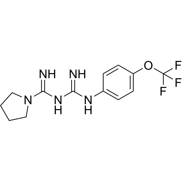
Lixumistat (IM156 free base) is a potent and orally active AMPK activator and OXPHOS inhibitor. Lixumistat strongly activates AMPK, while it lacks the systemic metabolic regulatory effects of classic metformin, such as hypoglycemic and weight-lowering activities. Lixumistat exhibits significant therapeutic effects on cognitive decline associated with brain aging and pulmonary fibrosis.
For research use only. We do not sell to patients.
- Purity : 99.33%
- CAS No.: 1422365-93-2
- Formula: C13H16F3N5O
- Molecular Weight:315.29
-
Storage:Powder -20°C, 3 years , 4°C, 2 years ; In solvent -80°C, 6 months , -20°C, 1 month

To place orders, for customer services and technical support, please contact: MedChemExpress USA
Tel: 609-228-6898 E-mail: [email protected] [email protected]


-
 Biological Activity
-
Chemical Information
-
Solvent & Solubility
- Protocols
- Purity & Documentation
- References
-
 Help & FAQs


-
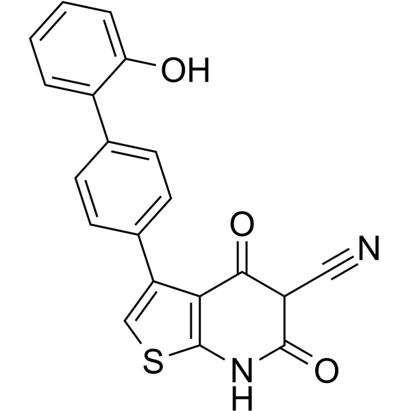
HY-50662
A-769662AMPK Activator -
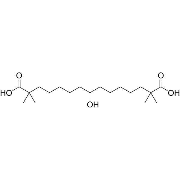
HY-12357
Bempedoic acidACL Inhibitor/AMPK Activator -
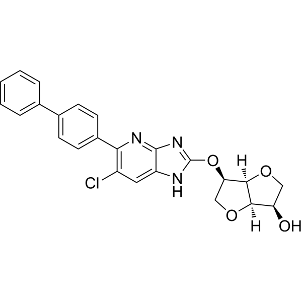
HY-111363
MK8722AMPK Activator -
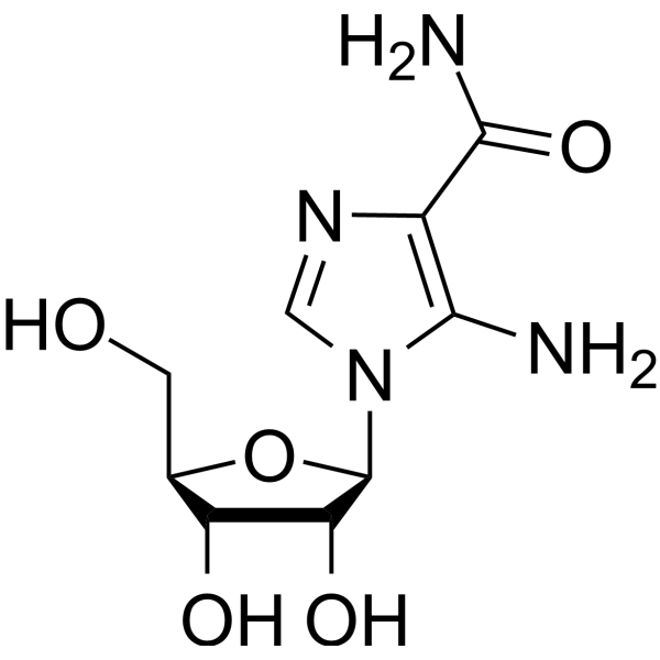
HY-13417
AICARAMPK Activator -
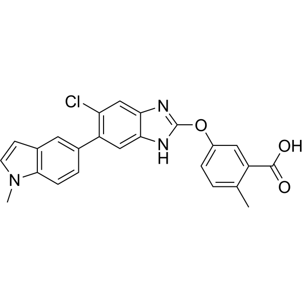
HY-112769
EX229AMPK Activator -
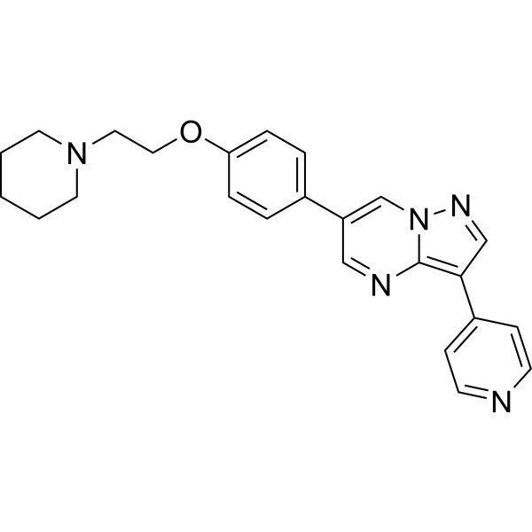
HY-13418A
DorsomorphinAMPK Inhibitor -
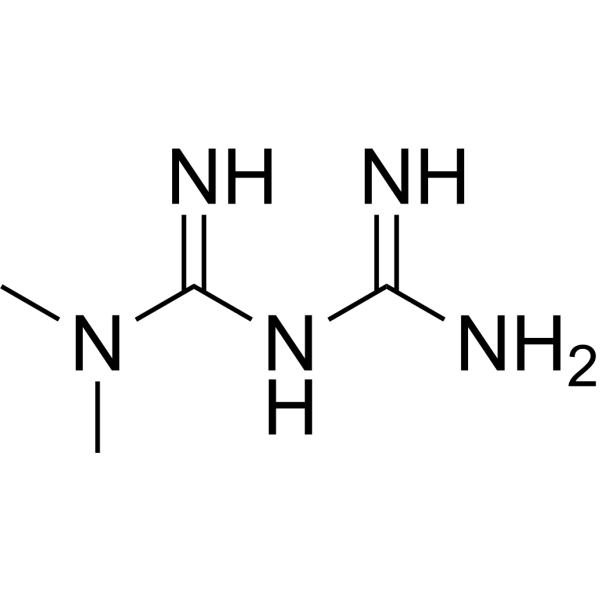
HY-B0627
MetforminAntihyperglycemic Agent -
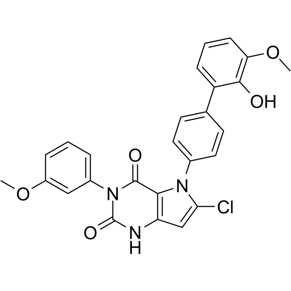
HY-100548
GSK621AMPK Activator -
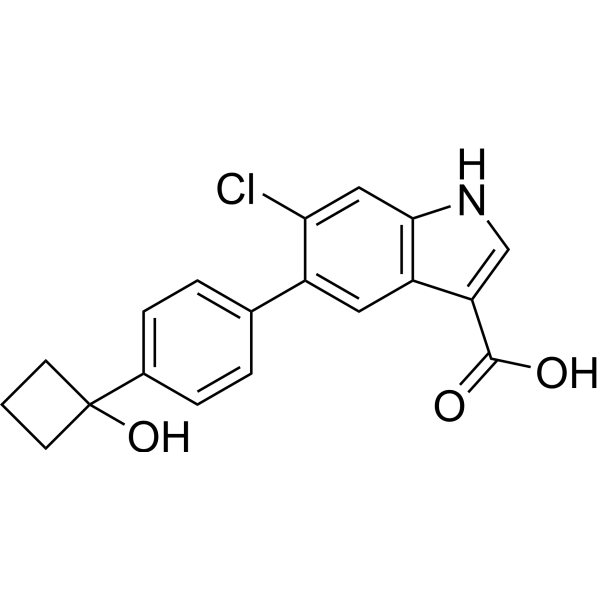
HY-103683
PF-06409577AMPK Activator -
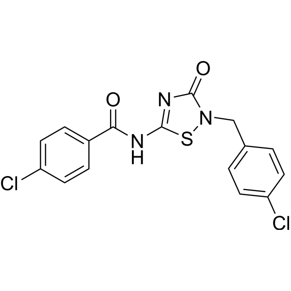
HY-112233
O-304AMPK Activator -
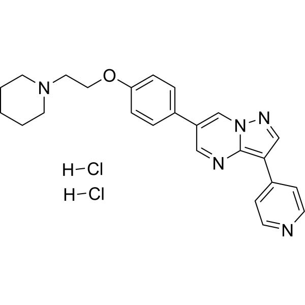
HY-13418
Dorsomorphin dihydrochlorideAMPK Inhibitor -
HY-136447
ASP4132AMPK Activator
Publications Citing Use of MedChemExpress (MCE) Lixumistat
MoreAll AMPK Isoforms
More

Biological Activity
Description
In Vitro
Lixumistat (15 μM; 26 h) potently inhibits TGFβ1-induced mitochondrial oxidative phosphorylation reprogramming in WI-38 human embryonic lung fibroblasts, with an IC50 of 14.7 μM, and does not alter TGFβ1-induced glycolytic changes[2].
Lixumistat (7.5-30 μM; 26-48 h) inhibits TGFβ1-induced myofibroblast differentiation (α-SMA expression, IC50 = 21.3 μM) and collagen deposition (IC50 = 13.2 μM) in a dose-dependent manner in WI-38 human embryonic lung fibroblasts without reducing cell viability[2].
Lixumistat (0.31-10 μM; 2-8 h) potently and rapidly induces the phosphorylation of AMPK at the Thr172 site in NIH3T3 mouse fibroblasts[3].
Lixumistat (10 μM; 0-4 h) promotes the proteasome-dependent degradation of PER2 and CRY1 proteins in NIH3T3 mouse fibroblasts when co-incubated with Cycloheximide (HY-12320)[3].
MedChemExpress (MCE) has not independently confirmed the accuracy of these methods. They are for reference only.
-
Cell Line:NIH3T3 mouse fibroblast cells
-
Concentration:0.31, 0.62, 1.25, 2.5, 5, 10 μM (dose-response); 10 μM (time-course)
-
Incubation Time:4 h (dose-response); 2, 4, 8 h (time-course)
-
Result:Dose-dependently stimulated AMPK phosphorylation, with significant increases observed at concentrations ≥ 1.25 μM after 4 h incubation.
Increased AMPK phosphorylation in a time-dependent manner, with significant elevations detected as early as 2 h after treatment with 10 μM.
-
Cell Line:NIH3T3 mouse fibroblast cells
-
Concentration:10 μM (with 30 μg/mL CHX); 10 μM (with 30 μg/mL CHX + 50 μM MG132)
-
Incubation Time:0, 1, 2, 4 h (with 30 μg/mL CHX); 0, 1, 2, 4 h (with 30 μg/mL CHX + 50 μM MG132)
-
Result:Enhanced the degradation of V5-tagged PER2 and FLAG-tagged CRY1 proteins in the presence of CHX, with protein levels decreasing more rapidly than in control cells.
Completely blocked this enhanced degradation when co-treated with MG132.
In Vivo
Lixumistat (10-30 mg/kg; i.g.; once daily for 14 consecutive days) dose-dependently alleviates pulmonary fibrosis and inflammatory responses in male C57BL/6 mice[2].
MedChemExpress (MCE) has not independently confirmed the accuracy of these methods. They are for reference only.
-
Animal Model:C57BL/6J (male and female, 20~22 months of age)[1]
-
Dosage:50 mg/kg
-
Administration:p.o.; daily; 2 months
-
Result:Significantly improved age-related cognitive decline in aged C57BL/6J mice, with a 23.2% increase in novel object recognition discrimination ratio, 27.4% increase in spatial working memory %SAP, 47.3% increase in contextual fear memory freezing level, and 91.6% increase in hippocampal pAMPK relative intensity compared to controls
-
Animal Model:C57BL/6 (male, 6-7 weeks of age, bleomycin-induced pulmonary fibrosis)[2]
-
Dosage:10 mg/kg; 30 mg/kg
-
Administration:i.g.; daily; 14 days
-
Result:Attenuated body weight loss, with significant improvements observed on study days 19 and 21.
Reduced bleomycin-induced increases in lung weight at both doses, with greater efficacy at 30 mg/kg.
Significantly reduced modified Ashcroft fibrosis scores.


Chemical Information
-
CAS No. 1422365-93-2
-
Appearance Solid
-
Molecular Weight 315.29
-
Formula C13H16F3N5O
-
Color White to off-white
-
SMILES
N=C(N1CCCC1)NC(NC2=CC=C(OC(F)(F)F)C=C2)=N
-
Synonyms
IM156 free base; HL156A free base; HL271 free base
-
Shipping
Room temperature in continental US; may vary elsewhere.
-
Storage
Powder -20°C 3 years 4°C 2 years In solvent -80°C 6 months -20°C 1 month


Publications (1)
-
Journal Impact Factor
-
Most Recent
-
Cell Death Discov
Long-term adaptation of lymphoma cell lines to hypoxia is mediated by diverse molecular mechanisms that are targetable with specific inhibitors. [Abstract]2025 Feb 18;11(1):65. PMID: 39966387


Solvent & Solubility
In Vitro:
DMSO : 100 mg/mL (317.17 mM; Need ultrasonic; Hygroscopic DMSO has a significant impact on the solubility of product, please use newly opened DMSO)
Please refer to the solubility information to select the appropriate solvent. Once prepared, please aliquot and store the solution to prevent product inactivation from repeated freeze-thaw cycles.
Storage method and period of stock solution: -80°C, 6 months; -20°C, 1 month. When stored at -80°C, please use it within 6 months. When stored at -20°C, please use it within 1 month.
Please refer to the solubility information to select the appropriate solvent. Once prepared, please aliquot and store the solution to prevent product inactivation from repeated freeze-thaw cycles.
Storage method and period of stock solution: -80°C, 6 months; -20°C, 1 month. When stored at -80°C, please use it within 6 months. When stored at -20°C, please use it within 1 month.
Concentration (start) × Volume (start) = Concentration (final) × Volume (final)
In Vivo:
Select the appropriate dissolution method based on your experimental animal and administration route.
- For the following dissolution methods, please ensure to first prepare a clear stock solution using an In Vitro approach and then sequentially add co-solvents:
- To ensure reliable experimental results, the clarified stock solution can be appropriately stored based on storage conditions. As for the working solution for In Vivo experiments, it is recommended to prepare freshly and use it on the same day.
- The percentages shown for the solvents indicate their volumetric ratio in the final prepared solution. If precipitation or phase separation occurs during preparation, heat and/or sonication can be used to aid dissolution.
Add each solvent one by one: 10% DMSO 40% PEG300 5% Tween-80 45% Saline
Solubility: ≥ 2.5 mg/mL (7.93 mM); Clear solution
This protocol yields a clear solution of ≥ 2.5 mg/mL (saturation unknown).
Taking 1 mL working solution as an example, add 100 μL DMSO stock solution (25.0 mg/mL) to 400 μL PEG300, and mix evenly; then add 50 μL Tween-80 and mix evenly; then add 450 μL Saline to adjust the volume to 1 mL.
Preparation of Saline: Dissolve 0.9 g sodium chloride in ddH₂O and dilute to 100 mL to obtain a clear Saline solution.
Add each solvent one by one: 10% DMSO 90% (20% SBE-β-CD in Saline)
Solubility: ≥ 2.5 mg/mL (7.93 mM); Clear solution
This protocol yields a clear solution of ≥ 2.5 mg/mL (saturation unknown).
Taking 1 mL working solution as an example, add 100 μL DMSO stock solution (25.0 mg/mL) to 900 μL 20% SBE-β-CD in Saline, and mix evenly.
Preparation of 20% SBE-β-CD in Saline (4°C, storage for one week): 2 g SBE-β-CD powder is dissolved in 10 mL Saline, completely dissolve until clear.
In Vivo Dissolution Calculator
Please enter the basic information of animal experiments:
-
-
-
-
Recommended: Prepare an additional quantity of animals to account for potential losses during experiments.
Please enter your animal formula composition:
-
%DMSO +
Recommended: Keep the proportion of DMSO in working solution below 2% if your animal is weak.
-
%+
-
+%Tween-80 + +
-
%Saline +
The co-solvents required include: DMSO, . All of co-solvents are available by MedChemExpress (MCE). , Tween 80. All of co-solvents are available by MedChemExpress (MCE).
Working solution concentration: 0.22 mg/mL
Method for preparing stock solution: mg drug dissolved in μL DMSO. Stock solution concentration: mg/mL.
1. Take μL DMSO stock solution;
2. Add μL .
μL , mix evenly;
3. Then add μL Tween 80, mix evenly;
4. Then add μL
Please ensure that the stock solution in the first step is dissolved to a clear state, and add co-solvents in sequence. You can use ultrasonic heating (ultrasonic cleaner, recommended frequency 20-40 kHz), vortexing, etc. to assist dissolution.


Purity & Documentation
-
Data Sheet (278 KB)
-
SDS (393 KB)
- English - EN (393 KB)
- Français - FR (393 KB)
- Deutsch - DE (393 KB)
- Norwegian - NO (393 KB)
- Español - ES (393 KB)
- Swedish - SV (393 KB)
- Italian - IT (393 KB)
- Korean - KR (393 KB)
- Portuguese - PT (393 KB)
-
Handling Instructions (2659 KB)
References
[1]. Bang E, et al. The Improving Effect of HL271, a Chemical Derivative of Metformin, a Popular Drug for Type II Diabetes Mellitus, on Aging-induced Cognitive Decline. Exp Neurobiol. 2018;27(1):45-56. [Content Brief]
[2]. Willette RN, et al. Modulation of Oxidative Phosphorylation with IM156 Attenuates Mitochondrial Metabolic Reprogramming and Inhibits Pulmonary Fibrosis. J Pharmacol Exp Ther. 2021;379(3):290-300. [Content Brief]
[3]. Row H, et al. HL271, a novel chemical compound derived from metformin, differs from metformin in its effects on the circadian clock and metabolism. Biochem Biophys Res Commun. 2016;469(3):783-789. [Content Brief]


Complete Stock Solution Preparation Table
Please refer to the solubility information to select the appropriate solvent. Once prepared, please aliquot and store the solution to prevent product inactivation from repeated freeze-thaw cycles.
Storage method and period of stock solution: -80°C, 6 months; -20°C, 1 month. When stored at -80°C, please use it within 6 months. When stored at -20°C, please use it within 1 month.
| Optional Solvent | Concentration Solvent Mass | 1 mg | 5 mg | 10 mg | 25 mg |
|---|---|---|---|---|---|
| DMSO | 1 mM | 3.1717 mL | 15.8584 mL | 31.7168 mL | 79.2921 mL |
| 5 mM | 0.6343 mL | 3.1717 mL | 6.3434 mL | 15.8584 mL | |
| 10 mM | 0.3172 mL | 1.5858 mL | 3.1717 mL | 7.9292 mL | |
| 15 mM | 0.2114 mL | 1.0572 mL | 2.1145 mL | 5.2861 mL | |
| 20 mM | 0.1586 mL | 0.7929 mL | 1.5858 mL | 3.9646 mL | |
| 25 mM | 0.1269 mL | 0.6343 mL | 1.2687 mL | 3.1717 mL | |
| 30 mM | 0.1057 mL | 0.5286 mL | 1.0572 mL | 2.6431 mL | |
| 40 mM | 0.0793 mL | 0.3965 mL | 0.7929 mL | 1.9823 mL | |
| 50 mM | 0.0634 mL | 0.3172 mL | 0.6343 mL | 1.5858 mL | |
| 60 mM | 0.0529 mL | 0.2643 mL | 0.5286 mL | 1.3215 mL | |
| 80 mM | 0.0396 mL | 0.1982 mL | 0.3965 mL | 0.9912 mL | |
| 100 mM | 0.0317 mL | 0.1586 mL | 0.3172 mL | 0.7929 mL |


Lixumistat Related Classifications
HY-136093B Related Classifications
 Powered by Bioz