



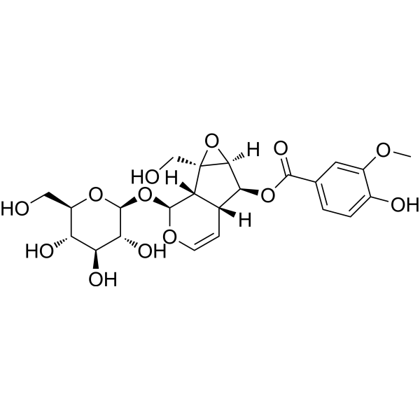
Picroside II
Based on 4 publication(s) in Google Scholar
Picroside II is an antioxidant with oral activity that can reduce the production of ROS and protect the blood-brain barrier (BBB) after CI/R injury, offering neuroprotective effects. Picroside II has antioxidant, immune-regulating, antiviral properties, and inhibits apoptosis. Picroside II alleviates the inflammatory response in sepsis by suppressing the activation of the NLRP3 inflammasome and NF-κB pathways.
商品は「研究用試薬」です。人や動物の医療用・臨床診断用・食品用の製品ではありません。
研究用途以外に使用した場合、当社は一切の責任を負いかねます。
- 純度 : 99.91%
- CAS 番号: 39012-20-9
- 分子式: C23H28O13
- 分子量:512.46
-
保管条件:
4°C, protect from light
* In solvent : -80°C, 6 months; -20°C, 1 month (protect from light)

To place orders, for customer services and technical support, please contact: MedChemExpress USA
Tel: 609-228-6898 E-mail: [email protected] [email protected]


-
 生物活性
生物活性
-
化学情報
-
溶剤 & 溶解度
- プロトコル
- 純度とドキュメンテーション
- 参考文献
-
 Help & FAQs


-
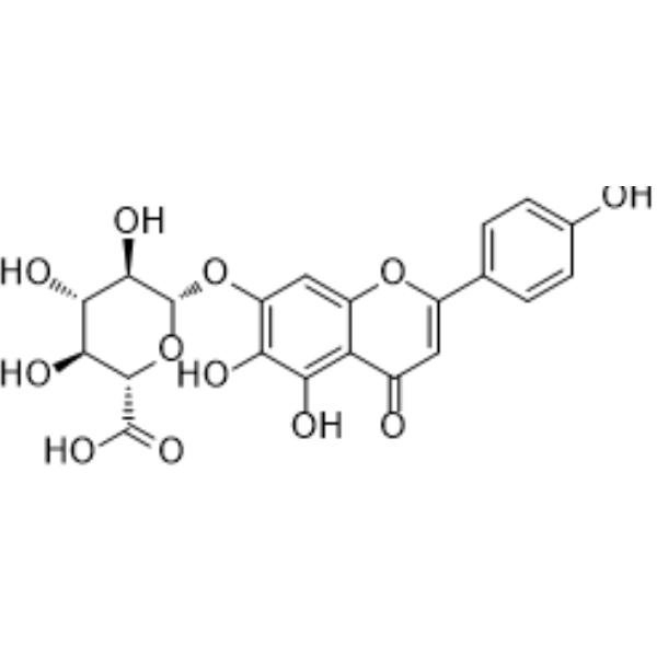
HY-N0751
ScutellarinSTAT Inhibitor -
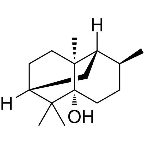
HY-N0207
Patchouli alcoholHelicobacter Pylori Inhibitor -
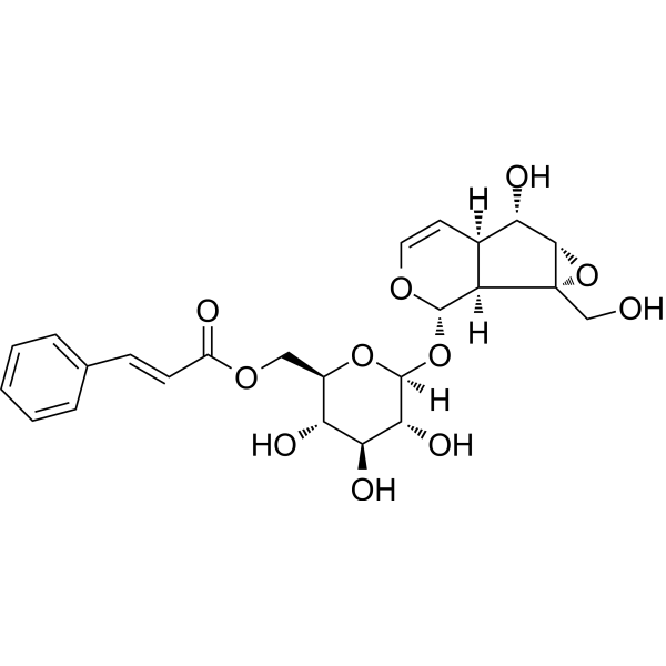
HY-N0407
Picroside ISTAT Inhibitor -

HY-N0408R
Picroside II (Standard)Apoptosis Inducer -
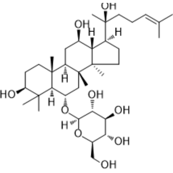
HY-N0604
Ginsenoside Rh1Endogenous Metabolite -
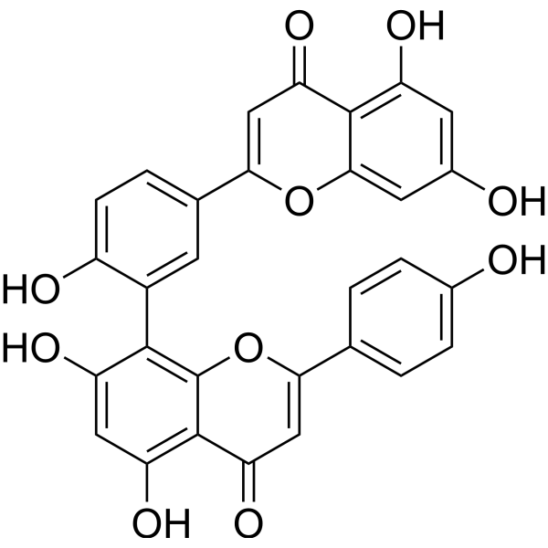
HY-N0662
AmentoflavoneGABA(A) Negative Modulator -
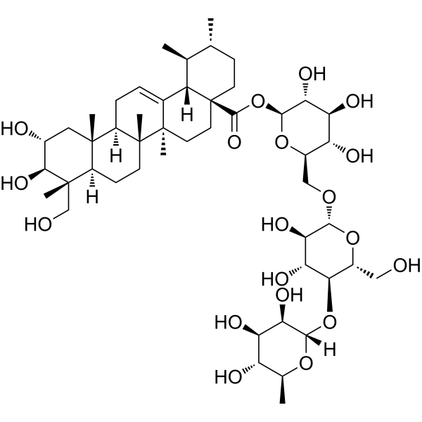
HY-N0439
AsiaticosideTriterpene Glycoside -
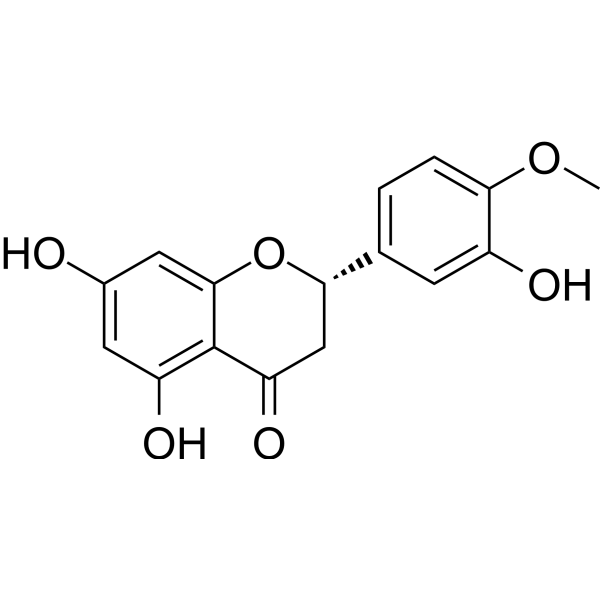
HY-N0168
HesperetinUGT Inhibitor -

HY-B0215
AcetylcysteineMucolytic Agent -
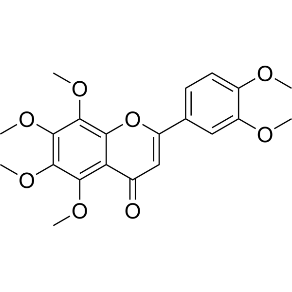
HY-N0155
NobiletinROR Agonist -
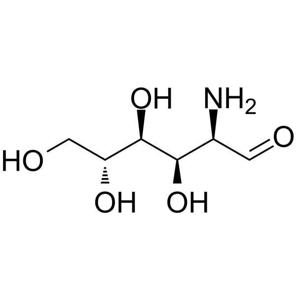
HY-B1125
GlucosamineFood Additive -
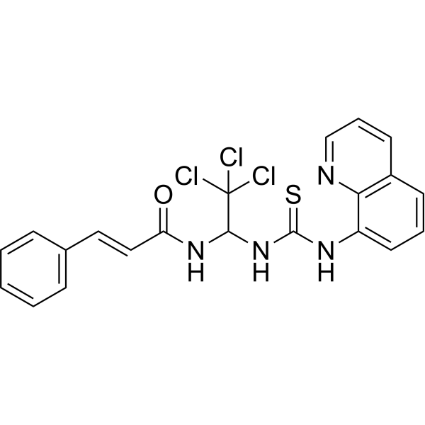
HY-15486
SalubrinaleIF2α Dephosphorylation Inhibitor
MedChemExpress(MCE)の使用を引用している文献 Picroside II
More Customer Validation & Images

-
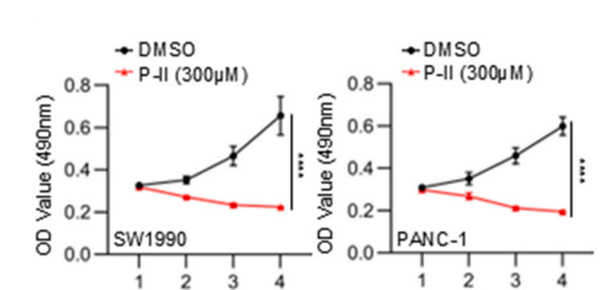
Cell Proliferation/Viability Assay
-
Cell Migration/Invasion Assay
-
In Vivo Efficacy Study
-
WB


生物活性
製品説明
体外実験
Picroside II (5-20 μM, 24 h) upregulates the expression of the bcl-2 gene in primary rat liver cells[1].
Picroside II (0-300 μM, 24 h) inhibits the growth of HepG2 cells[2].
Picroside II (10 μM, 20 h) reduces the transcription of genes involved in fatty acid uptake and synthesis in HepG2 cells[2].
MedChemExpress (MCE) has not independently confirmed the accuracy of these methods. They are for reference only.
-
Cell Line:Primary rat hepatocytes
-
Concentration:5, 10, 20 μM
-
Incubation Time:24 h
-
Result:Upregulated bcl-2.
-
Cell Line:HepG2
-
Concentration:0, 3, 10, 30, 100, 300 μM
-
Incubation Time:24 h
-
Result:Showed the inhibition rate of cell growth was 24%.
-
Cell Line:HepG2
-
Concentration:10 μM
-
Incubation Time:20 h
-
Result:Increased expression of SREBP-1 protein.
-
Cell Line:HepG2
-
Concentration:10 μM
-
Incubation Time:20 h
-
Result:Down-regulated the expression of FATP5 and increased the expression of SCD mRNA.
体内実験
Picroside II (20 mg/kg, intravenous injection, given in four doses) inhibits the activation of NLRP3 inflammasome and NF-κB pathway in mice, reducing the inflammatory response in sepsis and enhancing immune function[3].
Picroside II (20 mg/kg, intraperitoneal injection, a single dose) protects the blood-brain barrier by inhibiting oxidative signaling pathways in a rat model of ischemia-reperfusion injury[4].
Picroside II (5-20 mg/kg, i.g., once a day for seven days) has anti-lipid peroxidation effects and protects against mitochondrial damage in mice with liver injury[5].
MedChemExpress (MCE) has not independently confirmed the accuracy of these methods. They are for reference only.
-
Animal Model:DGalN and LPS(HY-D1056)-induced acute liver injury in mice[1]
-
Dosage:0, 5, 10, 20 mg/kg; administered 10 min before and 4 h after D-GalN and LPS administration
-
Administration:i.g.
-
Result:Reduced the levels of alanine aminotransferase and aspartate aminotransferase in the serum, decreased MDA content, had a dose-dependent effect on hepatocyte apoptosis, and upregulated the expression of the bcl-2 gene.
-
Animal Model:Sepsis mouse model[3]
-
Dosage:20 mg/kg; four times injected (2 h, 14 h, 26 h and 38 h after CLP)
-
Administration:Intravenous injection (i.v.)
-
Result:Improved survival, reduced sepsis-related infiltration and attenuated lung injury, enhanced bacterial clearance, reduced IL-6, IL-1β, and TNF-α levels, inhibited splenic lymphocyte apoptosis, reduced p-NF-κB (p65) expression, and reduced IL-1β and caspase-1 levels.
-
Animal Model:Rats with cerebral ischemia-reperfusion injury[4]
-
Dosage:20 mg/kg; single dose
-
Administration:Intraperitoneal injection (i.p.)
-
Result:Reduced the nervous system score, neuronal injury, BBB damage, ROS content and NADPH oxidase activity, down-regulated the protein levels of Rac-1, Nox2, ROCK, MLCK and MMP-2, and up-regulated the expression of claudin-5.
-
Animal Model:Mouse model of liver injury induced by CCl4, D-GalN and AP[5]
-
Dosage:5, 10, 20mg/kg; daily; 7 days
-
Administration:i.g.
-
Result:Reduced the high levels of ALT and AST in serum caused by CCl4, D-GalN and AP, significantly alleviated liver cell damage, reduced MDA, and increased SOD and GSH-Px content in a dose-dependent manner, inhibited AP-induced liver toxicity in mice, enhanced ATPase activity, and improved mitochondrial swelling.
- Research Protocol for Infectious Diseases
- Detection of Apoptosis
- TUNEL staining for apoptotic DNA fragmentation
- LPS-Induced Endotoxemia/Systemic Inflammation
- Annexin V plus membrane-impermeant dye apoptosis staining
- ROS/oxidative-stress fluorescent staining
- Apoptosis Solutions
- Research Protocol for Inflammation-related Diseases
- Pyroptosis Solutions


化学情報
-
CAS 番号 39012-20-9
-
性状 Solid
-
分子量 512.46
-
分子式 C23H28O13
-
Color White to yellow
-
SMILES
O[C@H]([C@H]([C@@H]([C@@H](CO)O1)O)O)[C@@H]1O[C@H]2[C@@]3([H])[C@@]([C@H](OC(C4=CC=C(O)C(OC)=C4)=O)[C@@]5([H])[C@]3(CO)O5)([H])C=CO2
-
Structure Classification
-
輸送条件
Room temperature in continental US; may vary elsewhere.
-
保管条件
4°C, protect from light
* In solvent : -80°C, 6 months; -20°C, 1 month (protect from light)


Publications (4)
-
Journal Impact Factor
-
Most Recent
-
ACS Nano
Picroside II-Encapsulated Nanoformulations as Pyroptosis Inhibitor Alleviate Cytokine Storms and Remodel Gut Microbiota Disturbances. [Abstract]2025 Sep 16;19(36):32569-32584. PMID: 40889346 -
Redox Biol
Targeting MYOF suppresses pancreatic ductal adenocarcinoma progression by inhibiting ILF3-LCN2 signaling through disrupting OTUB1-mediated deubiquitination of ILF3. [Abstract]2025 Jul:84:103665. PMID: 40381229
Picroside II purchased from MedChemExpress. Usage Cited in: Redox Biol. 2025 Jul:84:103665. [Abstract]
MTS proliferation assay to detect the proliferative ability of cells in Picroside II (300 μM)-treated and untreated groups.
Picroside II purchased from MedChemExpress. Usage Cited in: Redox Biol. 2025 Jul:84:103665. [Abstract]
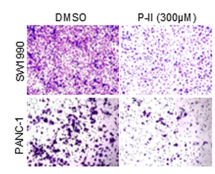
Transwell migration assay treated with Picroside II (300 μM).
Picroside II purchased from MedChemExpress. Usage Cited in: Redox Biol. 2025 Jul:84:103665. [Abstract]
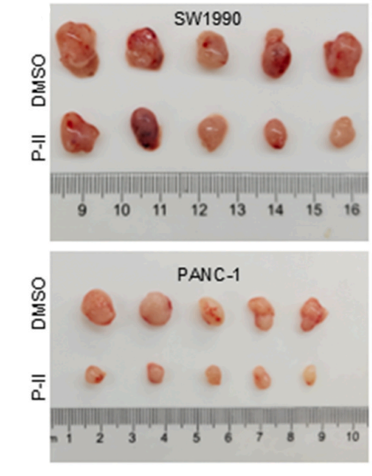
Images of subcutaneous xenografts from mice in Picroside II (0.4 mg/kg, i.p.)-treated and untreated groups.
Picroside II purchased from MedChemExpress. Usage Cited in: Redox Biol. 2025 Jul:84:103665. [Abstract]
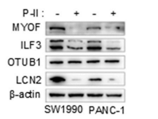
Expression of MYOF, ILF3, OTUB1, LCN2 after Picroside II (300 μM) treatment.
-
Eur J Pharmacol
The therapeutic effect of Picroside II in renal ischemia-reperfusion induced acute kidney injury: An experimental study. [Abstract]2024 Mar 15:967:176391. PMID: 38325794 -
Vet Microbiol
The Chinese medicine monomer Schisandrin C inhibits PRRSV infection by regulating the OGT-PI3K/AKT/mTOR signaling pathway. [Abstract]2026 May:316:110992. PMID: 41865607


溶剤 & 溶解度
体外:
DMSO : 100 mg/mL (195.14 mM; Need ultrasonic; Hygroscopic DMSO has a significant impact on the solubility of product, please use newly opened DMSO)
Please refer to the solubility information to select the appropriate solvent. Once prepared, please aliquot and store the solution to prevent product inactivation from repeated freeze-thaw cycles.
Storage method and period of stock solution: -80°C, 6 months; -20°C, 1 month (protect from light). When stored at -80°C, please use it within 6 months. When stored at -20°C, please use it within 1 month.
Please refer to the solubility information to select the appropriate solvent. Once prepared, please aliquot and store the solution to prevent product inactivation from repeated freeze-thaw cycles.
Storage method and period of stock solution: -80°C, 6 months; -20°C, 1 month (protect from light). When stored at -80°C, please use it within 6 months. When stored at -20°C, please use it within 1 month.
濃度 (開始) × 体積 (開始) = 濃度 (終了) × 体積 (終了)
体内:
Select the appropriate dissolution method based on your experimental animal and administration route.
- For the following dissolution methods, please ensure to first prepare a clear stock solution using an In Vitro approach and then sequentially add co-solvents:
- To ensure reliable experimental results, the clarified stock solution can be appropriately stored based on storage conditions. As for the working solution for In Vivo experiments, it is recommended to prepare freshly and use it on the same day.
- The percentages shown for the solvents indicate their volumetric ratio in the final prepared solution. If precipitation or phase separation occurs during preparation, heat and/or sonication can be used to aid dissolution.
Add each solvent one by one: 10% DMSO 40% PEG300 5% Tween-80 45% Saline
Solubility: ≥ 2.5 mg/mL (4.88 mM); Clear solution
This protocol yields a clear solution of ≥ 2.5 mg/mL (saturation unknown).
Taking 1 mL working solution as an example, add 100 μL DMSO stock solution (25.0 mg/mL) to 400 μL PEG300, and mix evenly; then add 50 μL Tween-80 and mix evenly; then add 450 μL Saline to adjust the volume to 1 mL.
Preparation of Saline: Dissolve 0.9 g sodium chloride in ddH₂O and dilute to 100 mL to obtain a clear Saline solution.
Add each solvent one by one: 10% DMSO 90% (20% SBE-β-CD in Saline)
Solubility: ≥ 2.5 mg/mL (4.88 mM); Clear solution
This protocol yields a clear solution of ≥ 2.5 mg/mL (saturation unknown).
Taking 1 mL working solution as an example, add 100 μL DMSO stock solution (25.0 mg/mL) to 900 μL 20% SBE-β-CD in Saline, and mix evenly.
Preparation of 20% SBE-β-CD in Saline (4°C, storage for one week): 2 g SBE-β-CD powder is dissolved in 10 mL Saline, completely dissolve until clear.
In Vivo Dissolution Calculator
Please enter the basic information of animal experiments:
-
-
-
-
Recommended: Prepare an additional quantity of animals to account for potential losses during experiments.
Please enter your animal formula composition:
-
%DMSO +
Recommended: Keep the proportion of DMSO in working solution below 2% if your animal is weak.
-
%+
-
+%Tween-80 + +
-
%Saline +
The co-solvents required include: DMSO, . All of co-solvents are available by MedChemExpress (MCE). , Tween 80. All of co-solvents are available by MedChemExpress (MCE).
Working solution concentration: 0.22 mg/mL
Method for preparing stock solution: mg drug dissolved in μL DMSO. Stock solution concentration: mg/mL. * In solvent : -80°C, 6 months; -20°C, 1 month (protect from light)
1. Take μL DMSO stock solution;
2. Add μL .
μL , mix evenly;
3. Then add μL Tween 80, mix evenly;
4. Then add μL
Please ensure that the stock solution in the first step is dissolved to a clear state, and add co-solvents in sequence. You can use ultrasonic heating (ultrasonic cleaner, recommended frequency 20-40 kHz), vortexing, etc. to assist dissolution.


純度とドキュメンテーション
-
データシート (285 KB)
-
SDS (393 KB)
- English - EN (393 KB)
- Français - FR (393 KB)
- Deutsch - DE (393 KB)
- Norwegian - NO (393 KB)
- Español - ES (393 KB)
- Swedish - SV (393 KB)
- Italian - IT (393 KB)
- Korean - KR (393 KB)
- Portuguese - PT (393 KB)
-
取扱説明書 (2659 KB)
参考文献
[1]. Hua Gao, et al. Inhibitory effect of picroside II on hepatocyte apoptosis. Acta Pharmacol Sin. 2005 Jun;26(6):729-36. [Content Brief]
[2]. Hiteshi Dhami-Shah, et al. Picroside II attenuates fatty acid accumulation in HepG2 cells via modulation of fatty acid uptake and synthesis. Clin Mol Hepatol. 2018 Mar;24(1):77-87. [Content Brief]
[3]. Huang Y, et al. Picroside II protects against sepsis via suppressing inflammation in mice. Am J Transl Res. 2016 Dec 15;8(12):5519-5531. eCollection 2016. [Content Brief]
[4]. Zhai L, et al. Picroside II protects the blood-brain barrier by inhibiting the oxidative signaling pathway in cerebral ischemia-reperfusion injury. PLoS One. 2017 Apr 7;12(4):e0174414. [Content Brief]
[5]. Hua Gao, et al. Anti-lipid peroxidation and protection of liver mitochondria against injuries by picroside II. World J Gastroenterol. 2005 Jun 28;11(24):3671-4. [Content Brief]


Complete Stock Solution Preparation Table
Please refer to the solubility information to select the appropriate solvent. Once prepared, please aliquot and store the solution to prevent product inactivation from repeated freeze-thaw cycles.
Storage method and period of stock solution: -80°C, 6 months; -20°C, 1 month (protect from light). When stored at -80°C, please use it within 6 months. When stored at -20°C, please use it within 1 month.
| Optional Solvent | Concentration Solvent Mass | 1 mg | 5 mg | 10 mg | 25 mg |
|---|---|---|---|---|---|
| DMSO | 1 mM | 1.9514 mL | 9.7569 mL | 19.5137 mL | 48.7843 mL |
| 5 mM | 0.3903 mL | 1.9514 mL | 3.9027 mL | 9.7569 mL | |
| 10 mM | 0.1951 mL | 0.9757 mL | 1.9514 mL | 4.8784 mL | |
| 15 mM | 0.1301 mL | 0.6505 mL | 1.3009 mL | 3.2523 mL | |
| 20 mM | 0.0976 mL | 0.4878 mL | 0.9757 mL | 2.4392 mL | |
| 25 mM | 0.0781 mL | 0.3903 mL | 0.7805 mL | 1.9514 mL | |
| 30 mM | 0.0650 mL | 0.3252 mL | 0.6505 mL | 1.6261 mL | |
| 40 mM | 0.0488 mL | 0.2439 mL | 0.4878 mL | 1.2196 mL | |
| 50 mM | 0.0390 mL | 0.1951 mL | 0.3903 mL | 0.9757 mL | |
| 60 mM | 0.0325 mL | 0.1626 mL | 0.3252 mL | 0.8131 mL | |
| 80 mM | 0.0244 mL | 0.1220 mL | 0.2439 mL | 0.6098 mL | |
| 100 mM | 0.0195 mL | 0.0976 mL | 0.1951 mL | 0.4878 mL |


Picroside II 関連分類
HY-N0408 関連分類
 Powered by Bioz