


PX-478 free base
Based on 109 publication(s) in Google Scholar
PX-478 free base is a multifunctional HIF-1α inhibitor with properties including radiosensitization, autophagy activation, and lipid accumulation inhibition. PX-478 free base also blocks hypoxia-induced VEGF production and regulates β-cell phenotypes under hypoxic conditions. PX-478 free base induces cell cycle arrest and DNA damage, restores autophagic function, reduces foam cell formation, and maintains glucose homeostasis. PX-478 free base is widely used in research on related diseases such as human tumors (e.g., prostate cancer), type 2 diabetes, and atherosclerosis.
For research use only. We do not sell to patients.
- CAS No.: 685847-78-3
- Formula: C13H18Cl2N2O3
- Molecular Weight:321.20
-
Storage:
Please store the product under the recommended conditions in the Certificate of Analysis.

To place orders, for customer services and technical support, please contact: MedChemExpress USA
Tel: 609-228-6898 E-mail: [email protected] [email protected]


-
 Biological Activity
-
Chemical Information
- Purity & Documentation
- References
-
 Help & FAQs


Publications Citing Use of MedChemExpress (MCE) PX-478 free base
More- Cancer Cell. 2025 May 12;43(5):937-954.e9. [Abstract]
- Cell. 2026 Jun 22:S0092-8674(26)00636-7. [Abstract]
- Circulation. 2020 Jul 28;142(4):384-400. [Abstract]
- Bioact Mater. 2025 Apr 8:50:152-170. [Abstract]
- Cancer Res. 2024 Oct 1;84(19):3141-3157. [Abstract]
- Nat Commun. 2025 May 20;16(1):4681. [Abstract]
- Nat Commun. 2024 Nov 8;15(1):9667. [Abstract]
- Sci Transl Med. 2020 Apr 8;12(538):eaay1620. [Abstract]
- J Nanobiotechnology. 2022 Sep 24;20(1):422. [Abstract]
- Acta Pharm Sin B. 2024 Jan;14(1):273-291. [Abstract]
- Acta Pharm Sin B. 2022 Apr;12(4):1899-1912. [Abstract]
- Adv Sci (Weinh). 2026 Feb;13(12):e10302. [Abstract]
- Sci Adv. 2025 Feb 14;11(7):eads4227. [Abstract]
- Research (Wash D C). 2025 Dec 23:8:1041. [Abstract]
- Chem Eng J. 2025 Aug 28;522:167831.
- J Control Release. 2025 Aug 25:387:114168. [Abstract]
- Cell Death Dis. 2025 Dec 10;16(1):889. [Abstract]
- Cell Death Dis. 2025 Jun 3;16(1):432. [Abstract]
- Cell Mol Biol Lett. 2025 May 7;30(1):57. [Abstract]
- Cell Death Dis. 2024 May 14;15(5):338. [Abstract]
- Cell Death Dis. 2023 Oct 21;14(10):694. [Abstract]
- Cancer Lett. 2024 May 8:593:216940. [Abstract]
- Phytomedicine. 2025 Oct:146:157135. [Abstract]
- Phytomedicine. 2024 Jul:129:155570. [Abstract]
- Phytomedicine. 2023 Sep:118:154966. [Abstract]
- J Pharm Anal. 2025 Nov 11.
- Cell Death Discov. 2026 Jan 6;12(1):81. [Abstract]
- Acta Pharmacol Sin. 2023 May;44(5):999-1013. [Abstract]
- Cell Death Discov. 2022 Jul 16;8(1):323. [Abstract]
- J Transl Med. 2023 Mar 27;21(1):224. [Abstract]
- Proc Natl Acad Sci U S A. 2025 Mar 11;122(10):e2404899122. [Abstract]
- Proc Natl Acad Sci U S A. 2024 Jan 9;121(2):e2315898120. [Abstract]
- Arterioscler Thromb Vasc Biol. 2025 Nov;45(11):1983-1996. [Abstract]
- Neural Regen Res. 2025 Feb 24. [Abstract]
- Antioxidants (Basel). 2022 Apr 7;11(4):728. [Abstract]
- Free Radic Biol Med. 2026 Mar 16:246:614-626. [Abstract]
- Free Radic Biol Med. 2024 Nov 20:225:910-924. [Abstract]
- J Anim Sci Biotechnol. 2025 Sep 20;16(1):127. [Abstract]
- Clin Transl Med. 2024 Apr;14(4):e1653. [Abstract]
- Cell Rep. 2025 Feb 25;44(3):115350. [Abstract]
- Environ Pollut. 2022 May 1:300:118937. [Abstract]
- Front Immunol. 2018 Jul 23:9:1667. [Abstract]
- Redox Rep. 2026 Dec 31;31(1):2663628. [Abstract]
- Mol Cancer Ther. 2025 May 2;24(5):772-783. [Abstract]
- JCI Insight. 2023 Aug 22;8(16):e166076. [Abstract]
- Biochem Pharmacol. 2024 Nov 9:116620. [Abstract]
- Neurobiol Dis. 2026 May 25:226:107459. [Abstract]
- Oncogenesis. 2026 Apr 2;15(1):20. [Abstract]
- Eur J Pharmacol. 2026 Jan 12:1011:178480. [Abstract]
- Eur J Pharmacol. 2025 Jun 5:996:177424. [Abstract]
- Int Immunopharmacol. 2026 Sep 1:184:116961. [Abstract]
- Int Immunopharmacol. 2026 May 14:182:116841. [Abstract]
- Int Immunopharmacol. 2026 Feb 15:171:116164. [Abstract]
- Int Immunopharmacol. 2025 Sep 26:166:115602. [Abstract]
- Int J Mol Sci. 2025 Mar 29;26(7):3182. [Abstract]
- Int Immunopharmacol. 2025 Apr 16:152:114400. [Abstract]
- Int J Mol Sci. 2024 Sep 30;25(19):10534. [Abstract]
- Int Immunopharmacol. 2024 Aug 1:140:112828. [Abstract]
- Int Immunopharmacol. 2023 Aug:121:110559. [Abstract]
- Int Immunopharmacol. 2023 May:118:110054. [Abstract]
- Mol Neurobiol. 2023 May;60(5):2355-2366. [Abstract]
- Inflammation. 2024 Oct 4. [Abstract]
- mBio. 2023 Oct 31;14(5):e0211023. [Abstract]
- Mol Med Rep. 2026 Jun;33(6):162. [Abstract]
- Mol Med Rep. 2026 May;33(5):141. [Abstract]
- Biochim Biophys Acta Mol Basis Dis. 2024 Jun;1870(5):167119. [Abstract]
- Sci Rep. 2025 Jul 2;15(1):23001. [Abstract]
- Oncol Rep. 2026 Apr;55(4):74. [Abstract]
- Hum Gene Ther. 2024 Oct;35(19-20):838-854. [Abstract]
- iScience. 2024 Jan 11;27(2):108869. [Abstract]
- Mol Nutr Food Res. 2018 Aug;62(16):e1800164. [Abstract]
- FASEB J. 2025 Mar 31;39(6):e70437. [Abstract]
- FASEB J. 2023 Sep;37(9):e23140. [Abstract]
- Front Biosci (Landmark Ed). 2025 Jan 8;30(1):27004. [Abstract]
- J Biol Chem. 2017 Jun 30;292(26):11009-11020. [Abstract]
- Metab Brain Dis. 2025 Oct 11;40(7):288. [Abstract]
- J Biomed Mater Res A. 2025 Jul;113(7):e37954. [Abstract]
- Biochim Biophys Acta Gene Regul Mech. 2025 Apr 4:195087. [Abstract]
- Arch Biochem Biophys. 2026 Apr:778:110747. [Abstract]
- Arch Biochem Biophys. 2019 Jul 30:670:94-103. [Abstract]
- Chem Biol Drug Des. 2026 Mar;107(3):e70279. [Abstract]
- Biol Reprod. 024 Dec 12;111(6):1220-1234. [Abstract]
- Iran J Basic Med Sci. 2020 Jun;23(6):819-825. [Abstract]
- J Neuroimmunol. 2026 Aug:417:578943. [Abstract]
- J Pharmacol Sci. 2026 Aug;161(4):119-129. [Abstract]
- Gene. 2024 Mar 10:898:148077. [Abstract]
- Mol Reprod Dev. 2025 May;92(5):e70025. [Abstract]
- Cardiovasc Innov Appl. 2023 May 23.
- Biochem Biophys Res Commun. 2026 Aug 27:828:154141. [Abstract]
- Biochem Biophys Res Commun. 2025 Aug 21:781:152500. [Abstract]
- Biochem Biophys Res Commun. 2021 Oct 1:572:72-79. [Abstract]
- bioRxiv. 2026 Jun 10:2026.06.09.731212. [Abstract]
- bioRxiv. 2026 Apr 1.
- bioRxiv. 2026 Mar 10.
- SSRN. 2025 Dec 19.
- Res Sq. 2025 Oct 5.
- Patent. US20250041255A1.
- Res Sq. 2024 Nov 11.
- bioRxiv. 2024 November 21.
- bioRxiv. 2024 July 06.
- bioRxiv. 2024 Jul 16:2024.07.10.602985. [Abstract]
- SSRN. 2023 Jun 29.
- SSRN. 2023 Jun 22.
- Oxid Med Cell Longev. 2022 Nov 18:2022:7862430. [Abstract]
- Oxid Med Cell Longev. 2022 Oct 18:2022:4938811. [Abstract]
- Research Square Preprint. 2022 Jul.
- Research Square Print. 2022 Jul.
- Research Square Preprint. 2021 Dec.
- Research Square Preprint. 2021 Dec.
 Customer Validation & Images

-
IHC
-
Cell Migration/Invasion Assay
-
IF
-
IHC
-
WB
All VEGFR Isoforms
More

Biological Activity
|
HIF-1α |
PX-478 free base (25 μM; 16 h) inhibits HIF-1α translation in human breast cancer MCF-7 cells under both normoxic and hypoxic conditions, with a more pronounced effect under hypoxic conditions; it also reduces global protein synthesis (especially under hypoxic conditions), yet does not alter the phosphorylation levels of hypoxia-induced translation pathway components eIF2α and 4E-BP, and suppresses the reporter gene activity driven by the 5'-UTR of HIF-1α and c-Myc[1].
PX-478 free base (20-50 μM; 20 h) inhibits HIF-1α protein in human prostate cancer cell lines PC3 and DU 145. Its activity in PC3 cells (with an IC50 of 20-25 μM under normoxic conditions) is stronger than that in DU 145 cells (with an IC50 of approximately 40-50 μM under normoxic conditions), and it also attenuates hypoxia-induced HIF-1α accumulation in both cell lines[2].
PX-478 free base (16-35 μM; 18-20 hr) reduces the clonogenic survival rate of PC3 and DU 145 human prostate cancer cells; PC3 cells show higher sensitivity under normoxic conditions (IC50 17 μM), while the sensitivity of DU 145 cells increases under hypoxic conditions (IC50 22 μM)[2].
PX-478 free base (15-20 μM; 24 hr) enhances the radiosensitivity of human PC3 prostate cancer cells under normoxic irradiation conditions, with an enhancement factor (EF) of 1.44 at 20 μM (24 hr incubation) and 1.19 at 15 μM (24 hr incubation)[2].
PX-478 free base (20-30 μM; 20 hr) enhances the radiosensitivity of hypoxic-irradiated human prostate cancer PC3 cells, with an enhancement factor (EF) of 1.56 at 20 μM (pretreated under normoxic conditions for 20 hours) and 1.78 at 30 μM (pretreated under normoxic conditions for 20 hours)[2].
Treatment with PX-478 free base (10-50 μM; 24 hr) induces S/G2M phase arrest in the human prostate cancer cell line PC3, but exerts no significant effect on the cell cycle distribution of the human prostate cancer cell line DU 145[2].
PX-478 free base (20 μM; 30 min-18 hr) induces sustained phosphorylation of γH2AX (a marker of DNA double-strand breaks) in human prostate cancer cell lines PC3 and DU 145, and prolongs radiation-induced γH2AX expression in PC3 cells even after only a short (30 min) treatment[2].
PX-478 free base improves pancreatic β-cell function in islets of mice with glucose metabolism disorders by inhibiting abnormal low-glucose intracellular Ca2+ oscillations and excessive basal insulin release[3].
PX-478 free base (100 μM; 48 h) restores autophagic function, inhibits ferroptosis and reduces lipid accumulation in ox-LDL-induced THP-1 macrophage foam cells by downregulating HIF-1α, and these effects depend on intact autophagy[4].
MedChemExpress (MCE) has not independently confirmed the accuracy of these methods. They are for reference only.
-
Cell Line:PC3 and DU 145 human prostate carcinoma cells
-
Concentration:20 μM (20 hr normoxia pretreatment followed by 1 hr hypoxia, PC3 cells); 50 μM (20 hr normoxia pretreatment followed by 1 hr hypoxia, DU 145 cells)
-
Incubation Time:20 hr (normoxia); 20 hr normoxia pretreatment followed by 1 hr hypoxia
-
Result:Inhibited HIF-1α protein with an IC50 of 20-25 μmol/L in PC3 cells under normoxia.
Inhibited HIF-1α protein with an IC50 of ~40-50 μmol/L in DU 145 cells under normoxia.
Reduced hypoxia-induced HIF-1α accumulation by 40% in PC3 cells treated with 20 μM.
Reduced hypoxia-induced HIF-1α accumulation by 35% in DU 145 cells treated with 50 μM.
-
Cell Line:PC3 and DU 145 human prostate carcinoma cells
-
Concentration:17 μM (20 hr normoxia, PC3 cells); 35 μM (20 hr normoxia, DU 145 cells); 16 μM (18 hr hypoxia, PC3 cells); 22 μM (18 hr hypoxia, DU 145 cells)
-
Incubation Time:18-20 hr (normoxia or hypoxia)
-
Result:Achieved an IC50 of 17 μmol/L for clonogenic survival in PC3 cells under normoxia.
Achieved an IC50 of 35 μmol/L for clonogenic survival in DU 145 cells under normoxia.
Achieved an IC50 of 16 μmol/L for clonogenic survival in PC3 cells under hypoxia.
Achieved an IC50 of 22 μmol/L for clonogenic survival in DU 145 cells under hypoxia.
Increased sensitivity of DU 145 cells to PX-478 under hypoxia compared to normoxia.
-
Cell Line:PC3 and DU 145 human prostate carcinoma cells
-
Concentration:20 μM
-
Incubation Time:30 min; 18 hr
-
Result:Increased γH2AX foci and protein levels in both cell lines when used alone, with the effect persisting at 24 hr after drug removal.
Resulted in significantly higher γH2AX foci and protein levels at 24 hr post-irradiation in PC3 cells when combined with radiation compared to radiation alone.
Increased γH2AX foci in irradiated PC3 cells at 24 hr post-irradiation even after 30-min exposure compared to radiation alone.
Resulted in γH2AX levels comparable to PX-478 alone at 24 hr post-irradiation in DU 145 cells when combined with radiation.
-
Cell Line:ox-LDL-induced THP-1 macrophage foam cells
-
Concentration:100 μM
-
Incubation Time:48 h
-
Result:Counteracted the increase in MitoROS-positive cells induced by ox-LDL.
Restored the reduced protein aggregation and expression levels of GPX4 and LC3II.
Increased autophagosome numbers.
Alleviated mitochondrial shrinkage.
Attenuated the upregulation of HIF-1α protein.
Reduced elevated iron content.
Reversed decreased GSH concentration.
Significantly reduced intracellular lipid accumulation.
Had all its effects abrogated by co-treatment with autophagy inhibitor 3-MA.
PX-478 (free base) exhibits in vivo antitumor activity against human tumor xenografts, with activity positively correlated with tumor HIF-1α levels and primary activity attributed to inhibition of glycolysis via prolonged Glut-1 reduction[2].
PX-478 (free base) (daily; 5 days) causes neutropenia as the primary acute toxicity in nonimmunodeficient C57BL/6 mice[2].
PX-478 (free base) prevents glycemia elevation and diabetes progression by sustaining elevated plasma insulin concentrations in db/db mice[3].
PX-478 (free base) improves glucose homeostasis recovery and enhances multiple markers of pancreatic β cell function and maturity in streptozotocin-induced diabetic mice[3].
PX-478 (5 mg/kg; i.p.; once every two days; two months) reduces atherosclerotic plaque, lipid lesion, and mucin areas by downregulating HIF-1α expression in HFD-fed ApoE-/- mice[4].
MedChemExpress (MCE) has not independently confirmed the accuracy of these methods. They are for reference only.
-
Animal Model:Apolipoprotein E knockout (ApoE-/-) (C57BL/6 background, 4-weeks-old, fed high-fat diet for five months)[4]
-
Dosage:5 mg/kg
-
Administration:i.p.; once every two days; two months
-
Result:Reduced relative plaque area, relative lipid lesion area, and relative mucin area in the aortic sinus compared to untreated high-fat diet-fed ApoE-/- mice.
Reduced enhanced fluorescence intensity of HIF-1α in high-fat diet-fed mice.
Significantly downregulated aortic HIF-1α protein expression compared to untreated high-fat diet-fed ApoE-/- mice.


Chemical Information
-
CAS No. 685847-78-3
-
Molecular Weight 321.20
-
Formula C13H18Cl2N2O3
-
SMILES
ClCC[N+](C1=CC=C(C=C1)C[C@H](N)C(O)=O)([O-])CCCl
-
Shipping
Room temperature in continental US; may vary elsewhere.
-
Storage
Please store the product under the recommended conditions in the Certificate of Analysis.


Publications (109)
-
Journal Impact Factor
-
Most Recent
-
Cancer Cell
2025 May 12;43(5):937-954.e9. PMID: 40054467
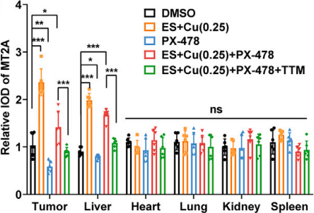
PX-478 free base purchased from MedChemExpress. Usage Cited in: Cancer Cell. 2025 May 12;43(5):937-954.e9. [Abstract]
IHC of MT2A in HCT116-bearing mice treated with ES + Cu (0.25 mg/kg, per day, i.p.) and PX-478 (20 mg/kg per day, i.p.).
-
Cell
2026 Jun 22:S0092-8674(26)00636-7. PMID: 42330950 -
Circulation
Single-Cell RNA Sequencing to Dissect the Immunological Network of Autoimmune Myocarditis. [Abstract]2020 Jul 28;142(4):384-400. PMID: 32431172 -
Bioact Mater
Bidirectional modulation of glycolysis using a multifunctional nanocomposite hydrogel promotes bone fracture healing in type 2 diabetes mellitus. [Abstract]2025 Apr 8:50:152-170. PMID: 40256330 -
Cancer Res
Hypoxic Memory Mediates Prolonged Tumor Intrinsic Type I Interferon Suppression to Promote Breast Cancer Progression. [Abstract]2024 Oct 1;84(19):3141-3157. PMID: 38990731 -
Nat Commun
Histone methyltransferase ASH1L primes metastases and metabolic reprogramming of macrophages in the bone niche. [Abstract]2025 May 20;16(1):4681. PMID: 40394007
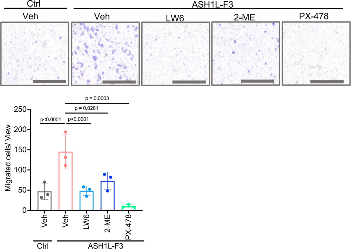
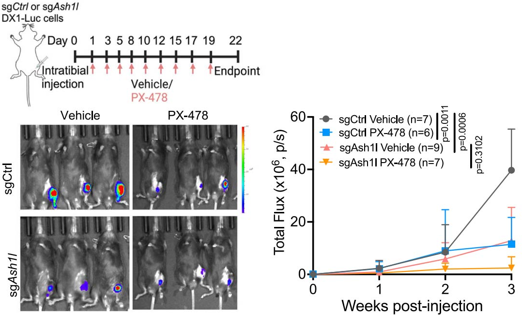
PX-478 free base purchased from MedChemExpress. Usage Cited in: Nat Commun. 2025 May 20;16(1):4681. [Abstract]
Control and ASH1L-F3-overexpressing LNCaP cells were treated with Vehicle (Veh) or HIF-1α inhibitors LW6 (15 μM), 2-MeOE2 (2-ME, 25 μM), or PX-478 (20 μM) for 48 h in the presence of CoCl2 (200uM), followed by migration assays. n = 3 biological replicates per group. Scale bar = 1000 μm.
PX-478 free base purchased from MedChemExpress. Usage Cited in: Nat Commun. 2025 May 20;16(1):4681. [Abstract]
PX-478 (40 mg/kg; i.p.; thrice weekly for three weeks) abrogates the significant inhibition of intraosseous tumor growth induced by ASH1L knockdown in C57BL/6J mice.
-
Nat Commun
FNDC4 alleviates cardiac ischemia/reperfusion injury through facilitating HIF1α-dependent cardiomyocyte survival and angiogenesis in male mice. [Abstract]2024 Nov 8;15(1):9667. PMID: 39516487 -
Sci Transl Med
2020 Apr 8;12(538):eaay1620. PMID: 32269165
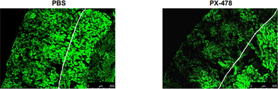
PX-478 free base purchased from MedChemExpress. Usage Cited in: Sci Transl Med. 2020 Apr 8;12(538):eaay1620. [Abstract]
Representative pimonidazole staining of kidney sections from 16-week-old MRL/lpr mice treated with PBS or PX-478, and quantification of pimonidazole-positive cortical tubular cells.
-
J Nanobiotechnology
Small extracellular vesicles of hypoxic endothelial cells regulate the therapeutic potential of adipose-derived mesenchymal stem cells via miR-486-5p/PTEN in a limb ischemia model. [Abstract]2022 Sep 24;20(1):422. PMID: 36153544 -
Acta Pharm Sin B
Adiponectin restores the obesity-induced impaired immunomodulatory function of mesenchymal stromal cells via glycolytic reprogramming. [Abstract]2024 Jan;14(1):273-291. PMID: 38261813 -
Acta Pharm Sin B
Disruption of adipocyte HIF-1 α improves atherosclerosis through the inhibition of ceramide generation. [Abstract]2022 Apr;12(4):1899-1912. PMID: 35847503 -
Adv Sci (Weinh)
Single-Nucleus Multi-Omics Reveals Hypoxia-Driven Angiogenic Programs and Their Epigenetic Control in Sinonasal Squamous Cell Carcinoma. [Abstract]2026 Feb;13(12):e10302. PMID: 41498635 -
Sci Adv
2025 Feb 14;11(7):eads4227. PMID: 39937892 -
Research (Wash D C)
UBE2V1 Promotes Hepatocellular Carcinoma Progression by Forming a Positive Feedback Loop with HIF-1α. [Abstract]2025 Dec 23:8:1041. PMID: 41446875 -
-
J Control Release
In situ formed chemo-immunotherapeutic hydrogel for suppression of postoperative glioma recurrence and intraoperative hemostasis. [Abstract]2025 Aug 25:387:114168. PMID: 40865879 -
Cell Death Dis
De-ubiquitinase USP35 promotes peritoneal dissemination of gastric cancer by regulating metabolic reprogramming. [Abstract]2025 Dec 10;16(1):889. PMID: 41372134 -
Cell Death Dis
Oncometabolite fumarate facilitates PD-L1 expression and immune evasion in clear cell renal cell carcinoma. [Abstract]2025 Jun 3;16(1):432. PMID: 40461489 -
Cell Mol Biol Lett
Immunodynamic axis of fibroblast-driven neutrophil infiltration in acute pancreatitis: NF-κB-HIF-1α-CXCL1. [Abstract]2025 May 7;30(1):57. PMID: 40335899 -
Cell Death Dis
METTL8 links mt-tRNA m3C modification to the HIF1α/RTK/Akt axis to sustain GBM stemness and tumorigenicity. [Abstract]2024 May 14;15(5):338. PMID: 38744809 -
Cell Death Dis
Sirtuin4 alleviates severe acute pancreatitis by regulating HIF-1α/HO-1 mediated ferroptosis. [Abstract]2023 Oct 21;14(10):694. PMID: 37865653 -
Cancer Lett
β-hydroxybutyrate restrains colitis-associated tumorigenesis by inhibiting HIF-1α-mediated angiogenesis. [Abstract]2024 May 8:593:216940. PMID: 38729554 -
Phytomedicine
Astragaloside IV alleviates radiation-induced heart disease by regulating energy metabolism. [Abstract]2025 Oct:146:157135. PMID: 40774010 -
Phytomedicine
Baicalin attenuates neuronal damage associated with SDH activation and PDK2-PDH axis dysfunction in early reperfusion. [Abstract]2024 Jul:129:155570. PMID: 38579645 -
Phytomedicine
Dan-Deng-Tong-Nao softgel capsule promotes angiogenesis of cerebral microvasculature to protect cerebral ischemia reperfusion injury via activating HIF-1α-VEGFA-Notch1 signaling pathway. [Abstract]2023 Sep:118:154966. PMID: 37487254 -
-
Cell Death Discov
Mitochondrial retrograde signaling initiates HIF-1α/BNIP3/NIX-mediated mitophagy in Tibetan high-altitude adaptation. [Abstract]2026 Jan 6;12(1):81. PMID: 41490888 -
Acta Pharmacol Sin
GDF11 promotes wound healing in diabetic mice via stimulating HIF-1ɑ-VEGF/SDF-1ɑ-mediated endothelial progenitor cell mobilization and neovascularization. [Abstract]2023 May;44(5):999-1013. PMID: 36347996 -
Cell Death Discov
Estrogens revert neutrophil hyperplasia by inhibiting Hif1α-cMyb pathway in zebrafish myelodysplastic syndromes models. [Abstract]2022 Jul 16;8(1):323. PMID: 35842445 -
J Transl Med
Cholesterol suppresses human iTreg differentiation and nTreg function through mitochondria-related mechanisms. [Abstract]2023 Mar 27;21(1):224. PMID: 36973679 -
Proc Natl Acad Sci U S A
2025 Mar 11;122(10):e2404899122. PMID: 40030031 -
Proc Natl Acad Sci U S A
Gp130-HIF1α axis-induced vascular damage is prevented by the short-term inhibition of IL-6 receptor signaling. [Abstract]2024 Jan 9;121(2):e2315898120. PMID: 38165930 -
Arterioscler Thromb Vasc Biol
2025 Nov;45(11):1983-1996. PMID: 41036559 -
Neural Regen Res
2025 Feb 24. PMID: 39995090 -
Antioxidants (Basel)
Hydroxysafflor Yellow A Blocks HIF-1 α Induction of NOX2 and Protects ZO-1 Protein in Cerebral Microvascular Endothelium. [Abstract]2022 Apr 7;11(4):728. PMID: 35453413 -
Free Radic Biol Med
HIF-1α translation mediated by PKCδ facilitates RSV-induced production of innate inflammatory cytokines in vitro and in vivo. [Abstract]2026 Mar 16:246:614-626. PMID: 41611036 -
Free Radic Biol Med
Influenza A virus-induced glycolysis facilitates virus replication by activating ROS/HIF-1α pathway. [Abstract]2024 Nov 20:225:910-924. PMID: 39491735 -
J Anim Sci Biotechnol
HIF1A regulates follicular atresia through O-GlcNAcylation-mediated VEZF1/ET-1/FOXO1/BAX signaling in porcine granulosa cells. [Abstract]2025 Sep 20;16(1):127. PMID: 40973947 -
Clin Transl Med
Mitochondrial dysfunction induced by HIF-1α under hypoxia contributes to the development of gastric mucosal lesions. [Abstract]2024 Apr;14(4):e1653. PMID: 38616702 -
Cell Rep
Hexokinase 2-mediated metabolic stress and inflammation burden of liver macrophages via histone lactylation in MASLD. [Abstract]2025 Feb 25;44(3):115350. PMID: 40014451 -
Environ Pollut
Oxygen sensors mediated HIF-1α accumulation and translocation: A pivotal mechanism of fine particles-exacerbated myocardial hypoxia injury. [Abstract]2022 May 1:300:118937. PMID: 35114305 -
Front Immunol
Hypoxia Exacerbates Inflammatory Acute Lung Injury via the Toll-Like Receptor 4 Signaling Pathway. [Abstract]2018 Jul 23:9:1667. PMID: 30083155
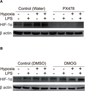
PX-478 free base purchased from MedChemExpress. Usage Cited in: Front Immunol. 2018 Jul 23:9:1667. [Abstract]
Manipulation of the protein level of hypoxia-inducible factor 1 alpha (HIF-1α) in NR8383. 50 µM PX478 treatment for 20 h is used to downregulate the HIF-1α protein level in NR8383 cells (A). 1 mM DMOG treatment for 8 h is used to upregulate the HIF-1α protein level in NR8383 cells (B).
-
Redox Rep
Rhaponticin alleviates pancreatic acinar cell necrosis by attenuating oxidative stress via modulation of the HIF-1α signaling pathway. [Abstract]2026 Dec 31;31(1):2663628. PMID: 42261783 -
Mol Cancer Ther
Heterogeneous Responses to High-Dose Testosterone in Castration-Resistant Prostate Cancer Tumors with Mixed Rb-Proficient and Rb-Deficient Cells. [Abstract]2025 May 2;24(5):772-783. PMID: 40116305 -
JCI Insight
2023 Aug 22;8(16):e166076. PMID: 37526979 -
Biochem Pharmacol
PX-478 induces apoptosis in acute myeloid leukemia under hypoxia by inhibiting the PI3K/AKT/mTOR pathway through downregulation of GBE1. [Abstract]2024 Nov 9:116620. PMID: 39528073 -
Neurobiol Dis
ROS-HIF1α-driven glycolytic reprogramming sustains ATP production in Huntington's disease. [Abstract]2026 May 25:226:107459. PMID: 42190991 -
Oncogenesis
GPX3 suppresses gallbladder cancer progression by modulating redox balance, glycolysis, and anti-tumor immunity. [Abstract]2026 Apr 2;15(1):20. PMID: 41927557 -
Eur J Pharmacol
Nobiletin ameliorates intervertebral disc degeneration by upregulating HIF-1α to inhibit endoplasmic reticulum stress-induced apoptosis. [Abstract]2026 Jan 12:1011:178480. PMID: 41397628 -
Eur J Pharmacol
Arctiin attenuated NASH by inhibiting glycolysis and inflammation via FGFR2/CSF1R signaling. [Abstract]2025 Jun 5:996:177424. PMID: 40010483 -
Int Immunopharmacol
Rheumatoid arthritis synovial fibroblasts promote the glycolysis of myeloid-derived suppressor cells via TREM1/mTOR axis. [Abstract]2026 Sep 1:184:116961. PMID: 42242136 -
Int Immunopharmacol
Albiflorin contributes to Xuebijing-mediated protection against sepsis-associated acute kidney injury by modulating the succinate-PFKFB3 immunometabolic axis. [Abstract]2026 May 14:182:116841. PMID: 42134292 -
Int Immunopharmacol
Icariin sensitizes glucocorticoid therapy in doxorubicin-induced fibrotic nephrotic syndrome via the HIF-1α/NF-κB/HDAC2 Axis. [Abstract]2026 Feb 15:171:116164. PMID: 41500175 -
Int Immunopharmacol
N-formyl methionine mediates NETosis of neutrophil to promote sepsis-induced cardiomyopathy via the FPR1 pathway. [Abstract]2025 Sep 26:166:115602. PMID: 41014772 -
Int J Mol Sci
Role of Hypoxia-Inducible Factors in Respiratory Syncytial Virus Infection-Associated Lung Disease. [Abstract]2025 Mar 29;26(7):3182. PMID: 40244000 -
Int Immunopharmacol
Unraveling the crucial role of SDF-1 in osteoarthritis progression: IL6/HIF-1α positive feedback and chondrocyte ferroptosis. [Abstract]2025 Apr 16:152:114400. PMID: 40058106 -
Int J Mol Sci
Nicotinamide Mononucleotide (NMN) Ameliorates Free Fatty Acid-Induced Pancreatic β-Cell Dysfunction via the NAD+/AMPK/SIRT1/HIF-1α Pathway. [Abstract]2024 Sep 30;25(19):10534. PMID: 39408861 -
Int Immunopharmacol
Isocitrate dehydrogenases 2-mediated dysfunctional metabolic reprogramming promotes intestinal cancer progression via regulating HIF-1A signaling pathway. [Abstract]2024 Aug 1:140:112828. PMID: 39094359 -
Int Immunopharmacol
Hypoxia induces the production of epithelial-derived cytokines in eosinophilic chronic rhinosinusitis with nasal polyps. [Abstract]2023 Aug:121:110559. PMID: 37364325 -
Int Immunopharmacol
Hypoxia disrupts the nasal epithelial barrier by inhibiting PTPN2 in chronic rhinosinusitis with nasal polyps. [Abstract]2023 May:118:110054. PMID: 36963262 -
Mol Neurobiol
Advanced Glycation End-Products (AGEs) Promote Endothelial Cell Pyroptosis Under Cerebral Ischemia and Hypoxia via HIF-1α-RAGE-NLRP3. [Abstract]2023 May;60(5):2355-2366. PMID: 36652049 -
Inflammation
Aquaporin-1 Facilitates Macrophage M1 Polarization by Enhancing Glycolysis Through the Activation of HIF1α in Lipopolysaccharide-Induced Acute Kidney Injury. [Abstract]2024 Oct 4. PMID: 39365391 -
mBio
Respiratory syncytial virus co-opts hypoxia-inducible factor-1α-mediated glycolysis to favor the production of infectious virus. [Abstract]2023 Oct 31;14(5):e0211023. PMID: 37796013 -
Mol Med Rep
Uremic toxin p‑cresyl sulfate enhances the calcification of aortic valvular interstitial cells via klotho/sirtuin‑1 signaling. [Abstract]2026 Jun;33(6):162. PMID: 41930468 -
Mol Med Rep
LDHA protects vascular endothelial cells from oxidative stress‑induced mitochondrial damage via HIF‑1α activation and glycolytic reprogramming. [Abstract]2026 May;33(5):141. PMID: 41891951 -
Biochim Biophys Acta Mol Basis Dis
Pulmonary succinate receptor 1 elevation in high-fat diet mice exacerbates lipopolysaccharides-induced acute lung injury via sensing succinate. [Abstract]2024 Jun;1870(5):167119. PMID: 38479484 -
Sci Rep
Circadian gene BMAL1 ameliorates renal ischaemia-reperfusion injury in diabetic mice by enhancing mitophagy via the HIF-1/BNIP3 pathway. [Abstract]2025 Jul 2;15(1):23001. PMID: 40594206 -
Oncol Rep
Hypoxia‑induced miR‑135b‑5p promotes neuroendocrine differentiation of prostate cancer cells through HIF1AN‑HIF1α axis. [Abstract]2026 Apr;55(4):74. PMID: 41716025 -
Hum Gene Ther
Matrix protein of vesicular stomatitis virus targets the mitochondria, reprograms glucose metabolism,and sensitizes to 2-deoxyglucose in glioblastoma. [Abstract]2024 Oct;35(19-20):838-854. PMID: 39001830 -
iScience
Heat-killed Mycobacterium tuberculosis induces trained immunity in vitro and in vivo administered systemically or intranasally. [Abstract]2024 Jan 11;27(2):108869. PMID: 38318361 -
Mol Nutr Food Res
Frataxin-Mediated PINK1-Parkin-Dependent Mitophagy in Hepatic Steatosis: The Protective Effects of Quercetin. [Abstract]2018 Aug;62(16):e1800164. PMID: 29935106 -
FASEB J
Uric acid promotes aortic valve calcification via mediating valve interstitial cell osteogenic differentiation and endothelial dysfunction. [Abstract]2025 Mar 31;39(6):e70437. PMID: 40100089 -
FASEB J
HIF-1α inhibition in macrophages preserves acute liver failure by reducing IL-1β production. [Abstract]2023 Sep;37(9):e23140. PMID: 37584647 -
Front Biosci (Landmark Ed)
HIF-1α and VEGF Immunophenotypes as Potential Biomarkers in the Prognosis and Evaluation of Treatment Efficacy of Atherosclerosis: A Systematic Review of the Literature. [Abstract]2025 Jan 8;30(1):27004. PMID: 39862086 -
J Biol Chem
The coccidian parasites Toxoplasma and Neospora dysregulate mammalian lipid droplet biogenesis. [Abstract]2017 Jun 30;292(26):11009-11020. PMID: 28487365 -
Metab Brain Dis
Promotion of angiogenesis in cerebral infarction by Tongqiao Huoxue Decoction through activation of glycolysis. [Abstract]2025 Oct 11;40(7):288. PMID: 41074933 -
J Biomed Mater Res A
Enhancing Bone Regeneration: The Role of Biomimetic Silicified Collagen Scaffold in Osteogenesis and Angiogenesis. [Abstract]2025 Jul;113(7):e37954. PMID: 40635198 -
Biochim Biophys Acta Gene Regul Mech
Hypoxia inducible factor HIF1α elevates expression of mRNA capping enzyme during cobalt chloride-induced hypoxia. [Abstract]2025 Apr 4:195087. PMID: 40189045 -
Arch Biochem Biophys
Exosomes derived from HISLA overexpressed-adipose stem cells accelerate wound healing in diabetic foot ulcers by regulating HIF-1α signal transduction. [Abstract]2026 Apr:778:110747. PMID: 41581639 -
Arch Biochem Biophys
Chaetocin attenuates gout in mice through inhibiting HIF-1α and NLRP3 inflammasome-dependent IL-1β secretion in macrophages. [Abstract]2019 Jul 30:670:94-103. PMID: 31255694 -
Chem Biol Drug Des
Tetrahydropalmatine Alleviates Osteoarthritis-Associated Pain and Inflammation by Suppressing KDM4A/MDM2/HIF-1α-Mediated M1 Macrophage Polarization. [Abstract]2026 Mar;107(3):e70279. PMID: 41857767 -
Biol Reprod
Fetal hypoxia exposure induces Hif1a activation and autophagy in adult ovarian granulosa cells†. [Abstract]024 Dec 12;111(6):1220-1234. PMID: 39361887 -
Iran J Basic Med Sci
Triiodothyronine potentiates angiogenesis-related factor expression through PI3K/AKT signaling pathway in human osteoarthritic osteoblasts. [Abstract]2020 Jun;23(6):819-825. PMID: 32695299 -
J Neuroimmunol
Establishment of a passive-transfer mouse model of anti-NMDAR encephalitis and delineation of the PI3K-AKT-HIF-1α Axis in antibody-induced neuronal injury. [Abstract]2026 Aug:417:578943. PMID: 42048740 -
J Pharmacol Sci
Dendrobine alleviates lung injury in septic mice by inhibiting mitochondrial-endoplasmic reticulum crosstalk-mediated NLRP3 inflammasome activation. [Abstract]2026 Aug;161(4):119-129. PMID: 42303347 -
Gene
HIF signaling overactivation inhibits lateral line neuromast development through Wnt in zebrafish. [Abstract]2024 Mar 10:898:148077. PMID: 38097093 -
Mol Reprod Dev
Decreased OGT Attenuates Endometrial Decidualization and Embryo Implantation by Affecting HIF-1α Stability. [Abstract]2025 May;92(5):e70025. PMID: 40342239 -
-
Biochem Biophys Res Commun
Dietary fatty acids activate FATP4 and lipid transport via HIF1α and KDM4B in the small intestinal enterocytes. [Abstract]2026 Aug 27:828:154141. PMID: 42284990 -
Biochem Biophys Res Commun
Isoquercitrin inhibits ferroptosis and ameliorates insulin resistance: Evidence from network pharmacology and in vitro studies. [Abstract]2025 Aug 21:781:152500. PMID: 40858065 -
Biochem Biophys Res Commun
Inhibition of hypoxia-inducible factor-1α alleviates acinar cell necrosis in a mouse model of acute pancreatitis. [Abstract]2021 Oct 1:572:72-79. PMID: 34358966 -
bioRxiv
Epigenetic de-repression of basal cell metaplasia in aging AT2 cells is a risk factor for idiopathic pulmonary fibrosis (IPF). [Abstract]2026 Jun 10:2026.06.09.731212. PMID: 42327275 -
-
-
-
-
-
-
-
-
bioRxiv
2024 Jul 16:2024.07.10.602985. PMID: 39071307 -
-
-
Oxid Med Cell Longev
Hypoxia Enhances Glioma Resistance to Sulfasalazine-Induced Ferroptosis by Upregulating SLC7A11 via PI3K/AKT/HIF-1 α Axis. [Abstract]2022 Nov 18:2022:7862430. PMID: 36439690 -
Oxid Med Cell Longev
MicroRNA 101 Attenuated NSCLC Proliferation through IDH2/HIF α Axis Suppression in the Warburg Effect. [Abstract]2022 Oct 18:2022:4938811. PMID: 36304962 -
-
-
-


Purity & Documentation
References
[1]. Koh MY, et al. Molecular mechanisms for the activity of PX-478, an antitumor inhibitor of the hypoxia-inducible factor-1alpha. Molecular cancer therapeutics. 2008 Jan;7(1):90-100. [Content Brief]
[2]. Palayoor ST, et al. PX-478, an inhibitor of hypoxia-inducible factor-1alpha, enhances radiosensitivity of prostate carcinoma cells. International journal of cancer. 2008 Nov 15;123(10):2430-7. [Content Brief]

Calculators
Concentration (start) × Volume (start) = Concentration (final) × Volume (final)


PX-478 free base Related Classifications
HY-Z16784 Related Classifications
 Powered by Bioz
- PX-478
- 685847-78-3
- PX478
- PX 478
- HIF/HIF Prolyl-Hydroxylase
- VEGFR
- Autophagy
- vascular endothelial growth factor
- MCF-7 human breast cancer cells
- THP-1 macrophage foam cells
- hypoxia-inducible factor-1α
- PC3 human prostate carcinoma cells
- DU 145 human prostate carcinoma cells
- pancreatic β-cell
- VEGF
- ApoE-/- mice
- HIF-1α
- Inhibitor
- inhibitor
- inhibit