



RIPK1-IN-7

Based on 1 Customer Validation
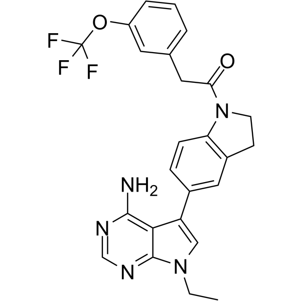
RIPK1-IN-7 is a potent and selective RIPK1 inhibitor with a Kd of 4 nM and an enzymatic IC50 of 11 nM. RIPK1-IN-7 exhibits excellent antimetastasis activity in the experimental B16 melanoma lung metastasis model.
For research use only. We do not sell to patients.
- Purity : 98.56%
- CAS No.: 2300982-44-7
- Formula: C25H22F3N5O2
- Molecular Weight:481.47
-
Storage:Powder -20°C, 3 years , 4°C, 2 years ; In solvent -80°C, 2 years , -20°C, 1 year

To place orders, for customer services and technical support, please contact: MedChemExpress USA
Tel: 609-228-6898 E-mail: [email protected] [email protected]


-
 Biological Activity
-
Chemical Information
-
Solvent & Solubility
- Protocols
- Purity & Documentation
- References
-
 Help & FAQs


-
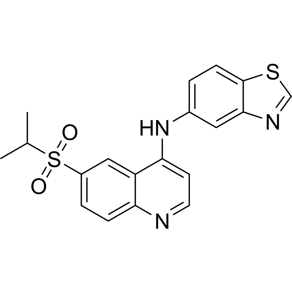
HY-101872
GSK-872RIPK3 Inhibitor -
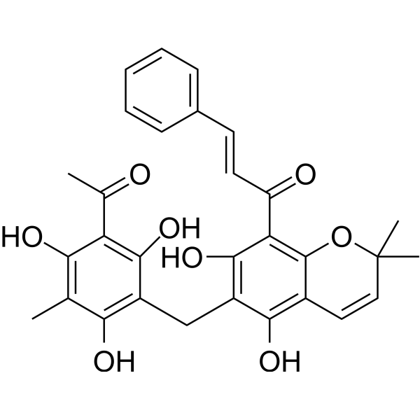
HY-18980
RottlerinPKC Inhibitor -
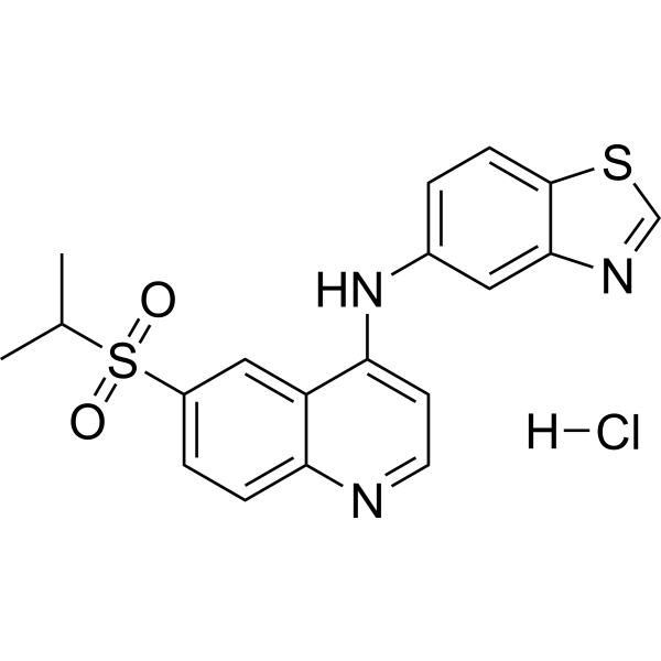
HY-101872A
GSK-872 hydrochlorideRIPK3 Inhibitor -
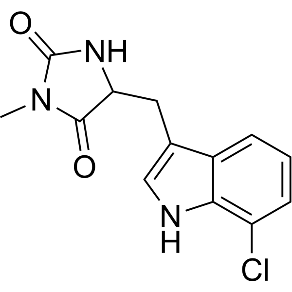
HY-14622A
Necrostatin 2 racemateRIPK1 Inhibitor -
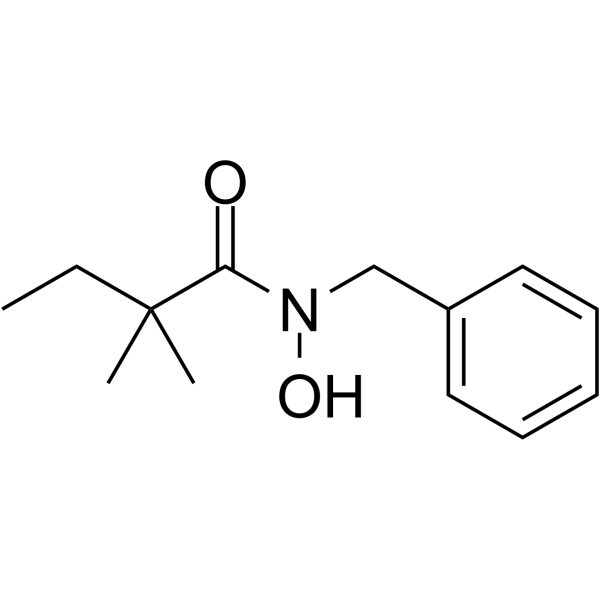
HY-101032
RIPA-56RIP1 Inhibitor -
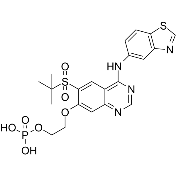
HY-112038
GSK2983559 free acidRIP2 Inhibitor -
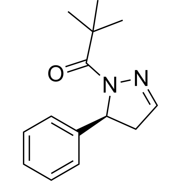
HY-103028A
GSK963RIP1 Kinase Inhibitor -
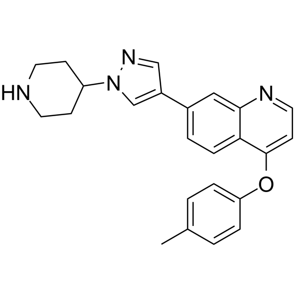
HY-114349
HS-1371RIP kinase Inhibitor -
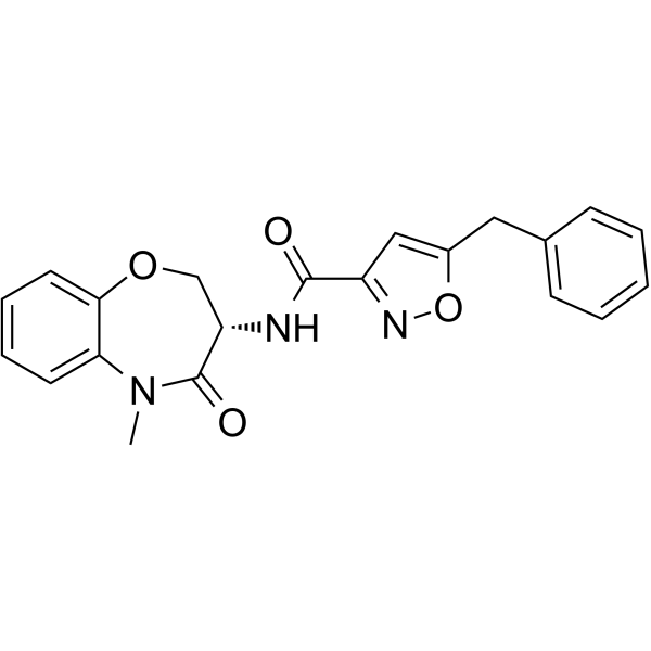
HY-100131
GSK481RIP1 Kinase Inhibitor -
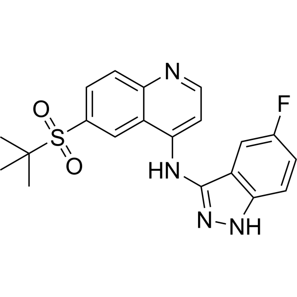
HY-100339
GSK583RIPK2 Inhibitor -
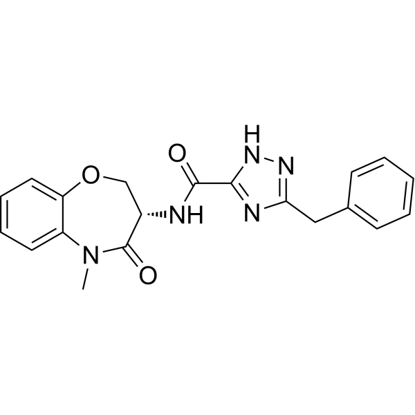
HY-101760
GSK2982772RIPK1 Inhibitor -
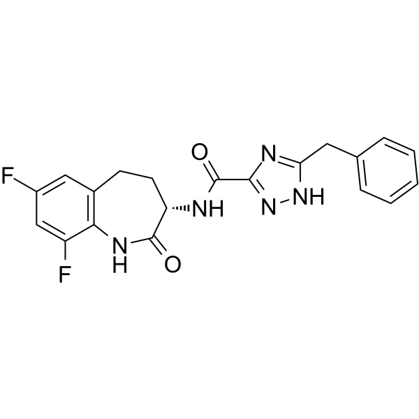
HY-111946
GSK3145095RIP1 Kinase Inhibitor


Biological Activity
Description
IC50 & Target
Cellular Effect
|
Cell Line

|
Type | Value | Description | References |
|---|---|---|---|---|
| J774.A1 | EC50 |
0.517 nM
Compound: 22b
|
Cytoprotection against TNFalpha/Smac-mimetic/z-VAD-FMK induced necroptosis in mouse J774A.1 cells after 24 hrs by CCK-8 assay
Cytoprotection against TNFalpha/Smac-mimetic/z-VAD-FMK induced necroptosis in mouse J774A.1 cells after 24 hrs by CCK-8 assay
|
[PMID: 30480444] |
| L929 | EC50 |
0.999 nM
Compound: 22b
|
Cytoprotection against TNFalpha/z-VAD-FMK induced necroptosis in mouse L929 cells after 24 hrs by CCK-8 assay
Cytoprotection against TNFalpha/z-VAD-FMK induced necroptosis in mouse L929 cells after 24 hrs by CCK-8 assay
|
[PMID: 30480444] |
| U-937 | EC50 |
11.55 nM
Compound: 22b
|
Cytoprotection against TNFalpha/Smac-mimetic/z-VAD-FMK induced necroptosis in human U937 cells after 24 hrs by CCK-8 assay
Cytoprotection against TNFalpha/Smac-mimetic/z-VAD-FMK induced necroptosis in human U937 cells after 24 hrs by CCK-8 assay
|
[PMID: 30480444] |
In Vitro
RIPK1-IN-7 shows potent cell protection effect in the TSZ-induced HT29 cell necroptosis model with an EC50 of 2nM[1].
RIPK1-IN-7 displays considerable activity against several other kinases, such as Flt4, TrkA, TrkB, TrkC, Axl, HRI, Mer, and MAP4K5 with IC50s of 20, 26, 8, 7, 35, 26, 29, and 27 nM, respectively[1].
MedChemExpress (MCE) has not independently confirmed the accuracy of these methods. They are for reference only. Further protocols information, click here.


Chemical Information
-
CAS No. 2300982-44-7
-
Appearance Solid
-
Molecular Weight 481.47
-
Formula C25H22F3N5O2
-
Color Light yellow to yellow
-
SMILES
NC1=C2C(N(CC)C=C2C3=CC(CCN4C(CC5=CC=CC(OC(F)(F)F)=C5)=O)=C4C=C3)=NC=N1
-
Shipping
Room temperature in continental US; may vary elsewhere.
-
Storage
Powder -20°C 3 years 4°C 2 years In solvent -80°C 2 years -20°C 1 year


Solvent & Solubility
In Vitro:
DMSO : 62.5 mg/mL (129.81 mM; Need ultrasonic; Hygroscopic DMSO has a significant impact on the solubility of product, please use newly opened DMSO)
Please refer to the solubility information to select the appropriate solvent. Once prepared, please aliquot and store the solution to prevent product inactivation from repeated freeze-thaw cycles.
Storage method and period of stock solution: -80°C, 2 years; -20°C, 1 year. When stored at -80°C, please use it within 2 years. When stored at -20°C, please use it within 1 year.
Please refer to the solubility information to select the appropriate solvent. Once prepared, please aliquot and store the solution to prevent product inactivation from repeated freeze-thaw cycles.
Storage method and period of stock solution: -80°C, 2 years; -20°C, 1 year. When stored at -80°C, please use it within 2 years. When stored at -20°C, please use it within 1 year.
Concentration (start) × Volume (start) = Concentration (final) × Volume (final)
In Vivo:
Select the appropriate dissolution method based on your experimental animal and administration route.
- For the following dissolution methods, please ensure to first prepare a clear stock solution using an In Vitro approach and then sequentially add co-solvents:
- To ensure reliable experimental results, the clarified stock solution can be appropriately stored based on storage conditions. As for the working solution for In Vivo experiments, it is recommended to prepare freshly and use it on the same day.
- The percentages shown for the solvents indicate their volumetric ratio in the final prepared solution. If precipitation or phase separation occurs during preparation, heat and/or sonication can be used to aid dissolution.
Add each solvent one by one: 10% DMSO 40% PEG300 5% Tween-80 45% Saline
Solubility: ≥ 2.08 mg/mL (4.32 mM); Clear solution
This protocol yields a clear solution of ≥ 2.08 mg/mL (saturation unknown).
Taking 1 mL working solution as an example, add 100 μL DMSO stock solution (20.8 mg/mL) to 400 μL PEG300, and mix evenly; then add 50 μL Tween-80 and mix evenly; then add 450 μL Saline to adjust the volume to 1 mL.
Preparation of Saline: Dissolve 0.9 g sodium chloride in ddH₂O and dilute to 100 mL to obtain a clear Saline solution.
In Vivo Dissolution Calculator
Please enter the basic information of animal experiments:
-
-
-
-
Recommended: Prepare an additional quantity of animals to account for potential losses during experiments.
Please enter your animal formula composition:
-
%DMSO +
Recommended: Keep the proportion of DMSO in working solution below 2% if your animal is weak.
-
%+
-
+%Tween-80 + +
-
%Saline +
The co-solvents required include: DMSO, . All of co-solvents are available by MedChemExpress (MCE). , Tween 80. All of co-solvents are available by MedChemExpress (MCE).
Working solution concentration: 0.22 mg/mL
Method for preparing stock solution: mg drug dissolved in μL DMSO. Stock solution concentration: mg/mL.
1. Take μL DMSO stock solution;
2. Add μL .
μL , mix evenly;
3. Then add μL Tween 80, mix evenly;
4. Then add μL
Please ensure that the stock solution in the first step is dissolved to a clear state, and add co-solvents in sequence. You can use ultrasonic heating (ultrasonic cleaner, recommended frequency 20-40 kHz), vortexing, etc. to assist dissolution.

Protocols
-
Cell Cytotoxicity Assay
Cytotoxicity assays are usually based on the assessment of cell membrane damage, which can also be indirectly detected by measuring cell viability. Detection methods include MTT assay, CKK-8 assay, LDH assay and ATP assay, etc.
-
Tail-Vein Experimental Metastasis Xenograft
Tail-vein experimental metastasis xenograft models assess the ability of injected tumor cells to survive circulation, arrest in vascular beds, extravasate, and colonize distant organs, most commonly lung after lateral tail-vein injection; this model bypasses primary-tumor formation, local invasion, and intravasation, so the readout reflects late metastatic colonization rather than the full metastatic cascade. The main readouts are metastatic burden measured by bioluminescence imaging, gross metastatic nodules, histology, organ weight, survival, or ex vivo tumor-cell quantification; luciferase-labeled tumor cells permit longitudinal noninvasive monitoring, while histology confirms organ colonization and tissue localization.
-
Splenic/Portal-Vein Liver Metastasis Xenograft
Splenic and portal-vein liver metastasis xenograft models deliver tumor cells into the portal circulation so that cells reach the liver first and form hepatic metastatic lesions; splenic injection uses the spleen as an access route to the portal system, while direct portal-vein injection introduces cells into the portal vein without requiring splenectomy. The assay detects liver colonization, intrahepatic tumor growth, tumor distribution, treatment response, survival, and liver-metastasis microenvironment changes; readouts include bioluminescence or fluorescence imaging, gross liver nodule counts, liver weight or tumor burden, histology, and survival.
-
Intracardiac/Intra-Arterial Metastasis Xenograft
Intracardiac xenograft metastasis models are based on the direct delivery of fluorescent or bioluminescent tumor cells into the left ventricle of immunocompromised mice, allowing systemic arterial dissemination that mimics hematogenous spread and enables colonization of distant organs such as bone, brain, and lung. Real-time bioluminescence imaging (BLI) is used to non-invasively track tumor cell seeding, survival, and metastatic outgrowth over time, reflecting early arrest in capillary beds followed by organ-specific colonization and proliferation.
-
Orthotopic Cell-Line Xenograft
Orthotopic cell-line xenograft models involve implantation of human cancer cell lines into the anatomically corresponding organ of immunodeficient mice to reproduce tumor growth within a native microenvironment, enabling more clinically relevant tumor behavior compared with subcutaneous models. These models are widely used because orthotopic placement better recapitulates tumor progression, including invasion and metastatic spread, which are often underrepresented in heterotopic implantation systems. Compared with conventional xenografts, orthotopic implantation is described as more technically complex but provides improved simulation of tumor-microenvironment interactions and metastatic behavior, making it particularly valuable for translational oncology research. Surgical orthotopic implantation approaches have been emphasized as enabling faithful reproduction of clinical cancer features, including metastasis and disease progression patterns that align with the tumor’s organ of origi


Purity & Documentation
-
Data Sheet (277 KB)
-
SDS (396 KB)
- English - EN (396 KB)
- Français - FR (396 KB)
- Deutsch - DE (396 KB)
- Norwegian - NO (396 KB)
- Español - ES (396 KB)
- Swedish - SV (396 KB)
- Italian - IT (396 KB)
- Korean - KR (396 KB)
- Portuguese - PT (396 KB)
-
Handling Instructions (2659 KB)
References


Complete Stock Solution Preparation Table
Please refer to the solubility information to select the appropriate solvent. Once prepared, please aliquot and store the solution to prevent product inactivation from repeated freeze-thaw cycles.
Storage method and period of stock solution: -80°C, 2 years; -20°C, 1 year. When stored at -80°C, please use it within 2 years. When stored at -20°C, please use it within 1 year.
| Optional Solvent | Concentration Solvent Mass | 1 mg | 5 mg | 10 mg | 25 mg |
|---|---|---|---|---|---|
| DMSO | 1 mM | 2.0770 mL | 10.3849 mL | 20.7697 mL | 51.9243 mL |
| 5 mM | 0.4154 mL | 2.0770 mL | 4.1539 mL | 10.3849 mL | |
| 10 mM | 0.2077 mL | 1.0385 mL | 2.0770 mL | 5.1924 mL | |
| 15 mM | 0.1385 mL | 0.6923 mL | 1.3846 mL | 3.4616 mL | |
| 20 mM | 0.1038 mL | 0.5192 mL | 1.0385 mL | 2.5962 mL | |
| 25 mM | 0.0831 mL | 0.4154 mL | 0.8308 mL | 2.0770 mL | |
| 30 mM | 0.0692 mL | 0.3462 mL | 0.6923 mL | 1.7308 mL | |
| 40 mM | 0.0519 mL | 0.2596 mL | 0.5192 mL | 1.2981 mL | |
| 50 mM | 0.0415 mL | 0.2077 mL | 0.4154 mL | 1.0385 mL | |
| 60 mM | 0.0346 mL | 0.1731 mL | 0.3462 mL | 0.8654 mL | |
| 80 mM | 0.0260 mL | 0.1298 mL | 0.2596 mL | 0.6491 mL | |
| 100 mM | 0.0208 mL | 0.1038 mL | 0.2077 mL | 0.5192 mL |


RIPK1-IN-7 Related Classifications
HY-119933 Related Classifications
 Powered by Bioz