



X15695

Based on 1 Customer Validation
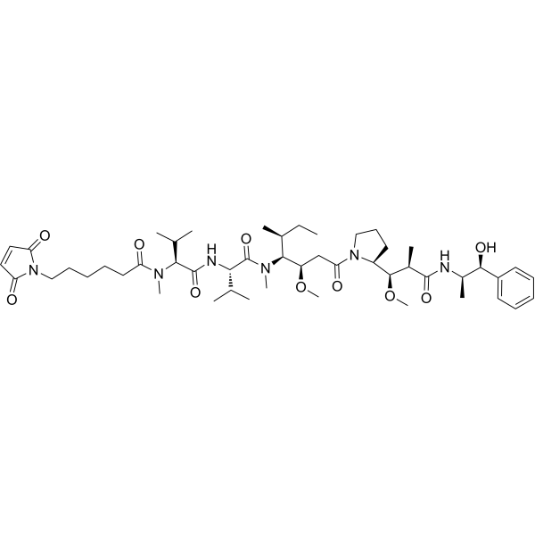
X15695 is selective and orally active estrogen receptor (ERα) degrader. X15695 is an aryl hydrocarbon receptor (AHR) ligand. X15695 enables AHR to form a complex with the ERα, promoting its proteasomal degradation. X15695 inhibits the breast cancer cells proliferation, promotes cell cycle block and induces apoptosis. X15695 can be used for the study of breast cancer.
For research use only. We do not sell to patients.
- Purity : 98.45%
- CAS No.: 353258-25-0
- Formula: C14H7ClF4N2
- Molecular Weight:314.67
-
Storage:Powder -20°C, 3 years , 4°C, 2 years ; In solvent -80°C, 6 months , -20°C, 1 month


To place orders, for customer services and technical support, please contact: MedChemExpress USA
Tel: 609-228-6898 E-mail: [email protected] [email protected]


-
 Biological Activity
-
Chemical Information
-
Solvent & Solubility
- Protocols
- Purity & Documentation
- References
-
 Help & FAQs



![2-Methyl-2-propanyl(1S,8R)-4-nitro-11-azatricyclo[6.2.1.02,7]undeca-2,4,6-triene-11-carboxylate](https://file.medchemexpress.com/product_pic/hy-77304.gif)


Biological Activity
Description
IC50 & Target
[1]|
ERα |
In Vitro
X15695 inhibits MCF-7, MC7D538G and MC7Y537S cells proliferation with IC50s of 0.7, 74.42 and 0.38 nM, and the IC50 values in the presence of estrogen (E2) are 10.28, 48.01 and 10.78 nM, respectively[1].
X15695 inhibits MCF-7 cells colony growth when the activity of AHR is blocked with CH223191 (HY-12684) [1].
X15695 (1-10 μM, 7 days) inhibits the growth of organoids from patient-derived xenograft tumours (PDxO) [1].
X15695 (1 μM, 48 h) blocks cell cycle and induces apoptosis in MCF-7 cells, T47D cells, LNCaP cells and LAPC-4 cells by activating wild-type p53 and disrupting the gain-of-function tumorigenic activity of mutant p53, respectively[2].
MedChemExpress (MCE) has not independently confirmed the accuracy of these methods. They are for reference only.
-
Cell Line:MCF-7 cells, T47D cells, LNCaP cells and LAPC-4 cells
-
Concentration:1 μM
-
Incubation Time:48 h
-
Result:Induced apoptosis in ER+ breast cancer cells, and the apoptotic effect was more significant in wild-type p53 cells.
-
Cell Line:MCF-7 cells, T47D cells, LNCaP cells and LAPC-4 cells
-
Concentration:1 μM
-
Incubation Time:48 h
-
Result:Significantly arrested the cells in the G1 phase in MCF-7 cells and G2/M phase in T47D cells.
Induced G1/S phase arrest in the LNCaP cells and a G2/M arrest in the LAPC-4 cells.
-
Cell Line:MCF-7 cells and T47D cells
-
Concentration:1 μM
-
Incubation Time:48 h
-
Result:Significantly increase the level of p53 protein in MCF-7 cells.
Had no significant impact in T47D cells.
In Vivo
MedChemExpress (MCE) has not independently confirmed the accuracy of these methods. They are for reference only.
-
Animal Model:MCF-7 and LAPC-4 cells induced xenograft model established in 6–7weeks old female(for MCF-7 cells) and male (forLAPC-4cells) athymic nude-Foxn1nu mice (Envigo 6901M)[2
-
Dosage:30 mg/kg for MCF-7 model; 15 and 30 mg/kg for LAPC-4 model
-
Administration:Oal gavage (i.g), once daily for 16 days (MCF-7 model) or 42 days (LAPC-4 model)
-
Result:Significantly inhibits tumor growth, without weight loss or other toxic signs in MCF-7 model.
Downregulated ERα and upregulated p53 in tumor tissue.
Inhibited the growth of prostate tumors (30 mg/kg), but the effect was relatively weak.
- Detection of Apoptosis
- TUNEL staining for apoptotic DNA fragmentation
- Flow cytometric DNA-content cell-cycle staining
- Annexin V plus membrane-impermeant dye apoptosis staining
- BrdU Incorporation Assay
- Research Protocol for Endocrine Diseases
- Apoptosis Solutions
- Protocol for Cell Cycle
- Breast Cancer Modeling
- Cell Viability Determination by MTT Colorimetric Assay


Chemical Information
-
CAS No. 353258-25-0
-
Appearance Solid
-
Molecular Weight 314.67
-
Formula C14H7ClF4N2
-
Color Off-white to light yellow
-
SMILES
FC(C1=CN2C(C(Cl)=C1)=NC(C3=CC=C(F)C=C3)=C2)(F)F
-
Shipping
Room temperature in continental US; may vary elsewhere.
-
Storage
Powder -20°C 3 years 4°C 2 years In solvent -80°C 6 months -20°C 1 month


Solvent & Solubility
In Vitro:
DMSO : 100 mg/mL (317.79 mM; Need ultrasonic; Hygroscopic DMSO has a significant impact on the solubility of product, please use newly opened DMSO)
Please refer to the solubility information to select the appropriate solvent. Once prepared, please aliquot and store the solution to prevent product inactivation from repeated freeze-thaw cycles.
Storage method and period of stock solution: -80°C, 6 months; -20°C, 1 month. When stored at -80°C, please use it within 6 months. When stored at -20°C, please use it within 1 month.
Please refer to the solubility information to select the appropriate solvent. Once prepared, please aliquot and store the solution to prevent product inactivation from repeated freeze-thaw cycles.
Storage method and period of stock solution: -80°C, 6 months; -20°C, 1 month. When stored at -80°C, please use it within 6 months. When stored at -20°C, please use it within 1 month.
Concentration (start) × Volume (start) = Concentration (final) × Volume (final)
In Vivo:
Select the appropriate dissolution method based on your experimental animal and administration route.
- For the following dissolution methods, please ensure to first prepare a clear stock solution using an In Vitro approach and then sequentially add co-solvents:
- To ensure reliable experimental results, the clarified stock solution can be appropriately stored based on storage conditions. As for the working solution for In Vivo experiments, it is recommended to prepare freshly and use it on the same day.
- The percentages shown for the solvents indicate their volumetric ratio in the final prepared solution. If precipitation or phase separation occurs during preparation, heat and/or sonication can be used to aid dissolution.
In Vivo Dissolution Calculator
Please enter the basic information of animal experiments:
-
-
-
-
Recommended: Prepare an additional quantity of animals to account for potential losses during experiments.
Please enter your animal formula composition:
-
%DMSO +
Recommended: Keep the proportion of DMSO in working solution below 2% if your animal is weak.
-
%+
-
+%Tween-80 + +
-
%Saline +
The co-solvents required include: DMSO, . All of co-solvents are available by MedChemExpress (MCE). , Tween 80. All of co-solvents are available by MedChemExpress (MCE).
Working solution concentration: 0.22 mg/mL
Method for preparing stock solution: mg drug dissolved in μL DMSO. Stock solution concentration: mg/mL.
1. Take μL DMSO stock solution;
2. Add μL .
μL , mix evenly;
3. Then add μL Tween 80, mix evenly;
4. Then add μL
Please ensure that the stock solution in the first step is dissolved to a clear state, and add co-solvents in sequence. You can use ultrasonic heating (ultrasonic cleaner, recommended frequency 20-40 kHz), vortexing, etc. to assist dissolution.


Purity & Documentation
-
Data Sheet (276 KB)
-
SDS (251 KB)
- English - EN (251 KB)
- Français - FR (251 KB)
- Deutsch - DE (251 KB)
- Norwegian - NO (251 KB)
- Español - ES (251 KB)
- Swedish - SV (251 KB)
- Italian - IT (251 KB)
- Korean - KR (251 KB)
- Portuguese - PT (251 KB)
-
Handling Instructions (2659 KB)
References


Complete Stock Solution Preparation Table
Please refer to the solubility information to select the appropriate solvent. Once prepared, please aliquot and store the solution to prevent product inactivation from repeated freeze-thaw cycles.
Storage method and period of stock solution: -80°C, 6 months; -20°C, 1 month. When stored at -80°C, please use it within 6 months. When stored at -20°C, please use it within 1 month.
| Optional Solvent | Concentration Solvent Mass | 1 mg | 5 mg | 10 mg | 25 mg |
|---|---|---|---|---|---|
| DMSO | 1 mM | 3.1779 mL | 15.8897 mL | 31.7793 mL | 79.4483 mL |
| 5 mM | 0.6356 mL | 3.1779 mL | 6.3559 mL | 15.8897 mL | |
| 10 mM | 0.3178 mL | 1.5890 mL | 3.1779 mL | 7.9448 mL | |
| 15 mM | 0.2119 mL | 1.0593 mL | 2.1186 mL | 5.2966 mL | |
| 20 mM | 0.1589 mL | 0.7945 mL | 1.5890 mL | 3.9724 mL | |
| 25 mM | 0.1271 mL | 0.6356 mL | 1.2712 mL | 3.1779 mL | |
| 30 mM | 0.1059 mL | 0.5297 mL | 1.0593 mL | 2.6483 mL | |
| 40 mM | 0.0794 mL | 0.3972 mL | 0.7945 mL | 1.9862 mL | |
| 50 mM | 0.0636 mL | 0.3178 mL | 0.6356 mL | 1.5890 mL | |
| 60 mM | 0.0530 mL | 0.2648 mL | 0.5297 mL | 1.3241 mL | |
| 80 mM | 0.0397 mL | 0.1986 mL | 0.3972 mL | 0.9931 mL | |
| 100 mM | 0.0318 mL | 0.1589 mL | 0.3178 mL | 0.7945 mL |


X15695 Related Classifications
HY-175785 Related Classifications
 Powered by Bioz