



ZINC69391
Based on 1 publication(s) in Google Scholar
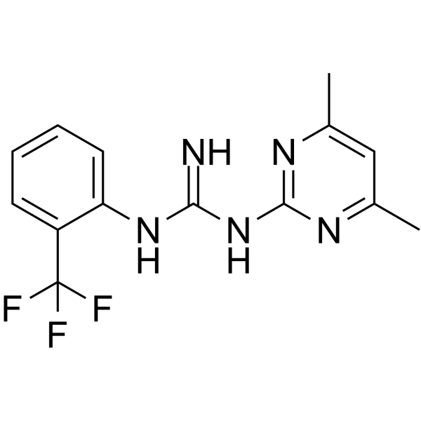
ZINC69391, a specific Rac1 inhibitor, interferes with Rac1-GEF interaction by masking Trp56 residue on Rac1 surface. ZINC69391 interferes with the interaction of Rac1 with Dock180 and reduces Rac1-GTP levels. ZINC69391 induces apoptosis, and shows antiproliferative and antimetastatic effects.
Nos produits utilisent uniquement pour la recherche. Nous ne vendons pas aux patients.
- Pureté : 99.12%
- CAS No.: 303094-67-9
- Formule: C14H14F3N5
- Masse moléculaire:309.29
-
Stockage:Powder -20°C, 3 years , 4°C, 2 years ; In solvent -80°C, 6 months , -20°C, 1 month

To place orders, for customer services and technical support, please contact: MedChemExpress USA
Tel: 609-228-6898 E-mail: [email protected] [email protected]


-
 Activité biologique
-
Chemical Information
-
Solvant et solubilité
- Protocole
- Pureté et documentation
- Références
-
 Help & FAQs


-
HY-104064
1A-116Rac1 Inhibitor -
HY-15723A
NSC 23766 trihydrochlorideRac Inhibitor -
HY-12646
Rhosin hydrochlorideRho GTPases Inhibitor -
HY-15723
NSC 23766Ras Inhibitor -
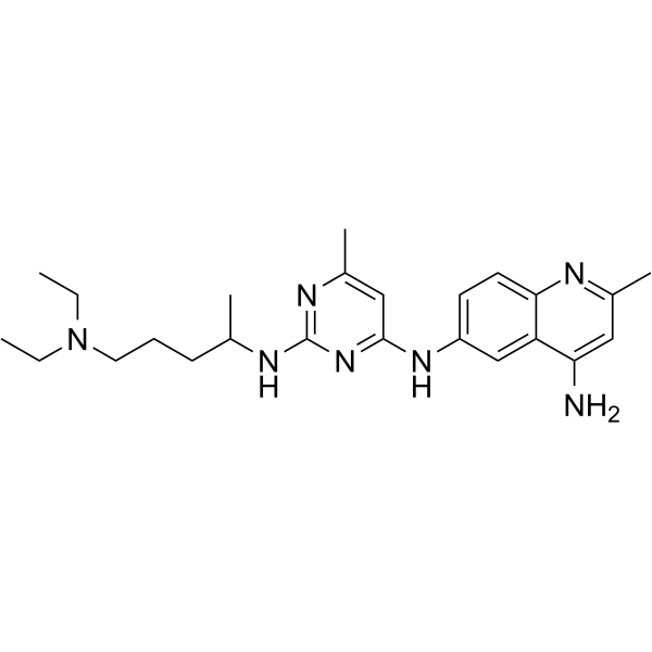
HY-115376
Z62954982Rac1 Inhibitor -
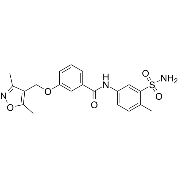
HY-12810
EHop-016Rac1/Rac3 Inhibitor -
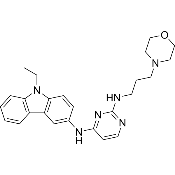
HY-118208
ML-097Ras-related GTPases Activator -

HY-102078R
ZINC69391 (Standard)Reference Standard -
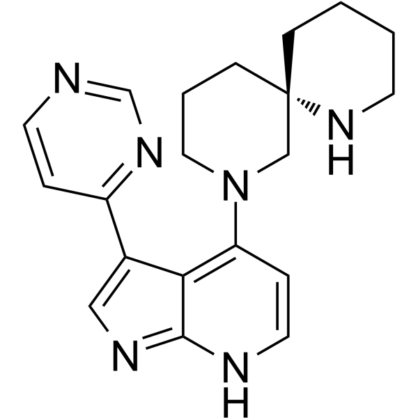
HY-111424
BDP9066MRCK Inhibitor -
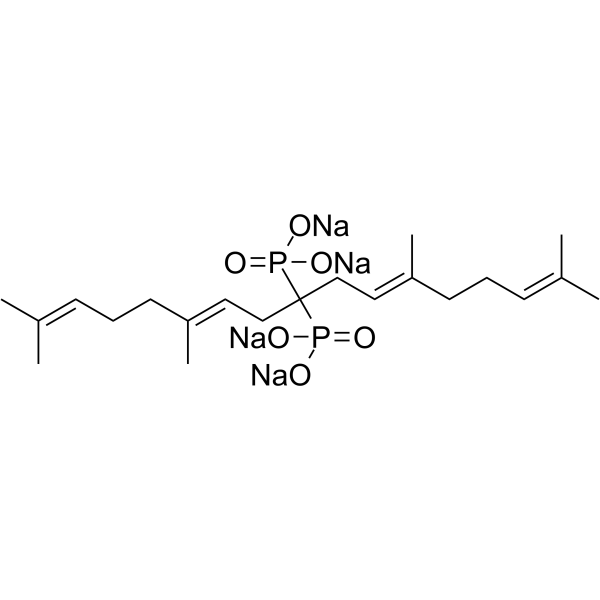
HY-U00145
Digeranyl bisphosphonate -

HY-104064R
1A-116 (Standard)Reference Standard
Publications Citing Use of MedChemExpress (MCE) ZINC69391
More

Activité biologique
Description
Cellular Effect
|
Cell Line

|
Type | Value | Description | References |
|---|---|---|---|---|
| MCF7 | IC50 |
31 μM
Compound: 13; ZINC69391
|
Antiproliferative activity against human MCF7 cells assessed as cell growth inhibition incubated for 72 hrs by MTT assay
Antiproliferative activity against human MCF7 cells assessed as cell growth inhibition incubated for 72 hrs by MTT assay
|
[PMID: 37253305] |
| MDA-MB-231 | IC50 |
48 μM
Compound: 13; ZINC69391
|
Antiproliferative activity against human MDA-MB-231 cells assessed as cell growth inhibition incubated for 72 hrs by MTT assay
Antiproliferative activity against human MDA-MB-231 cells assessed as cell growth inhibition incubated for 72 hrs by MTT assay
|
[PMID: 37253305] |
In Vitro
ZINC69391 inhibits growth of U937, HL-60, KG1A and Jurkat cells with IC50s ranging from 41 to 54 μM[1].
ZINC69391 (50-100 μM; 24 hours) augments the enzymatic activity of caspase 3 in a concentration dependent manner[1].
ZINC69391 (0-125 μM; 72h) reduces cell proliferation of human glioma cells[2].
ZINC69391 (50-100 μM; 48 hours) triggers cell cycle arrest[2].
ZINC69391 (50 μM; 24 hours) triggers apoptosis on human acute leukemic cells[1].
MedChemExpress (MCE) has not independently confirmed the accuracy of these methods. They are for reference only.
-
Cell Line:U-87 MG, LN229 cells
-
Concentration:0-125μM
-
Incubation Time:72 hours
-
Result:Reduced cell proliferation in a concentration-dependent manner.
-
Cell Line:LN229 cells
-
Concentration:50, 100 μM
-
Incubation Time:48 hours
-
Result:Resulted in a significant increased percentage of cells in the sub-G0/G1 phase in a concentration dependent manner.
-
Cell Line:HL-60, U937 and KG1A cell lines
-
Concentration:50 μM
-
Incubation Time:24 hours
-
Result:Led to a significant increase in apoptotic cells.
In Vivo
MedChemExpress (MCE) has not independently confirmed the accuracy of these methods. They are for reference only.
-
Animal Model:Specific pathogen-free female BALB/c inbred mice (bearing F3II cells)[3]
-
Dosage:25 mg/kg body weight
-
Administration:I.p; daily for 21 days
-
Result:Significantly reduced by about 60% the formation of total metastatic lung colonies.


Chemical Information
-
CAS No. 303094-67-9
-
Appearance Solid
-
Masse moléculaire 309.29
-
Formule C14H14F3N5
-
Color White to off-white
-
SMILES
N=C(NC1=CC=CC=C1C(F)(F)F)NC2=NC(C)=CC(C)=N2
-
Livraison
Room temperature in continental US; may vary elsewhere.
-
Stockage
Powder -20°C 3 years 4°C 2 years In solvent -80°C 6 months -20°C 1 month


Publications (1)
-
Journal Impact Factor
-
Most Recent
-
Cell Rep
LncRNA DGCR5-encoded polypeptide RIP aggravates SONFH by repressing nuclear localization of β-catenin in BMSCs. [Abstract]2023 Aug 12;42(8):112969. PMID: 37573506


Solvant et solubilité
In Vitro:
DMSO : 25 mg/mL (80.83 mM; ultrasonic and warming and heat to 60°C; Hygroscopic DMSO has a significant impact on the solubility of product, please use newly opened DMSO)
Please refer to the solubility information to select the appropriate solvent. Once prepared, please aliquot and store the solution to prevent product inactivation from repeated freeze-thaw cycles.
Storage method and period of stock solution: -80°C, 6 months; -20°C, 1 month. When stored at -80°C, please use it within 6 months. When stored at -20°C, please use it within 1 month.
Please refer to the solubility information to select the appropriate solvent. Once prepared, please aliquot and store the solution to prevent product inactivation from repeated freeze-thaw cycles.
Storage method and period of stock solution: -80°C, 6 months; -20°C, 1 month. When stored at -80°C, please use it within 6 months. When stored at -20°C, please use it within 1 month.
Concentration (start) × Volume (start) = Concentration (final) × Volume (final)


Pureté et documentation
-
Fiche technique (277 KB)
-
SDS (393 KB)
- English - EN (393 KB)
- Français - FR (393 KB)
- Deutsch - DE (393 KB)
- Norwegian - NO (393 KB)
- Español - ES (393 KB)
- Swedish - SV (393 KB)
- Italian - IT (393 KB)
- Korean - KR (393 KB)
- Portuguese - PT (393 KB)
-
Instruction de manipulation (2659 KB)
Références
[1]. Cabrera M, et al. Pharmacological Rac1 inhibitors with selective apoptotic activity in human acute leukemic cell lines. Oncotarget. 2017;8(58):98509‐98523. Published 2017 Oct 4. [Content Brief]
[2]. Cardama GA, et al. Proapoptotic and antiinvasive activity of Rac1 small molecule inhibitors on malignant glioma cells. Onco Targets Ther. 2014;7:2021‐2033. Published 2014 Oct 30. [Content Brief]
[3]. Cardama GA, et al. Preclinical development of novel Rac1-GEF signaling inhibitors using a rational design approach in highly aggressive breast cancer cell lines. Anticancer Agents Med Chem. 2014;14(6):840‐851. [Content Brief]


Complete Stock Solution Preparation Table
Please refer to the solubility information to select the appropriate solvent. Once prepared, please aliquot and store the solution to prevent product inactivation from repeated freeze-thaw cycles.
Storage method and period of stock solution: -80°C, 6 months; -20°C, 1 month. When stored at -80°C, please use it within 6 months. When stored at -20°C, please use it within 1 month.
| Optional Solvent | Concentration Solvent Mass | 1 mg | 5 mg | 10 mg | 25 mg |
|---|---|---|---|---|---|
| DMSO | 1 mM | 3.2332 mL | 16.1661 mL | 32.3321 mL | 80.8303 mL |
| 5 mM | 0.6466 mL | 3.2332 mL | 6.4664 mL | 16.1661 mL | |
| 10 mM | 0.3233 mL | 1.6166 mL | 3.2332 mL | 8.0830 mL | |
| 15 mM | 0.2155 mL | 1.0777 mL | 2.1555 mL | 5.3887 mL | |
| 20 mM | 0.1617 mL | 0.8083 mL | 1.6166 mL | 4.0415 mL | |
| 25 mM | 0.1293 mL | 0.6466 mL | 1.2933 mL | 3.2332 mL | |
| 30 mM | 0.1078 mL | 0.5389 mL | 1.0777 mL | 2.6943 mL | |
| 40 mM | 0.0808 mL | 0.4042 mL | 0.8083 mL | 2.0208 mL | |
| 50 mM | 0.0647 mL | 0.3233 mL | 0.6466 mL | 1.6166 mL | |
| 60 mM | 0.0539 mL | 0.2694 mL | 0.5389 mL | 1.3472 mL | |
| 80 mM | 0.0404 mL | 0.2021 mL | 0.4042 mL | 1.0104 mL |


ZINC69391 Related Classifications
HY-102078 Related Classifications
 Powered by Bioz