



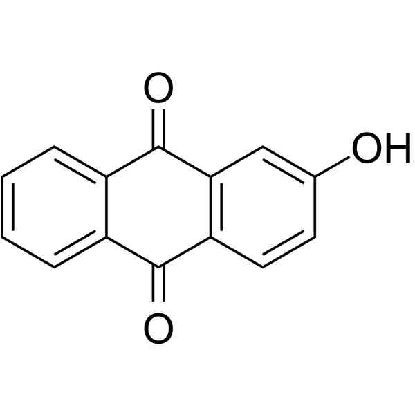
2-Hydroxyanthraquinone

Based on 1 Customer Validation
2-Hydroxyanthraquinone is a product generated by the photochemical oxidation of Anthracene (ANT) (HY-Y0299). 2-Hydroxyanthraquinone induces ferroptosis in cardiomyocytes by depleting GSH and inhibiting GPX4, leading to cardiac developmental malformations. 2-Hydroxyanthraquinone causes damage to the cerebrovascular system and blood-brain barrier in zebrafish by downregulating the Wnt/β-catenin signaling pathway, as well as inducing inflammation and neuronal apoptosis. 2-Hydroxyanthraquinone can be used in studies related to cerebrovascular diseases and cardiotoxicity.
For research use only. We do not sell to patients.
- Purity : 98.0%
- CAS No.: 605-32-3
- Formula: C14H8O3
- Molecular Weight:224.22
-
Storage:
4°C, stored under nitrogen
* In solvent : -80°C, 6 months; -20°C, 1 month (stored under nitrogen)

To place orders, for customer services and technical support, please contact: MedChemExpress USA
Tel: 609-228-6898 E-mail: [email protected] [email protected]


-
 Biological Activity
-
Chemical Information
-
Solvent & Solubility
- Protocols
- Purity & Documentation
- References
-
 Help & FAQs


-
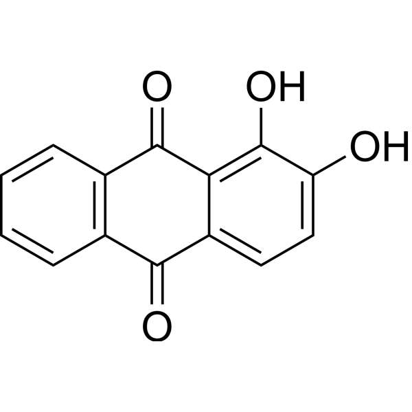
HY-N0563
AlizarinNatural Dye -

HY-W012352R
2-Hydroxyanthraquinone (Standard)Standard of 2-Hydroxyanthraquinone -
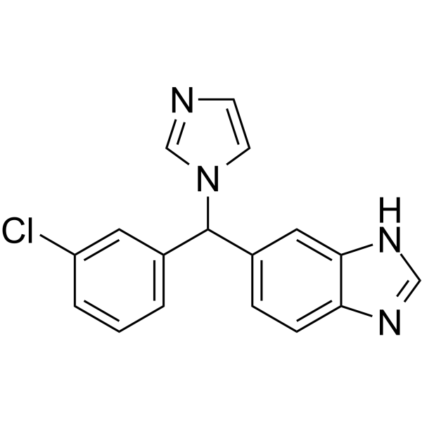
HY-106019
LiarozoleRAMBA -
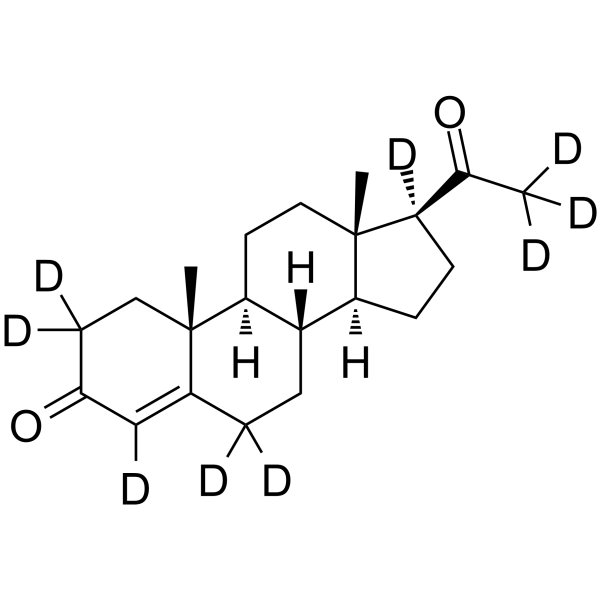
HY-N0437S
Progesterone-d9Stable Isotope -

HY-W738639
18-Hydroxy-11-deoxy corticosteroneMineralocorticoid -
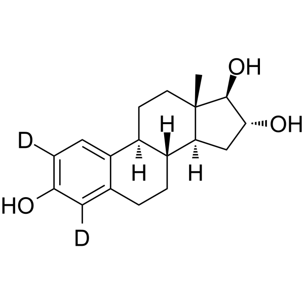
HY-B0412S
Estriol-d2Stable Isotope -
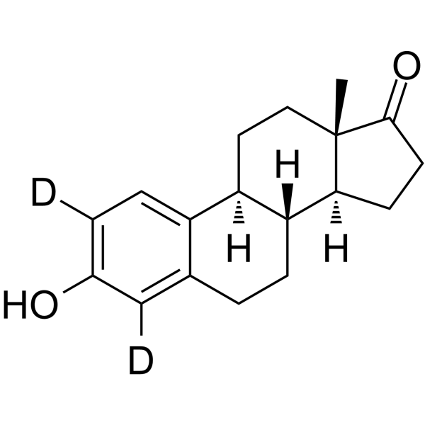
HY-B0234S1
Estrone-d2Stable Isotope -
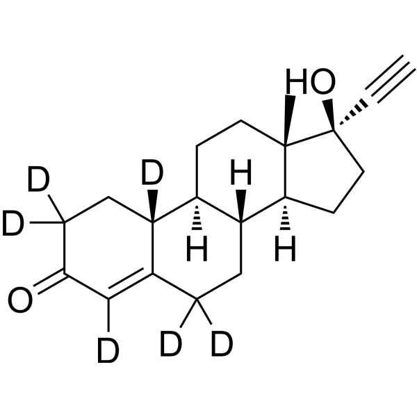
HY-B0554S
Norethindrone-d6Stable Isotope -
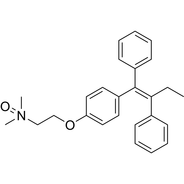
HY-133010
Tamoxifen N-oxideTamoxifen Metabolite -
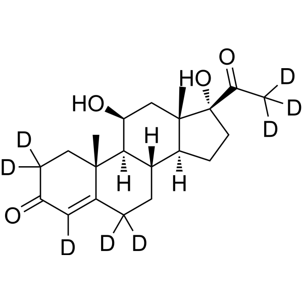
HY-113405S1
21-Deoxycortisol-d8Stable Isotope -
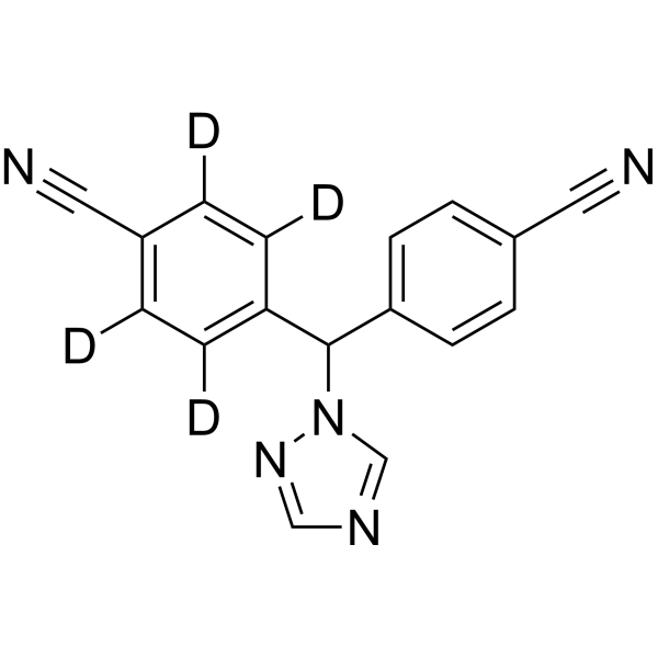
HY-14248S
Letrozole-d4Stable Isotope -
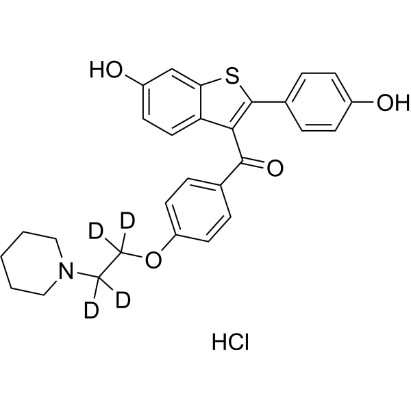
HY-13738S2
Raloxifene-d4 hydrochlorideStable Isotope


Biological Activity
Description
Cellular Effect
|
Cell Line

|
Type | Value | Description | References |
|---|---|---|---|---|
| DU-145 | IC50 |
72 μM
Compound: 5
|
Cytotoxicity against Homo sapiens (human) DU145 cells assessed as inhibition of cell survival after 96 hr by MTT assay
Cytotoxicity against Homo sapiens (human) DU145 cells assessed as inhibition of cell survival after 96 hr by MTT assay
|
10.1007/s00044-012-0197-5 |
| MCF7 | IC50 |
69 μM
Compound: 5
|
Cytotoxicity against Homo sapiens (human) MCF7 cells assessed as inhibition of cell survival after 96 hr by MTT assay
Cytotoxicity against Homo sapiens (human) MCF7 cells assessed as inhibition of cell survival after 96 hr by MTT assay
|
10.1007/s00044-012-0197-5 |
| NCI-H460 | IC50 |
66 μM
Compound: 5
|
Cytotoxicity against Homo sapiens (human) H460 cells assessed as inhibition of cell survival after 96 hr by MTT assay
Cytotoxicity against Homo sapiens (human) H460 cells assessed as inhibition of cell survival after 96 hr by MTT assay
|
10.1007/s00044-012-0197-5 |
In Vitro
2-Hydroxyanthraquinone binds stably to zebrafish GPX4 protein with a binding energy of −8.3 kcal/mol, mediated by amino acid residues Pro-140, Lys-143, and Arg-168[2].
MedChemExpress (MCE) has not independently confirmed the accuracy of these methods. They are for reference only. Further protocols information, click here.
In Vivo
2-Hydroxyanthraquinone (0.4-1.2 mg/L; waterborne exposure; 6-96 h) induces concentration-dependent cardiac developmental toxicity in zebrafish embryos via ferroptosis, including reduced cardiomyocyte proliferation, oxidative stress, lipid metabolism disorder, iron accumulation, and cardiac structural/functional defects[2].
2-Hydroxyanthraquinone (300-400 µg/L; waterborne exposure; 28 days) induces cardiac injury in adult zebrafish, including ventricular atrophy, cardiomyocyte degeneration, fibrosis, and vacuolization[2].
MedChemExpress (MCE) has not independently confirmed the accuracy of these methods. They are for reference only.
-
Animal Model:wild-type (AB); transgenic lines Tg (kdrl:mCherry), Tg (mpeg1:GFP), Tg (fli:GFP), Tg (lyz:DsRed), Tg (elavl3:GFP) (embryos at 6 hours post-fertilization)[1]
-
Dosage:0.7 mg/L; 1.3 mg/L; 1.9 mg/L
-
Administration:waterborne; daily renewal; 6 hpf to 24 hpf or 60 hpf
-
Result:Reduced cerebrovascular area by ~40% relative to controls in medium (1.3 mg/L) and high (1.9 mg/L) dose groups at 24 hpf.
Reduced cerebrovascular area by 50% relative to controls in high dose group at 60 hpf; diminished head size by ~25% in high dose group at 60 hpf; shortened distance between intersegmental vessels at 60 hpf.
Downregulated genes involved in vascular/lymphatic development (itgb4, kdr, kdrl, vegfc, vegfa, flt4) at 60 hpf.
Increased brain microglia by ~40% relative to controls in high dose group.
Increased ROS content by over fourfold relative to controls in high dose group.
Upregulated pro-inflammatory cytokines (il1β, tnf-α, nf-κb, il6, nos2, rela) and downregulated anti-inflammatory il10.
Increased TUNEL-positive brain cells by ~twofold relative to controls in high dose group; upregulated p53 gene expression.
Increased BBB permeability in medium and high dose groups; altered expression of genes related to BBB development (spock1, abcc1, abcg2, abcb4, abcb5).
Reduced β-catenin protein expression to ~one-third of control levels; downregulated pathway target genes (lef1, β-catenin) and upregulated inhibitory genes (dkk1, wif1).
Reduced total movement distance, average swimming speed, movement time, and continuous movement duration by ~75% relative to controls in all dose groups; decreased activity frequency and cumulative activity duration, and increased immobility duration in a concentration-dependent manner.
-
Animal Model:wild-type (AB); transgenic Tg (myl7:GFP) (embryos, 6 hours post-fertilization)[3]
-
Dosage:0.4 mg/L; 0.8 mg/L; 1.2 mg/L
-
Administration:waterborne; continuous; 6 hpf to 96 hpf
-
Result:Caused concentration-dependent increases in zebrafish embryo mortality at 96 hpf.
Reduced body length to ~3500 μm (0.4 mg/L), ~3200 μm (0.8 mg/L), ~2500 μm (1.2 mg/L) vs control ~4000 μm at 96 hpf.
Increased yolk sac area at 96 hpf.
Enlarged pericardial area to ~25000 μm2 (0.4 mg/L), ~45000 μm2 (0.8 mg/L), ~65000 μm2 (1.2 mg/L) vs control ~15000 μm2 at 96 hpf.
Decreased heart rate over 20 seconds to ~45 beats (0.4 mg/L), ~35 beats (0.8 mg/L), ~30 beats (1.2 mg/L) vs control ~50 beats at 96 hpf.
Increased sinus venosus-bulbus arteriosus (SV-BA) distance to ~170 μm (0.4 mg/L), ~220 μm (0.8 mg/L), ~250 μm (1.2 mg/L) vs control ~120 μm in transgenic Tg (myl7:GFP) embryos at 96 hpf.
Reduced cardiomyocyte numbers and caused failed cardiac cyclization revealed by H&E staining.
Downregulated expression of cardiac developmental genes myl7, tbx2b, hand2, and upregulated gata4 and nppa.
Reduced cardiomyocyte proliferation by ~50% at 0.8 and 1.2 mg/L, with downregulated expression of proliferation-related genes cdk2, ccne1, and ccnd1.
Significantly elevated ROS levels in the heart, with downregulated expression of antioxidant genes nrf2, cat, sod, and gclm; decreased CAT and GSH activities, and increased SOD and MDA activities.
Increased cardiac lipid content revealed by Oil Red O staining, elevated total cholesterol (TC) at 1.2 mg/L, and dysregulated expression of lipid metabolism genes (scd, fasn, elovl4, elovl5 upregulated; cd36 downregulated).
Increased iron accumulation revealed by FerroOrange staining and tissue iron measurements, with dysregulated ferroptosis-related gene expression: gpx4, slc7a11, slc3a2 downregulated; acsl4 upregulated; iron metabolism genes fht1, tfr1, fpn1, dmt1 altered.
Significantly reduced pericardial area and restored gpx4 gene expression when co-exposed with 0.6 μM Fer-1 compared to 0.8 mg/L 2-Hydroxyanthraquinone alone.
-
Animal Model:wild-type (3-month-old adult)[3]
-
Dosage:300 µg/L; 400 µg/L
-
Administration:waterborne; daily; 28 days
-
Result:Caused ventricular atrophy with ventricular area ~650000 μm2 (300 µg/L), ~550000 μm2 (400 µg/L) vs control ~700000 μm2.
Induced partial cardiomyocyte degeneration in the 300 µg/L group revealed by H&E staining.
Induced partial fibrosis and vacuole formation in the 400 µg/L group revealed by H&E staining.


Chemical Information
-
CAS No. 605-32-3
-
Appearance Solid
-
Molecular Weight 224.22
-
Formula C14H8O3
-
Color Yellow to green yellow
-
SMILES
OC1=CC2=C(C(C3=CC=CC=C3C2=O)=O)C=C1
-
Structure Classification
-
Initial Source
-
Shipping
Room temperature in continental US; may vary elsewhere.
-
Storage
4°C, stored under nitrogen
* In solvent : -80°C, 6 months; -20°C, 1 month (stored under nitrogen)


Solvent & Solubility
In Vitro:
DMSO : 100 mg/mL (445.99 mM; Need ultrasonic; Hygroscopic DMSO has a significant impact on the solubility of product, please use newly opened DMSO)
Please refer to the solubility information to select the appropriate solvent. Once prepared, please aliquot and store the solution to prevent product inactivation from repeated freeze-thaw cycles.
Storage method and period of stock solution: -80°C, 6 months; -20°C, 1 month (stored under nitrogen). When stored at -80°C, please use it within 6 months. When stored at -20°C, please use it within 1 month.
Please refer to the solubility information to select the appropriate solvent. Once prepared, please aliquot and store the solution to prevent product inactivation from repeated freeze-thaw cycles.
Storage method and period of stock solution: -80°C, 6 months; -20°C, 1 month (stored under nitrogen). When stored at -80°C, please use it within 6 months. When stored at -20°C, please use it within 1 month.
Concentration (start) × Volume (start) = Concentration (final) × Volume (final)
In Vivo:
Select the appropriate dissolution method based on your experimental animal and administration route.
- For the following dissolution methods, please ensure to first prepare a clear stock solution using an In Vitro approach and then sequentially add co-solvents:
- To ensure reliable experimental results, the clarified stock solution can be appropriately stored based on storage conditions. As for the working solution for In Vivo experiments, it is recommended to prepare freshly and use it on the same day.
- The percentages shown for the solvents indicate their volumetric ratio in the final prepared solution. If precipitation or phase separation occurs during preparation, heat and/or sonication can be used to aid dissolution.
Add each solvent one by one: 10% DMSO 40% PEG300 5% Tween-80 45% Saline
Solubility: ≥ 2.5 mg/mL (11.15 mM); Clear solution
This protocol yields a clear solution of ≥ 2.5 mg/mL (saturation unknown).
Taking 1 mL working solution as an example, add 100 μL DMSO stock solution (25.0 mg/mL) to 400 μL PEG300, and mix evenly; then add 50 μL Tween-80 and mix evenly; then add 450 μL Saline to adjust the volume to 1 mL.
Preparation of Saline: Dissolve 0.9 g sodium chloride in ddH₂O and dilute to 100 mL to obtain a clear Saline solution.
Add each solvent one by one: 10% DMSO 90% (20% SBE-β-CD in Saline)
Solubility: 2.5 mg/mL (11.15 mM); Suspended solution; Need ultrasonic
This protocol yields a suspended solution of 2.5 mg/mL. Suspended solution can be used for oral and intraperitoneal injection.
Taking 1 mL working solution as an example, add 100 μL DMSO stock solution (25.0 mg/mL) to 900 μL 20% SBE-β-CD in Saline, and mix evenly.
Preparation of 20% SBE-β-CD in Saline (4°C, storage for one week): 2 g SBE-β-CD powder is dissolved in 10 mL Saline, completely dissolve until clear.
In Vivo Dissolution Calculator
Please enter the basic information of animal experiments:
-
-
-
-
Recommended: Prepare an additional quantity of animals to account for potential losses during experiments.
Please enter your animal formula composition:
-
%DMSO +
Recommended: Keep the proportion of DMSO in working solution below 2% if your animal is weak.
-
%+
-
+%Tween-80 + +
-
%Saline +
The co-solvents required include: DMSO, . All of co-solvents are available by MedChemExpress (MCE). , Tween 80. All of co-solvents are available by MedChemExpress (MCE).
Working solution concentration: 0.22 mg/mL
Method for preparing stock solution: mg drug dissolved in μL DMSO. Stock solution concentration: mg/mL. * In solvent : -80°C, 6 months; -20°C, 1 month (stored under nitrogen)
1. Take μL DMSO stock solution;
2. Add μL .
μL , mix evenly;
3. Then add μL Tween 80, mix evenly;
4. Then add μL
Please ensure that the stock solution in the first step is dissolved to a clear state, and add co-solvents in sequence. You can use ultrasonic heating (ultrasonic cleaner, recommended frequency 20-40 kHz), vortexing, etc. to assist dissolution.

Protocols
-
Apoptosis
Apoptosis, also called programmed cell death, is generally characterized by distinct morphological characteristics.
-
TUNEL staining for apoptotic DNA fragmentation
TUNEL staining detects DNA strand breaks by using terminal deoxynucleotidyl transferase to add labeled nucleotides to exposed 3′-OH DNA termini, generating either microscopic staining in fixed cells or tissue sections, or fluorescence/cytometric signal in cell suspensions. TUNEL positivity reflects DNA fragmentation but should not be interpreted alone as definitive apoptosis, because TUNEL can also label necrotic, autolytic, mechanically damaged, or DNA-repair-associated DNA breaks.
-
Annexin V plus membrane-impermeant dye apoptosis staining
Annexin V-based apoptosis assays rely on the detection of phosphatidylserine (PS) externalization from the inner leaflet of the plasma membrane to the outer leaflet, an early biochemical hallmark of apoptosis. Fluorescently labeled Annexin V binds PS in a calcium-dependent manner, enabling identification of early apoptotic cells by flow cytometry or fluorescence microscopy. When combined with a membrane-impermeant DNA-binding dye (e. g. , propidium iodide), this approach allows discrimination between viable (Annexin V−/dye−), early apoptotic (Annexin V+/dye−), and late apoptotic or necrotic (Annexin V+/dye+) cell populations by assessing membrane integrity and PS exposure.
-
Apoptosis Solutions
Apoptosis is a regulated, generally non-lytic cell-death pathway that removes unwanted, damaged, infected, or abnormal cells through coordinated morphological changes, caspase activation, DNA fragmentation, and membrane remodeling. The intrinsic apoptosis pathway is controlled mainly by mitochondrial outer membrane permeabilization, BCL-2 family proteins, cytochrome c release, apoptosome formation, caspase-9 activation, and downstream executioner caspase-3/7 activation. The extrinsic apoptosis pathway is initiated by death receptors such as Fas, TNFR, and TRAIL receptors, which recruit adaptor proteins and activate caspase-8 before engaging executioner caspases or mitochondrial amplification through BID cleavage. Apoptosis is linked to many phenotypes, including cancer cell killing, tissue homeostasis, immune regulation, neurodegeneration, infection response, and treatment-induced cytotoxicity; unresolved questions include how apoptosis interacts with necroptosis, pyroptosis, ferroptos
-
Ferroptosis Solutions
Ferroptosis is an iron-dependent, non-apoptotic form of regulated cell death characterized by lethal lipid peroxidation and sensitivity to suppression by iron chelators or lipophilic radical-trapping antioxidants. The core pathway links cystine uptake through system Xc−, glutathione availability, GPX4-dependent detoxification of phospholipid hydroperoxides, iron-dependent oxidative reactions, and polyunsaturated-phospholipid metabolism into a cell-death program that is biochemically and morphologically distinct from apoptosis, necrosis, and autophagy. The ferroptosis pathway is experimentally linked to phenotype through chemical and genetic perturbation. Erastin induces ferroptosis by inhibiting cystine uptake through system Xc− and weakening antioxidant defenses, while GPX4 inhibition or depletion causes lipid peroxide accumulation and ferroptotic cancer-cell death. ACSL4 and oxidizable arachidonoyl- or adrenoyl-containing phosphatidylethanolamines shape ferroptosis sensitivity by con
-
Research Protocol for Inflammation-related Diseases
The NLRP3 inflammasome is a cytosolic innate immune signaling platform that integrates priming signals and danger-signal activation to promote caspase-1 activation, maturation of IL-1β and IL-18, and gasdermin D-mediated pyroptotic cell death. The core experimental logic is to determine whether inflammatory disease phenotypes are driven by increased NLRP3 expression, ASC-containing inflammasome assembly, caspase-1 cleavage, GSDMD cleavage, and extracellular release of IL-1β/IL-18 rather than by nonspecific cell injury alone. The pathway is strongly linked to inflammation-related disease phenotypes because monosodium urate crystals activate NALP3/NLRP3 inflammasome signaling in gout-like crystal inflammation, cholesterol crystals activate NLRP3 inflammasomes in atherogenesis models, and DSS-induced intestinal inflammation has been reported to involve NLRP3 inflammasome activity. However, experimental colitis studies also show context-dependent protective effects of NLRP3 inflammasome co


Purity & Documentation
-
Data Sheet (292 KB)
-
SDS (759 KB)
- English - EN (759 KB)
- Français - FR (759 KB)
- Deutsch - DE (759 KB)
- Norwegian - NO (759 KB)
- Español - ES (759 KB)
- Swedish - SV (759 KB)
- Italian - IT (759 KB)
- Korean - KR (759 KB)
- Portuguese - PT (759 KB)
-
Handling Instructions (2659 KB)
References
[1].
Li H, et al. 2-Hydroxyanthraquinone exposure causes the damage of cerebrovascular and blood brain barrier in zebrafish via inducing inflammation and downregulation of the Wnt/β-catenin signaling pathway. Comp Biochem Physiol C Toxicol Pharmacol. 2026 Mar;301:110432.
[Content Brief]
[2].
Wang Z, et al. 2-Hydroxyanthraquinone induces cardiotoxicity in zebrafish via ferroptosis. Aquat Toxicol. 2025 Sep;286:107485.
[Content Brief]


Complete Stock Solution Preparation Table
Please refer to the solubility information to select the appropriate solvent. Once prepared, please aliquot and store the solution to prevent product inactivation from repeated freeze-thaw cycles.
Storage method and period of stock solution: -80°C, 6 months; -20°C, 1 month (stored under nitrogen). When stored at -80°C, please use it within 6 months. When stored at -20°C, please use it within 1 month.
| Optional Solvent | Concentration Solvent Mass | 1 mg | 5 mg | 10 mg | 25 mg |
|---|---|---|---|---|---|
| DMSO | 1 mM | 4.4599 mL | 22.2995 mL | 44.5991 mL | 111.4976 mL |
| 5 mM | 0.8920 mL | 4.4599 mL | 8.9198 mL | 22.2995 mL | |
| 10 mM | 0.4460 mL | 2.2300 mL | 4.4599 mL | 11.1498 mL | |
| 15 mM | 0.2973 mL | 1.4866 mL | 2.9733 mL | 7.4332 mL | |
| 20 mM | 0.2230 mL | 1.1150 mL | 2.2300 mL | 5.5749 mL | |
| 25 mM | 0.1784 mL | 0.8920 mL | 1.7840 mL | 4.4599 mL | |
| 30 mM | 0.1487 mL | 0.7433 mL | 1.4866 mL | 3.7166 mL | |
| 40 mM | 0.1115 mL | 0.5575 mL | 1.1150 mL | 2.7874 mL | |
| 50 mM | 0.0892 mL | 0.4460 mL | 0.8920 mL | 2.2300 mL | |
| 60 mM | 0.0743 mL | 0.3717 mL | 0.7433 mL | 1.8583 mL | |
| 80 mM | 0.0557 mL | 0.2787 mL | 0.5575 mL | 1.3937 mL | |
| 100 mM | 0.0446 mL | 0.2230 mL | 0.4460 mL | 1.1150 mL |


2-Hydroxyanthraquinone Related Classifications
HY-W012352 Related Classifications
 Powered by Bioz
Keywords
- 2-Hydroxyanthraquinone
- 605-32-3
- Glutathione Peroxidase
- Ferroptosis
- Wnt
- β-catenin
- Apoptosis
- zebrafish embryos
- blood-brain barrier
- MCF-7 human breast cancer cells
- microglia
- DLD-1 human colon cancer cells
- Wnt/β-catenin signaling pathway
- cerebrovascular diseases
- adult zebrafish
- Survivin
- ferroptosis
- Inhibitor
- inhibitor
- inhibit