



IMPDH2 Antibody (YA2091)
(Synonyms: IMPD2; IMPDH 2; IMPDH II; Impdh2)
Based on 1 Customer Validation
IMPDH2 Antibody (YA2091) is a Rabbit-derived and non-conjugated IgG monoclonal antibody, targeting to IMPDH2.
-
Host:
Rabbit
-
Isotype:
IgG
-
Application:
WB, IHC-P, ICC/IF, IP, FC
-
Reactivity :
Human, Mouse, Rat
-
Formulation:
Supplied in Rabbit IgG in 10mM phosphate buffered saline , pH 7.4, 150mM sodium chloride, 0.05% BSA, 0.02% sodium azide and 50% glycerol.
-
Conjugation:
Non-conjugated


To place orders, for customer services and technical support, please contact: MedChemExpress USA
Tel: 609-228-6898 E-mail: [email protected] [email protected]





Applications
| Application |
WB

WB: Western Blot
|
IHC-P
IHC-P: Immunohistochemistry-Paraffin
|
ICC/IF
ICC/IF: Immunocytochemistry/
Immunofluorescence |
IP
IP: Immunoprecipitation
|
FC
FC: Flow Cytometry
|
|---|---|---|---|---|---|
| Dilution Ratio | 1:500-1:1000 | 1:50-1:100 | 1:50-1:200 | 1:50 | 1:50-1:100 |


Product Details
IMPDH2 Antibody (YA2091) is a Rabbit-derived and non-conjugated IgG monoclonal antibody, targeting to IMPDH2.
-
Host Rabbit

-
Clonality Recombinant,Monoclonal

-
Species ReactivityHuman, Mouse, Rat
-
Observed Molecular WeightObserved band size: 56 kDa
Note: Due to possible protein modifications or aggregation, the molecular weight should be confirmed by actual measurement, and the predicted value is for reference only.

-
Calculated Molecular Weight Predicted band size: 56 kDa

Entrez Gene: 3615 Human ; 23918 Mouse ; 301005 Rat
SwissProt: P12268 Human ; P24547 Mouse ; E9PU28 Rat
OMIM: 146691 Human
A synthesized peptide derived from human IMPDH2 aa120-175/514.
Endogenous
Affinity Chromatography
Non-conjugated
Unmodified
IgG


Product Properties
-
Appearance
Solution
-
Formulation
Supplied in Rabbit IgG in 10mM phosphate buffered saline , pH 7.4, 150mM sodium chloride, 0.05% BSA, 0.02% sodium azide and 50% glycerol.
-
Concentration
Batch-dependent, Please check the COA for the concentration of each lot. Check Lot Concentration
-
Storage & Stability
Stored at -20°C for 1 year. Avoid repeated freeze / thaw cycles.
-
Shipping
Shipping with blue ice.


Verification Images
-
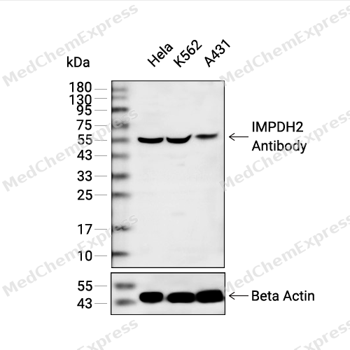 Western blot analysis of extracts from Hela(lane 2(20ug) , K562(lane 3(20ug) and A431(lane 4(20ug) using IMPDH2 Antibody (HY-P82346) Rabbit mAb. Proteins were transferred to a PVDF membrane and blocked with 5% non-fat milk in TBST for 2 hour at room temperature. The primary antibody (1/1000) and Loading control antibody (Beta Actin, HY-P80438, 1/10000) was used in 5% non-fat milk in TBST at 4°C overnight. Goat Anti-Mouse/Rabbit IgG-HRP Secondary Antibody (1/10000) was used for 1 hour at room temperature.
 -
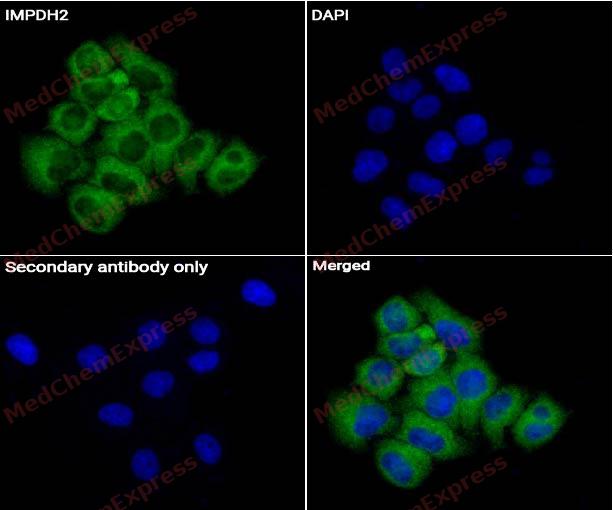 Immunocytochemistry analysis of hela cells labeling IMPDH2 Antibody (HY-P82346) at 1/50 dilution. Cells were fixed in 4% paraformaldehyde for 15 minutes at room temperature, permeabilized with 0.1% Triton X-100 for 10 minutes at room temperature, then blocked with QuickBlock™ Blocking Buffer for Immunol Staining for 10 min at room temperature. Cells were then incubated with IMPDH2 Antibody (HY-P82346) at 1/50 dilution in QuickBlock™ Blocking Buffer for Immunol Staining at 4 ℃. Alexa Fluor® 488-conjugated AffiniPure Goat Anti-Rabbit IgG H&L(HY-P8002, Green) was used as the secondary antibody at 1/1,000 dilution. PBS instead of the primary antibody was used as the secondary antibody only control. The Nuclear counterstain was DAPI (Blue).
 -
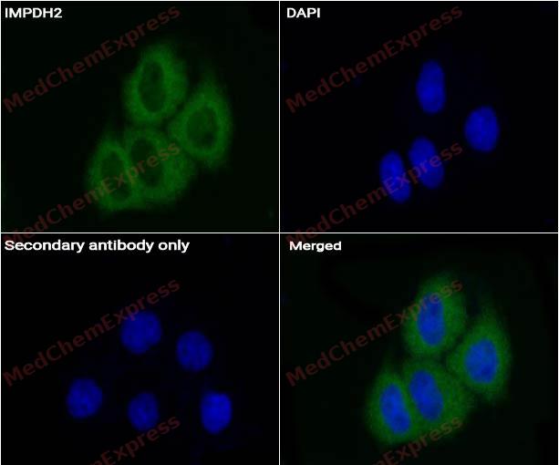 Immunocytochemistry analysis of hela cells labeling IMPDH2 Antibody (HY-P82346) at 1/200 dilution. Cells were fixed in 4% paraformaldehyde for 15 minutes at room temperature, permeabilized with 0.1% Triton X-100 for 10 minutes at room temperature, then blocked with QuickBlock™ Blocking Buffer for Immunol Staining for 10 min at room temperature. Cells were then incubated withMPDH2 Antibody (HY-P82346) at 1/50 dilution in QuickBlock™ Blocking Buffer for Immunol Staining at 4 ℃. Alexa Fluor® 488-conjugated AffiniPure Goat Anti-Rabbit IgG H&L(HY-P8002, Green) was used as the secondary antibody at 1/1,000 dilution. PBS instead of the primary antibody was used as the secondary antibody only control. The Nuclear counterstain was DAPI (Blue).
 -
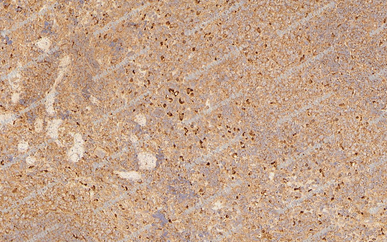 Immunohistochemical analysis of paraffin-embedded Rat spleen tissue using IMPDH2 Antibody (YA2091). The section was pre-treated using heat mediated antigen retrieval with Tris-EDTA buffer (pH 9.0) for 8 minutes. The tissues were blocked in QuickBlock for 20 minutes at room temperature, washed with ddH2O and PBS, and then probed with the primary antibody (HY-P82346, 1/100) in 4℃ overnight. The detection was performed using an HRP conjugated compact polymer system. DAB was used as the chromogen. Tissues were counterstained with hematoxylin and mounted with DPX.
 -
Immunohistochemical analysis of paraffin-embedded Rat spleen tissue using IMPDH2 Antibody (YA2091). The section was pre-treated using heat mediated antigen retrieval with Tris-EDTA buffer (pH 9.0) for 8 minutes. The tissues were blocked in QuickBlock for 20 minutes at room temperature, washed with ddH2O and PBS, and then probed with the primary antibody (HY-P82346, 1/100) in 4℃ overnight. The detection was performed using an HRP conjugated compact polymer system. DAB was used as the chromogen. Tissues were counterstained with hematoxylin and mounted with DPX.
-
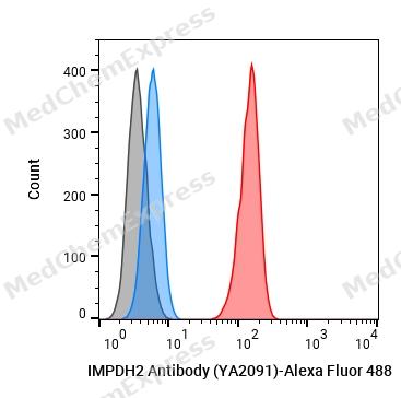 Flow cytometric analysis of 1X10^6 K562 cells labeling IMPDH2 Antibody (YA2091) (HY-P82346, red). Cells were fixed with 4% paraformaldehyde and permeabilised with 90% methanol. Then stained with the primary antibody at 1/50 dilution for an hour at 4℃. Alexa Fluor® 488-conjugated AffiniPure Goat Anti-Rabbit IgG H&L (HY-P8002) was used as the secondary antibody at 1/1,000 dilution for 30 minutes at 4℃. Rabbit IgG Isotype Control (HY-P80879, blue) was used as the isotype control, cells without incubation with primary antibody were used as the unlabeled control (black).


Background
-
Function
IMPDH2 (inosine-5'-monophosphate dehydrogenase 2) is a rate-limiting enzyme in de novo guanine nucleotide biosynthesis that catalyzes the NAD-dependent conversion of inosine monophosphate (IMP) to xanthosine monophosphate (XMP), thereby maintaining intracellular guanine nucleotide pools required for DNA synthesis, RNA synthesis, signal transduction, and cellular proliferation[1][2]. The enzyme occupies a central position in purine metabolism and directly regulates GTP availability, linking nucleotide biosynthesis to cell growth and metabolic demand[1][2]. Mechanistically, increased IMPDH2 expression is associated with proliferating cells and neoplastic tissues, where enhanced guanine nucleotide production supports tumor growth and cellular expansion[3][4]. In disease contexts, dysregulated IMPDH2 expression has been reported across multiple cancer models, and elevated IMPDH2 activity has been linked to malignant progression, therapy response, and metabolic reprogramming dependent on GTP synthesis[4][5]. Compared with the closely related isoform IMPDH1, which shares approximately 84% sequence identity and is generally considered a constitutively expressed housekeeping enzyme, IMPDH2 is preferentially upregulated during proliferation and tumorigenesis, making it the dominant isoform in many rapidly growing cells[3][6]. This isoform-specific regulation provides an important rationale for experimental targeting of IMPDH2 in cancer and immunology research[3][5]. For research applications, IMPDH inhibitors such as mycophenolic acid (MPA) suppress guanine nucleotide synthesis through direct enzyme inhibition and are widely used to investigate nucleotide metabolism, cell-cycle regulation, immune-cell activation, and IMPDH-dependent signaling pathways[7][8].
-
Subcellular Localization
Cytoplasm; Nucleus; Cytoplasm, cytosol
-
Expression
Tissue_specificity:IMPDH1 is the main species in normal leukocytes and IMPDH2 predominates over IMPDH1 in the tumor
Induction:Selectively up-regulated in neoplastic and replicating cells -
Subunit
Homotetramer (PubMed:7903306, Ref.28, Ref.29). Interacts with CLOCK; in a circadian manner (PubMed:28985504). Interacts with ANKRD9; leading to its ubiquitination and degradation by the proteasome (PubMed:30293565)
-
SwissProt ID
-
Synonyms
IMPD2; IMPDH 2; IMPDH II; Impdh2
-
Research Field
Signal Transduction


Documentation
-
Data Sheet (261 KB)
-
SDS (251 KB)
- English - EN (251 KB)
- Français - FR (251 KB)
- Deutsch - DE (251 KB)
- Norwegian - NO (251 KB)
- Español - ES (251 KB)
- Swedish - SV (251 KB)
- Italian - IT (251 KB)
- Korean - KR (251 KB)
- Portuguese - PT (251 KB)
-
User Guide for Antibodies (1077 KB)
References
[1]. Li Z, et al. IMPDH2's Central Role in Cellular Growth and Diseases: A Potential Therapeutic Target. Cell Prolif. 2025 Jun;58(6):e70031. [Content Brief]
[2]. Hedstrom L. IMP dehydrogenase: structure, et al. IMP dehydrogenase: structure, mechanism, and inhibition. Chem Rev. 2009 Jul;109(7):2903-28. [Content Brief]
[3]. Burrell AL. IMPDH dysregulation in disease: a mini review. Biochem Soc Trans. 2022 Feb 28;50(1):71-82. [Content Brief]
[4]. Kofuji S, et al. GTP metabolic reprogramming by IMPDH2: unlocking cancer cells' fuelling mechanism. J Biochem. 2020 Oct 1;168(4):319-328. [Content Brief]
[5]. IMPDH2 gene information from NCBI.
[6]. Jia X, et al. Inosine monophosphate dehydrogenase type1 sustains tumor growth in hepatocellular carcinoma. J Clin Lab Anal. 2022 May;36(5):e24416. [Content Brief]
[7]. Sintchak MD, et al. Structure and mechanism of inosine monophosphate dehydrogenase in complex with the immunosuppressant mycophenolic acid. Cell. 1996 Jun 14;85(6):921-30. [Content Brief]
[8]. Naffouje R, et al. Anti-Tumor Potential of IMP Dehydrogenase Inhibitors: A Century-Long Story. Cancers (Basel). 2019 Sep 11;11(9):1346. [Content Brief]

 Powered by Bioz
