



AZ12601011
Based on 4 publication(s) in Google Scholar
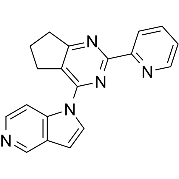
AZ12601011 is an orally active, selective TGFBR1 kinase inhibitor with an IC50 of 18 nM and a Kd of 2.9 nM. AZ12601011 inhibits phosphorylation of SMAD2 via selectively inhibiting ALK4, TGFBR1, and ALK7. AZ12601011 inhibits mammary tumor growth .
For research use only. We do not sell to patients.
- Purity : 99.78%
- CAS No.: 2748337-86-0
- Formula: C19H15N5
- Molecular Weight:313.36
-
Storage:Powder -20°C, 3 years , 4°C, 2 years ; In solvent -80°C, 2 years , -20°C, 1 year

To place orders, for customer services and technical support, please contact: MedChemExpress USA
Tel: 609-228-6898 E-mail: [email protected] [email protected]


-
 Biological Activity
-
Chemical Information
-
Solvent & Solubility
- Protocols
- Purity & Documentation
- References
-
 Help & FAQs


-

HY-10431
SB-431542TGF-β Receptor Kinase Inhibitor -
HY-P0299
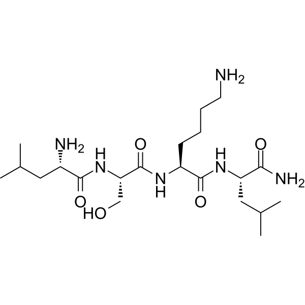
LSKL, Inhibitor of Thrombospondin (TSP-1)TGF-β1 Antagonist -
HY-12704
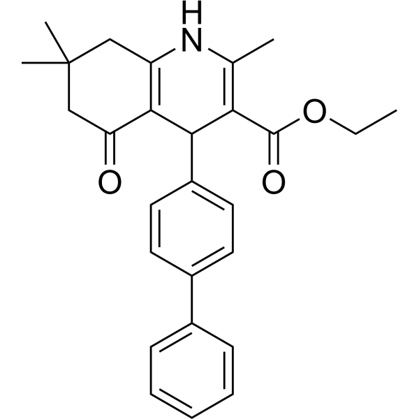
ITD-1TGF-β Receptor Inhibitor -
HY-151427
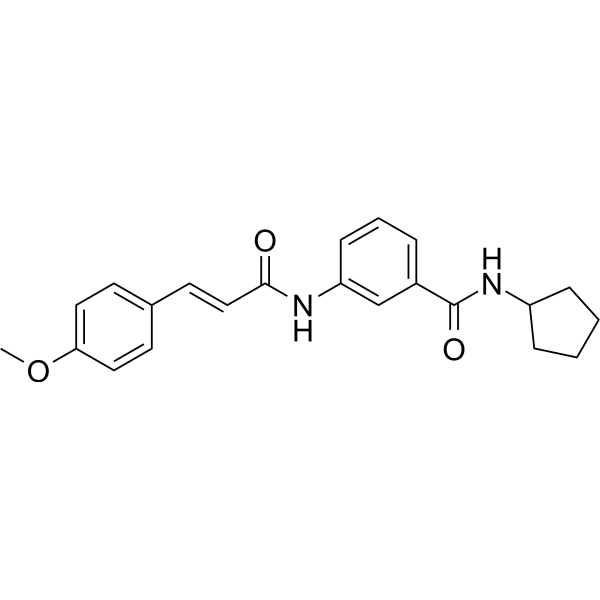
TGFβ1-IN-1TGFβ1 Inhibitor -
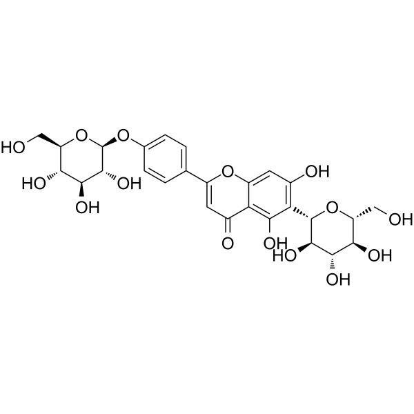
HY-N2589
IsosaponarinCollagen Synthesis Activator -
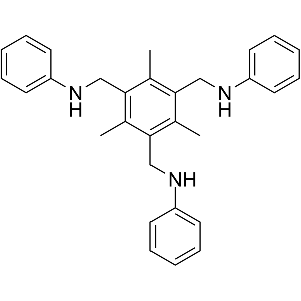
HY-146693
CJJ300TGF-β Inhibitor -
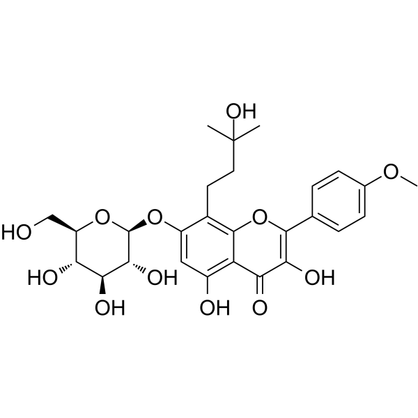
HY-N4019
Maohuoside A -
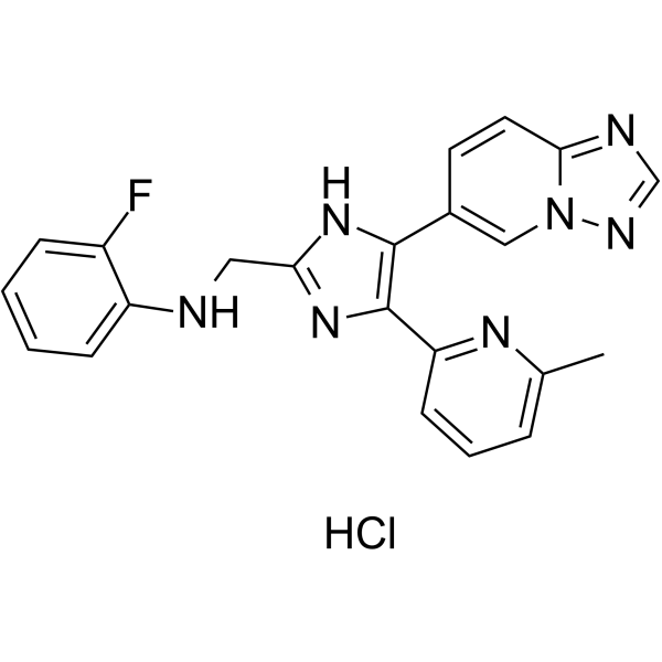
HY-19928A
Vactosertib HydrochlorideALK5 Inhibitor -
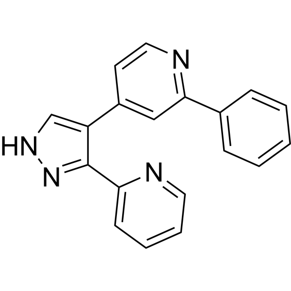
HY-10327
GW-6604ALK5 Inhibitor -
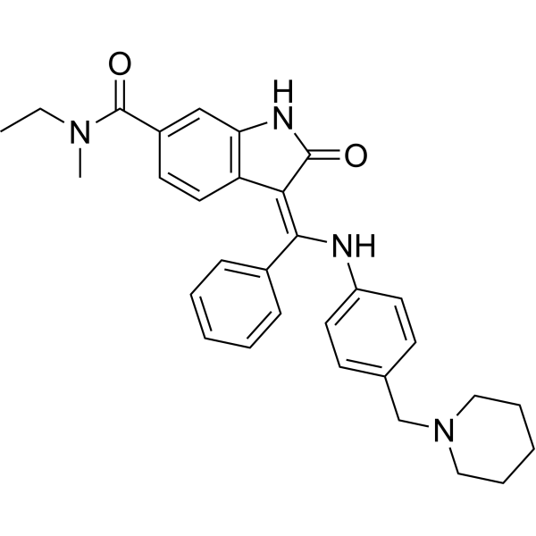
HY-13783
BIBF0775TGF-β Receptor Inhibitor -

HY-10327R
GW-6604 (Standard)Reference Standard
Publications Citing Use of MedChemExpress (MCE) AZ12601011
More

Biological Activity
Description
IC50 & Target
[1]|
ALK4 |
ALK7 |
In Vitro
AZ12601011 (0.01-10 μM; for 20 minutes) completely inhibits Phosphorylation of SMAD2 [1].
AZ12601011 (0.01 µM-10 µM) inhibits the activity of ALK4, ALK7 and TGFBR1 [1].
AZ12601011 inhibits 4T1 cells growth in vitro (IC50=0.4µM) [1].
MedChemExpress (MCE) has not independently confirmed the accuracy of these methods. They are for reference only.
-
Cell Line:NIH3T3, HaCaT, C2C12, T47D cells
-
Concentration:0.01, 0.03, 0.1, 0.3, 1, 3, 10 μM
-
Incubation Time:20 minutes
-
Result:Completely inhibited Phosphorylation of SMAD2.
In Vivo
MedChemExpress (MCE) has not independently confirmed the accuracy of these methods. They are for reference only.
-
Animal Model:Female BALB/c mice at greater than 18g with tumour[1]
-
Dosage:50mg/kg
-
Administration:Oral gavage; twice daily; for 25 days
-
Result:Inhibited tumour growth and metastasis in vivo.


Chemical Information
-
CAS No. 2748337-86-0
-
Appearance Solid
-
Molecular Weight 313.36
-
Formula C19H15N5
-
Color Off-white to light yellow
-
SMILES
C1(C2=NC=CC=C2)=NC(N3C=CC4=C3C=CN=C4)=C(CCC5)C5=N1
-
Shipping
Room temperature in continental US; may vary elsewhere.
-
Storage
Powder -20°C 3 years 4°C 2 years In solvent -80°C 2 years -20°C 1 year


Publications (4)
-
Journal Impact Factor
-
Most Recent
-
Adv Sci (Weinh)
Stabilization of TGF-β Receptor 1 by a Receptor-Associated Adaptor Dictates Feedback Activation of the TGF-β Signaling Pathway to Maintain Liver Cancer Stemness and Drug Resistance. [Abstract]2024 Sep;11(34):e2402327. PMID: 38981014 -
Cell Death Dis
TGF-β1 facilitates gallbladder carcinoma metastasis by regulating FOXA1 translation efficiency through m6A modification. [Abstract]2024 Jun 17;15(6):422. PMID: 38886389 -
Rheumatology (Oxford)
Asporin regulated by miR-26b-5p mediates chondrocyte senescence and exacerbates osteoarthritis progression via TGF-β1/Smad2 pathway. [Abstract]2022 May 30;61(6):2631-2643. PMID: 34559207 -
Biomed Pharmacother
Transforming growth factor-β receptor I kinase plays a crucial role in oligodendrocyte regeneration after demyelination. [Abstract]2025 Jun:187:118094. PMID: 40315672


Solvent & Solubility
In Vitro:
DMSO : 5 mg/mL (15.96 mM; Need ultrasonic; Hygroscopic DMSO has a significant impact on the solubility of product, please use newly opened DMSO)
Please refer to the solubility information to select the appropriate solvent. Once prepared, please aliquot and store the solution to prevent product inactivation from repeated freeze-thaw cycles.
Storage method and period of stock solution: -80°C, 2 years; -20°C, 1 year. When stored at -80°C, please use it within 2 years. When stored at -20°C, please use it within 1 year.
Please refer to the solubility information to select the appropriate solvent. Once prepared, please aliquot and store the solution to prevent product inactivation from repeated freeze-thaw cycles.
Storage method and period of stock solution: -80°C, 2 years; -20°C, 1 year. When stored at -80°C, please use it within 2 years. When stored at -20°C, please use it within 1 year.
Concentration (start) × Volume (start) = Concentration (final) × Volume (final)


Purity & Documentation
-
Data Sheet (280 KB)
-
SDS (251 KB)
- English - EN (251 KB)
- Français - FR (251 KB)
- Deutsch - DE (251 KB)
- Norwegian - NO (251 KB)
- Español - ES (251 KB)
- Swedish - SV (251 KB)
- Italian - IT (251 KB)
- Korean - KR (251 KB)
- Portuguese - PT (251 KB)
-
Handling Instructions (2659 KB)
References


Complete Stock Solution Preparation Table
Please refer to the solubility information to select the appropriate solvent. Once prepared, please aliquot and store the solution to prevent product inactivation from repeated freeze-thaw cycles.
Storage method and period of stock solution: -80°C, 2 years; -20°C, 1 year. When stored at -80°C, please use it within 2 years. When stored at -20°C, please use it within 1 year.
| Optional Solvent | Concentration Solvent Mass | 1 mg | 5 mg | 10 mg | 25 mg |
|---|---|---|---|---|---|
| DMSO | 1 mM | 3.1912 mL | 15.9561 mL | 31.9122 mL | 79.7804 mL |
| 5 mM | 0.6382 mL | 3.1912 mL | 6.3824 mL | 15.9561 mL | |
| 10 mM | 0.3191 mL | 1.5956 mL | 3.1912 mL | 7.9780 mL | |
| 15 mM | 0.2127 mL | 1.0637 mL | 2.1275 mL | 5.3187 mL |


AZ12601011 Related Classifications
HY-122856 Related Classifications
 Powered by Bioz