



BADAN
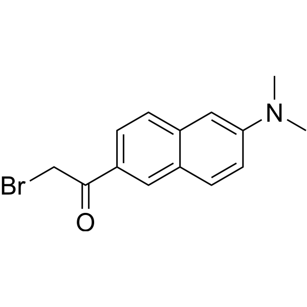
BADAN is a thiol-reactive, environment-sensitive fluorescent probe used for site-specific labeling of protein cysteine residues to detect protein topology, local polarity, protein-lipid interactions, and conformational changes. The bromoacetyl group of BADAN covalently binds to cysteine thiols, and its fluorescence originates from the intramolecular charge transfer (ICT) excited state. The emission peak position varies with the polarity of the surrounding medium: it blue-shifts to approximately 488 nm in nonpolar environments (such as the hydrophobic core of lipid bilayers) and red-shifts to approximately 496-507 nm in polar environments (such as aqueous phases), independent of enzymes, pH, or membrane potential. Ex = 380-402 nm; Em = 400-600 nm.
For research use only. We do not sell to patients.
- CAS No.: 210832-86-3
- Formula: C14H14BrNO
- Molecular Weight:292.17
-
Storage:
Please store the product under the recommended conditions in the Certificate of Analysis.

To place orders, for customer services and technical support, please contact: MedChemExpress USA
Tel: 609-228-6898 E-mail: [email protected] [email protected]


-
 Biological Activity
-
Chemical Information
- Purity & Documentation
- References
-
 Help & FAQs




Biological Activity
Guidelines (The following is a recommended experimental protocol for guidance only; adjust according to actual requirements)
1. Stock Solution Preparation
1.1 Solvent: 1:1 acetone:methanol.
1.2 Recommended Concentration: It is generally recommended to prepare a high-concentration stock solution of 1-10 mM.
2. Working Solution Preparation
2.1 Diluents: Optional options include:
2.1.1 100 mM Na-Hepes (pH 7.4) containing 10% glycerol, 0.5% CHAPS, 0.5 M KCl (for CYP3A4 labeling);
2.1.2 8 M guanidine hydrochloride, 100 mM Taps, 100 mM NaCl (pH 8.5) (for HlyA labeling).
2.2 Working Concentration: The protein concentration is 10 µM (CYP3A4) or 0.1 mM (HlyA); BADAN is added at a 1.2-fold molar excess (CYP3A4) or at least a 10-fold molar excess (HlyA).
2.3 Notes: Adjust the working solution concentration as needed; prepare fresh before use.
3. Staining Procedure
3.1 Description of Sample Types
3.1.1 Protein samples: Suitable for purified proteins containing cysteine residues.
3.2 Incubation Conditions
3.2.1 CYP3A4 labeling: 10 µM protein, 1.2-fold molar excess of BADAN, incubated at 25 °C under argon saturation with continuous stirring, protected from light for 20-30 min.
3.2.2 HlyA labeling: 0.1 mM protein, BADAN to a final concentration of 0.1 mM (≥10-fold molar excess), incubated at room temperature protected from light for 1 h.
3.3 Washing Steps
4. Control Setup
4.1 Unlabeled Protein Control: Use unlabeled wild-type protein at the same concentration to subtract the intrinsic fluorescence of the protein (e.g., tryptophan fluorescence) and background signals.
4.2 Positive Control: Use ligands or environmental changes known to induce changes in BADAN fluorescence as positive controls, such as the significant decrease in fluorescence intensity caused by the binding of α-naphthoflavone (ANF) to CYP3A4-BADAN (S50 = 18.2 µM); membrane association (e.g., liposomes or erythrocyte ghosts) causes a blue shift in the BADAN emission peak.
5. Detection and Analysis
5.1 Instrument Type: Fluorescence spectrophotometer
5.2.1 Excitation wavelength: 365-402 nm; time-resolved fluorescence uses 400 nm vertically polarized excitation (~200 µW).
5.3 Result Analysis
5.3.1 Changes in fluorescence intensity: BADAN fluorescence intensity is sensitive to the local environment. In CYP3A4, ANF binding causes a significant decrease in BADAN fluorescence: when BADAN is adjacent to tryptophan (Trp) or tyrosine (Tyr) residues, fluorescence quenching occurs via photoinduced electron transfer (PET), and the fluorescence decay follows tri-exponential kinetics (lifetimes of approximately 100 ps, 700-800 ps, and 3 ns).
5.3.2 Fluorescence localization: BADAN is labeled at specific sites on proteins, and its spectrum reflects the local environment of the labeled site. In membrane protein studies, BADAN located in the aqueous phase shows a red-shifted emission peak (~500 nm), while BADAN located in the hydrophobic core of the lipid bilayer shows a blue-shifted emission peak (~480 nm), which can be used to determine the topological structure and insertion depth of the protein in the membrane.
5.3.3 The position of the BADAN emission peak varies with medium polarity: In non-polar environments (inside the lipid bilayer), the emission peak is approximately 488 nm (blue shift); in polar environments (aqueous phase), the emission peak is approximately 496-507 nm (red shift). A blue shift of the emission peak indicates that the probe enters a hydrophobic environment (e.g., membrane insertion), while a red shift indicates that the probe is exposed to the aqueous phase.
MedChemExpress (MCE) has not independently confirmed the accuracy of these methods. They are for reference only.


Chemical Information
-
CAS No. 210832-86-3
-
Molecular Weight 292.17
-
Formula C14H14BrNO
-
SMILES
O=C(CBr)C1=CC=C2C=C(N(C)C)C=CC2=C1
-
Shipping
Room temperature in continental US; may vary elsewhere.
-
Storage
Please store the product under the recommended conditions in the Certificate of Analysis.


Purity & Documentation
References

Calculators
Concentration (start) × Volume (start) = Concentration (final) × Volume (final)


BADAN Related Classifications
HY-D1470 Related Classifications
 Powered by Bioz